Bioinspired Materials Science and Engineering
Edited by
Guang Yang, Lin Xiao, and Lallepak Lamboni
Huazhong University of Science and Technology, Wuhan, China
This edition first published 2018 © 2018 by John Wiley & Sons, Inc.
All rights reserved. No part of this publication may be reproduced, stored in a retrieval system, or transmitted, in any form or by any means, electronic, mechanical, photocopying, recording or otherwise, except as permitted by law. Advice on how to obtain permission to reuse material from this title is available at http://www.wiley.com/go/permissions.
The right of Guang Yang, Lin Xiao, and Lallepak Lamboni to be identified as the editors of this work has been asserted in accordance with law.
Registered Offices
John Wiley & Sons, Inc., 111 River Street, Hoboken, NJ 07030, USA
Editorial Office
111 River Street, Hoboken, NJ 07030, USA
For details of our global editorial offices, customer services, and more information about Wiley products visit us at www.wiley.com.
Wiley also publishes its books in a variety of electronic formats and by print‐on‐demand. Some content that appears in standard print versions of this book may not be available in other formats.
Limit of Liability/Disclaimer of Warranty
The publisher and the authors make no representations or warranties with respect to the accuracy or completeness of the contents of this work and specifically disclaim all warranties, including without limitation any implied warranties of fitness for a particular purpose. This work is sold with the understanding that the publisher is not engaged in rendering professional services. The advice and strategies contained herein may not be suitable for every situation. In view of ongoing research, equipment modifications, changes in governmental regulations, and the constant flow of information relating to the use of experimental reagents, equipment, and devices, the reader is urged to review and evaluate the information provided in the package insert or instructions for each chemical, piece of equipment, reagent, or device for, among other things, any changes in the instructions or indication of usage and for added warnings and precautions. The fact that an organization or website is referred to in this work as a citation and/or potential source of further information does not mean that the author or the publisher endorses the information the organization or website may provide or recommendations it may make. Further, readers should be aware that websites listed in this work may have changed or disappeared between when this works was written and when it is read. No warranty may be created or extended by any promotional statements for this work. Neither the publisher nor the author shall be liable for any damages arising herefrom.
Library of Congress Cataloging‐in‐Publication Data
Names: Yang, Guang, 1968 April 18– editor. | Xiao, Lin, 1986– editor. | Lamboni, L allepak, 1988– editor.
Title: Bioinspired materials science and engineering / edited by Guang Yang, Lin Xiao, L allepak Lamboni.
Description: First edition. | Hoboken, NJ : John Wiley & Sons, 2018. | Includes bibliographical references and index. |
Identifiers: LCCN 2018002997 (print) | LCCN 2018009868 (ebook) | ISBN 9781119390336 (pdf) | ISBN 9781119390343 (epub) | ISBN 9781119390329 (cloth)
Subjects: LCSH: Biomimetics. | Materials–Biotechnology. | Materials science. | Engineering.
Classification: LCC QP517.B56 (ebook) | LCC QP517.B56 B4796 2018 (print) | DDC 610.28–dc23
LC record available at https://lccn.loc.gov/2018002997
Cover Design: Wiley
Cover Images: © SergeOstroverhoff/Getty Images; © TonyBaggett/Getty Images; ©vitstudio/Shutterstock; © me4o/Getty Images; Shamrock logo courtesy of Guang Yang
Set in 10/12pt Warnock by SPi Global, Pondicherry, India
Printed in the United States of America
10 9 8 7 6 5 4 3 2 1
Contents
List of Contributors xiii
Foreword xvii
Preface xix
Introduction to Science and Engineering Principles for the Development of Bioinspired Materials 1 Muhammad Wajid Ullah, Zhijun Shi, Sehrish Manan, and Guang Yang
I.1 Bioinspiration 1
I.2 Bioinspired Materials 1
I.3 Biofabrication 2
I.3.1 Summary of Part I Biofabrication 2
I.4 Biofabrication Strategies 3
I.4.1 Conventional Biofabrication Strategies 3
I.4.2 Advanced Biofabrication Strategies 3
I.5 Part II Biomacromolecules 5
I.5.1 Summary of Part II Biomacromolecules 5
I.5.2 Carbohydrates 5
I.5.3 Proteins 8
I.5.4 Nucleic Acids 9
I.6 Part III Biomaterials 11
I.6.1 Summary of Part III Biomaterials 11
I.6.2 Features of Biomaterials 12
I.6.3 Current Advances in Biomaterials Science 13
I.7 Scope of the Book 13 Acknowledgments 14 References 14
Part I Biofabrication 17
1 Biotemplating Principles 19
Cordt Zollfrank and Daniel Van Opdenbosch
1.1 Introduction 19
1.2 Mineralization in Nature 20
1.2.1 Biomineralization 20
1.2.2 Geological Mineralization 21
1.3 Petrified Wood in Construction and Technology 23
1.4 Structural Description and Emulation 24
1.4.1 Antiquity 24
1.4.2 Modern Age: Advent of the Light Microscope 24
1.4.3 Aqueous Silicon Dioxide, Prime Mineralization Agent 25
1.4.4 Artificial Petrifaction of Wood 25
1.5 Characteristic Parameters 28
1.5.1 Hierarchical Structuring 28
1.5.2 Specific Surface Areas 32
1.5.3 Pore Structures 32
1.6 Applications 34
1.6.1 Mechanoceramics 34
1.6.2 Nanoparticle Substrates 35
1.6.3 Filter and Burner Assemblies 35
1.6.4 Photovoltaic and Sensing Materials 36
1.6.5 Wettability Control 37
1.6.6 Image Plates 38
1.7 Limitations and Challenges 38
1.7.1 Particle Growth 38
1.7.2 Comparison with Alternating Processing Principles 40
1.7.3 Availability 40
1.8 Conclusion and Future Topics 42
Acknowledgments 42
Notes 42
References 43
2 Tubular Tissue Engineering Based on Microfluidics 53
Lixue Tang, Wenfu Zheng, and Xingyu Jiang
2.1 Introduction 53
2.2 Natural Tubular Structures 53
2.2.1 Blood Vessels 53
2.2.2 Lymphatic Vessels 53
2.2.3 Vessels in the Digestive System 54
2.2.4 Vessels in the Respiratory System 54
2.2.5 The Features of the Natural Tubular Structures 54
2.3 Microfluidics 54
2.3.1 An Introduction to Microfluidics 54
2.3.2 Microf luidics to Manipulate Cells 55
2.4 Fabrication of Tubular Structures by Microfluidics 58
2.4.1 Angiogenesis 58
2.4.2 Tissue Engineering of Natural Tubes 58
2.4.3 Tissue Engineering of Other Tubular Structures 62
2.5 Conclusion 64
Acknowledgments 64 References 64
3 Construction of Three‐Dimensional Tissues with Capillary Networks by Coating of Nanometer‐ or Micrometer‐Sized Film on Cell Surfaces 67
Michiya Matsusaki, Akihiro Nishiguchi, Chun‐Yen Liu, and Mitsuru Akashi
3.1 Introduction 67
3.2 Fabrication of Nanometer‐ and Micrometer‐Sized ECM Layers on Cell Surfaces 68
3.2.1 Control of Cell Surface by FN Nanofilms 68
3.2.2 Control of Cell Surface by Collagen Microfilms 72
3.3 3D‐Tissue with Various Thicknesses and Cell Densities 75
3.4 Fabrication of Vascularized 3D‐Tissues and Their Applications 77
3.5 Conclusion 80
Acknowledgments 80 References 80
4 Three‐dimensional Biofabrication on Nematic Ordered Cellulose Templates 83
Tetsuo Kondo
4.1 Introduction 83
4.2 What Is Nematic Ordered Cellulose (NOC)? 84
4.2.1 Nematic Ordered Cellulose 84
4.2.2 Various Nematic Ordered Templates and Modified Nematic Ordered Cellulose 87
4.3 Exclusive Surface Properties of NOC and Its Unique Applications 89
4.3.1 Bio‐Directed Epitaxial Nano‐Deposition on Molecular Tracks of the NOC Template 89
4.3.2 Critical Factors in Bio‐Directed Epitaxial Nano‐Deposition on Molecular Tracks 90
4.3.3 Regulated Patterns of Bacterial Movements Based on Their Secreted Cellulose Nanofibers Interacting Interfacially with Ordered Chitin and Honeycomb Cellulose Templates 93
4.3.4 NOC Templates Mediating Order‐Patterned Deposition Accompanied by Synthesis of Calcium Phosphates as Biomimic Mineralization 97
4.3.5 Three‐Dimensional Culture of Epidermal Cells on NOC Scaffolds 98
4.4 Conclusion 100 References 101
5 Preparation and Application of Biomimetic Materials Inspired by Mussel Adhesive Proteins 103 Heng Shen, Zhenchao Qian, Ning Zhao, and Jian Xu
5.1 Introduction 103
5.2 Various Research Studies 104
5.3 Conclusion 116 References 116
6 Self‐assembly of Polylactic Acid‐based Amphiphilic Block Copolymers and Their Application in the Biomedical Field 119
Lin Xiao, Lixia Huang, Li Liu, and Guang Yang
6.1 Introduction 119
6.2 Micellar Structures from PLA‐based Amphiphilic Block Copolymers 119
6.2.1 Preparation and Mechanism of Micellar Structures 120
6.2.2 Stability and Stimuli‐Responsive Properties: Molecular Design and Biomedical Applications 122
6.3 Hydrogels from PLA‐based Amphiphilic Block Copolymers 125
6.3.1 Mechanism of Hydrogel Formation from PLA‐based Amphiphilic Block Copolymers 125
6.3.2 Properties and Biomedical Applications of Hydrogel from PLA‐based Amphiphilic Block Copolymers 126
6.4 Conclusion 127
Acknowledgments 127
References 127
Part II Biomacromolecules 131
7 Electroconductive Bioscaffolds for 2D and 3D Cell Culture 133
Zhijun Shi, Lin Mao, Muhammad Wajid Ullah, Sixiang Li, Li Wang, Sanming Hu, and Guang Yang
7.1 Introduction 133
7.2 Electrical Stimulation 133
7.3 Electroconductive Bioscaffolds 135
7.3.1 Conductive Polymers‐based Electroconductive Bioscaffolds 135
7.3.2 Carbon Nanotubes‐based Electroconductive Bioscaffolds 137
7.3.3 Graphene‐based Electroconductive Bioscaffolds 140
7.4 Conclusion 145 Acknowledgments 145
References 145
8 Starch and Plant Storage Polysaccharides 149
Francisco Vilaplana, Wei Zou, and Robert G. Gilbert
8.1 Starch and Other Seed Polysaccharides: Availability, Molecular Structure, and Heterogeneity 149
8.1.1 Molecular Structure and Composition of Seeds and Cereal Grains 149
8.1.2 Starch Hierarchical Structure from Bonds to the Granule 149
8.1.3 Crystalline Structure 149
8.1.4 Granular Structure 150
8.1.5 Mannans, Galactomannans, and Glucomannans 150
8.1.6 Xyloglucans 151
8.1.7 Xylans . Arabinoxylans, Glucuronoxylans, and Glucuronoarabinoxylans 153
8.2 Effect of the Molecular Structure of Starch and Seed Polysaccharides on the Macroscopic Properties of Derived Carbohydrate‐based Materials 154
8.2.1 Factors Affecting Starch Digestibility 154
8.2.2 Structural Aspects of Seed Polysaccharides Affecting Configuration and Macroscopic Properties 158
8.3 Chemo‐enzymatic Modification Routes for Starch and Seed Polysaccharides 160
8.4 Conclusion 161 References 162
9 Conformational Properties of Polysaccharide Derivatives 167
Ken Terao and Takahiro Sato
9.1 Introduction 167
9.2 Theoretical Backbone to Determine the Chain Conformation of Linear and Cyclic Polymers from Dilute Solution Properties 169
9.3 Chain Conformation of Linear Polysaccharides Carbamate Derivatives in Dilute Solution 171
9.3.1 Effects of the Main Chain Linkage of the Polysaccharides Phenylcarbamate Derivatives 171
9.3.2 Effects of Hydrogen Bonds to Stabilize the Helical Structure 172
9.3.3 Enantiomeric Composition Dependent Chain Dimensions: ATBC and ATEC in d‐, dl‐, l-ethyl lactates 175
9.3.4 Solvent‐Dependent Helical Structure and the Chain Stiffness of Amylose Phenylcarbamates in Polar Solvents 176
9.4 Lyotropic Liquid Crystallinity of Polysaccharide Carbamate Derivatives 177
9.5 Cyclic Amylose Carbamate Derivatives: An Application to Rigid Cyclic Polymers 178
9.6 Conclusion 180 Appendix: Wormlike Chain Parameters for Polysaccharide Carbamate Derivatives 181 References 182
10 Silk Proteins: A Natural Resource for Biomaterials 185
Lallepak Lamboni, Tiatou Souho, Amarachi Rosemary Osi, and Guang Yang
10.1 Introduction 185
10.2 Bio‐synthesis of Silk Proteins 186
10.2.1 Silkworm Silk Glands 186
10.2.2 Regulation of Silk Proteins Synthesis 186
10.2.3 Synthesis of Fibroin 187
10.2.4 Synthesis of Sericin 187
10.2.5 Silk Filament Assembly 187
10.3 Extraction of Silk Proteins 188
10.3.1 Silk Degumming 188
10.3.2 Fibroin Regeneration 188
10.3.3 Sericin Recovery 189
10.4 Structure and Physical Properties of Silk Proteins 189
10.4.1 Silk Fibroin 189
10.4.2 Silk Sericin 189
10.5 Properties of Silk Proteins in Biomedical Applications 190
10.5.1 Silk Fibroin 190
10.5.2 Biomedical Uses of Silk Sericin 190
10.6 Processing Silk Fibroin for the Preparation of Biomaterials 192
10.6.1 Fabrication of 3D Matrices 193
10.6.2 Fabrication of SF‐based Films 193
10.6.3 Preparation of SF‐based Particulate Materials 194
10.7 Processing Silk Sericin for Biomaterials Applications 194
10.8 Conclusion 194
Acknowledgments 195
Abbre viations 195
References 195
11 Polypeptides Synthesized by Ring‐opening Polymerization of N‐Carboxyanhydrides: Preparation, Assembly, and Applications 201
Yuan Yao, Yongfeng Zhou, and Deyue Yan
11.1 Introduction 201
11.2 Living Polymerization of NCAs 201
11.2.1 Transition Metal Complexes 201
11.2.2 Active Initiators Based on Amines 203
11.2.3 Recent Advances in Living NCA ROP Polymerization, 2013‐2016 204
11.3 Synthesis of Traditional Copolypeptides and Hybrids 204
11.3.1 Random Copolypeptides 205
11.3.2 Hybrid Block Polypeptides 205
11.3.3 Block Copolypeptides 206
11.3.4 Non‐linear Polypeptides and Copolypeptides 206
11.4 New Monomers and Side‐Chain Functionalized Polypeptides 208
11.4.1 New NC A Monomers 208
11.4.2 Glycopolypeptides 208
11.4.3 Water‐soluble Polypeptides with Stable Helical Conformation 209
11.4.4 Stimuli‐responsive Polypeptides 210
11.5 The Self‐assembly of Polypeptides 212
11.5.1 Chiral Self‐assembly 212
11.5.2 Self‐assembly with Inorganic Sources 213
11.5.3 Microphase Separation of Polypeptides 214
11.5.4 Self‐assembly in Solution 214
11.5.5 Polypeptide Gels 215
11.6 Novel Bio‐related Applications of Polypeptides 216
11.6.1 Drug Delivery 216
11.6.2 Gene Delivery 216
11.6.3 Membrane Active and Antimicrobial Polypeptides 217
11.6.4 Tissue Engineering 217
11.7 Conclusion 219
References 219
12 Preparation of Gradient Polymeric Structures and Their Biological Applications 225
Tao Du, Feng Zhou, and Shutao Wang
12.1 Introduction 225
12.2 Gradient Polymeric Structures 225
12.2.1 Gradient Hydrogels 225
12.2.2 Gradient Polymer Brushes 230
12.3 Gradient Polymeric Structures Regulated Cell Behavior 241
12.3.1 Gradient Cell Adhesion 241
12.3.2 Cell Migration 244
12.4 Conclusion 247
References 247
Part III Biomaterials 251
13 Bioinspired Materials and Structures: A Case Study Based on Selected Examples 253
Tom Masselter, Georg Bold, Marc Thielen, Olga Speck, and Thomas Speck
13.1 Introduction 253
13.2 Fiber‐reinforced Structures Inspired by Unbranched and Branched Plant Stems 253
13.2.1 Technical Plant Stem 254
13.2.2 Branched Fiber‐reinforced Structures 254
13.3 Pomelo Peel as Inspiration for Biomimetic Impact Protectors 255
13.3.1 Hierarchical Structuring and its Influence on the Mechanical Properties 256
13.3.2 Functional Principles for Biomimetic Impact Protectors 258
13.4 Self‐repair in Technical Materials Inspired by Plants’ Solutions 258
13.4.1 Plant Latex: Self‐Sealing, Self‐Healing and More 258
13.4.2 Wound Sealing in the Dutchmen’s Pipe: Concept Generator for Self‐Sealing Pneumatic Systems 259
13.5 Elastic Architecture: Lessons Learnt from Plant Movements 261
13.5.1 Plant Movements: A Treasure Trove for Basic and Applied Research 261
13.5.2 Flectofin®: a Biomimetic Façade‐Shading System Inspired by the Deformation Principle of the “Perch” of the Bird of Paradise Flower 262
13.6 Conclusions 264
Acknowledgments 264
References 264
14 Thermal‐ and Photo‐deformable Liquid Crystal Polymers and Bioinspired Movements 267
Yuyun Liu, Jiu‐an Lv, and Yanlei Yu
14.1 Introduction 267
14.2 Thermal‐responsive CLCPs 267
14.2.1 Thermal‐responsive Deformation of CLCPs 267
14.2.2 Bioinspired Thermal‐responsive Nanostructure CLCP Surfaces 271
14.3 Photothermal‐responsive CLCPs 276
14.4 Light‐responsive CLCPs 278
14.4.1 Light‐responsive Deformation of CLCPs 278
14.4.2 Bioinspired Soft Actuators 282
14.4.3 Bioinspired Light‐responsive Microstructured CLCP Surfaces 285
14.4 Conclusion 290
References 291
15 Tuning Mechanical Properties of Protein Hydrogels: Inspirations from Nature and Lessons from Synthetic Polymers 295
Xiao‐Wei Wang, Dong Liu, Guang‐Zhong Yin, and Wen‐Bin Zhang
15.1 Introduction 295
15.2 What Are Different about Proteins? 296
15.2.1 Protein Structure and Function 296
15.2.2 Protein Synthesis 297
15.3 Protein Cross‐linking 298
15.3.1 Chemical Cross‐linking of Proteins 298
15.3.2 Physical Cross‐linking of Proteins 299
15.4 Strategies for Mechanical Reinforcement 300
15.4.1 Lessons from Synthetic Polymers 302
15.4.2 Inspirations from Nature 305
15.5 Conclusion 306
References 307
16 Dendritic Polymer Micelles for Drug Delivery 311
Mosa Alsehli and Mario Gauthier
16.1 Introduction 311
16.2 Dendrimers 312
16.2.1 Dendrimer Synthesis: Divergent and Convergent Methods 312
16.3 Hyperbranched Polymers 319
16.4 Dendrigraft Polymers 323
16.4.1 Divergent Grafting Onto Strategy 323
16.4.2 Divergent Grafting from Strategy 328
16.4.3 Convergent Grafting Through Strategy 332
16.5 Conclusion 333
References 334
17 Bone‐inspired Biomaterials 337 Frank A. Müller
17.1 Introduction 337
17.2 Bone 337
17.3 Bone‐like Materials 340
17.3.1 Biomimetic Apatite 340
17.3.2 Bone‐inspired Hybrids 343
17.4 Bone‐like Scaffolds 344
17.4.1 Additive Manufacturing 344
17.4.2 Ice Templating 346
17.5 Conclusion 349
References 349
18 Research Progress in Biomimetic Materials for Human Dental Caries Restoration 351 Yazi Wang, Fengwei Liu, Eric Habib, Ruili Wang, Xiaoze Jiang, X.X. Zhu, and Meifang Zhu
18.1 Introduction 351
18.2 Tooth Structure 351
18.3 The Formation Mechanism of Dental Caries 352
18.4 HA‐filled Biomimetic Resin Composites 352
18.4.1 Particulate HA as Filler in Dental Restorative Resin Composites 352
18.4.2 Novel Shapes of HA as Fillers in Dental Restorative Resin Composites 354
18.4.3 Challenges and Future Developments 355
18.5 Biomimetic Synthesis of Enamel Microstructure 356
18.5.1 Amelogenins‐containing Systems 356
18.5.2 Peptides‐containing Systems 357
18.5.3 Biopolymer G el Systems 359
18.5.4 Dendrimers‐containing Systems 360
18.5.5 Surfactants/Chelators‐containing Systems 360
18.5.6 Challenges and Future Developments 360 Acknowledgments 362
References 362
Index 365
List of Contributors
Mitsuru Akashi Osaka University Osaka, Japan
Mosa Alsehli Taibah University Madina, Saudi Arabia
Georg Bold Freiburg Institute for Interactive Materials and Bioinspired Technologies Freiburg, Germany
Tao Du Lanzhou Institute of Chemical Physics Lanzhou, China
Mario Gauthier Waterloo University Ontario, Canada
Robert G. Gilbert Queensland University Brisbane, Australia
Eric Habib Université de Montréal Québec, Canada
Sanming Hu Huazhong University of Science and Technology Wuhan, China
Lixia Huang
Huazhong University of Science and Technology Wuhan, China
Xiaoze Jiang Donghua University Shanghai, China
Xingyu Jiang National Center for NanoScience and Technology Beijing, China
Tetsuo Kondo Kyushu University Fukuoka, Japan
Lallepak Lamboni Huazhong University of Science and Technology Wuhan, China
Sixiang Li Huazhong University of Science and Technology Wuhan, China
Chun‐Yen Liu Osaka University Osaka, Japan
Dong Liu Peking University Beijing, China
Fengwei Liu Donghua University Shanghai, China
Li Liu
Huazhong University of Science and Technology Wuhan, China
Yuyun Liu Fudan University Shanghai, China
Jiu‐an Lv Fudan University Shanghai, China
Sehrish Manan
Huazhong Agricultural University Wuhan, China
Lin Mao
Huazhong University of Science and Technology Wuhan, China
Tom Masselter University of Freiburg Freiburg, Germany
Michiya Matsusaki Osaka University Osaka, Japan
Frank A. Müller
Friedrich Schiller University Jena Jena, Germany
Akihiro Nishiguchi Osaka University Osaka, Japan
Daniel Van Opdenbosch Technische Universität München Munchen, Germany
Zhenchao Qian
Beijing National Laboratory for Molecular Sciences Beijing, China
Amarachi Rosemary Osi Ningbo Institute of Material Technology and Engineering Ningbo, China
Takahiro Sato Osaka University Osaka, Japan
Heng Shen
Beijing National Laboratory for Molecular Sciences Beijing, China
Zhijun Shi Huazhong University of Science and Technology Wuhan, China
Tiatou Souho University of Kara Kara, Togo Huazhong University of Science and Technology Wuhan, China
Olga Speck University of Freiburg Freiburg, Germany
Thomas Speck University of Freiburg Freiburg, Germany
Lixue Tang National Center for NanoScience and Technology Beijing, China
Ken Terao Osaka University Osaka, Japan
Marc Thielen Freiburg Materials Research Centre Freiburg, Germany
Francisco Vilaplana KTH Royal Institute of Technology Stockholm, Sweden
Muhammad Wajid Ullah Huazhong University of Science and Technology Wuhan, China
Li Wang
Huazhong University of Science and Technology Wuhan, China
Ruili Wang Donghua University Shanghai, China
Shutao Wang
Technical Institute of Physics and Chemistry Beijing, China
Xiao‐Wei Wang Peking University Beijing, China
Yazi Wang Donghua University Shanghai, China
Lin Xiao
Huazhong University of Science and Technology Wuhan, China
Jian Xu
Beijing National Laboratory for Molecular Sciences Beijing, China
Deyue Yan Shanghai Jiaotong University Shanghai, China
Guang Yang Huazhong University of Science and Technology Wuhan, China
Yuan Yao East China University of Science and Technology Shanghai, China
Guang‐Zhong Yin Peking University Beijing, China
Yanlei Yu Fudan University Shanghai, China
Ning Zhao
Beijing National Laboratory for Molecular Sciences Beijing, China
Wen‐Bin Zhang Peking University Beijing, China
Wenfu Zheng National Center for NanoScience and Technology Beijing, China
Feng Zhou Lanzhou Institute of Chemical Physics Lanzhou, China
Yongfeng Zhou Shanghai Jiaotong University Shanghai, China
Meifang Zhu Donghua University Shanghai, China
X.X. Zhu Université de Montréal Québec, Canada
Cordt Zollfrank Technische Universität München Munchen, Germany
Wei Zou Yangzhou University Yangzhou, China
Foreword
Centered on materials, this book explores the full scope of products inspired by nature. The process of learning from biological structures and principles for the development of advanced and multifunctional materials as novel resources that revolutionize human life is discussed, presenting fundamental concepts and methods of biofabrication. Examples are offered that showcase currently trending compounds and macromolecules with their properties, their potential, and their contribution to the fabrication of bioinspired materials. Concrete applications are discussed as well with an accent on biomedically engineered materials, that will take the reader into the realm of such seductive biomaterials.
With currently captivating topics such as biotemplating, microfluidics, self‐assembly, mussel‐inspired surface modification, 3D biofabrication and more, this book represents a source of inspiration for the design of novel materials, and an important tool for updating active researchers. Additionally, its comprehensive approach will be of great interest to the beginner in the field who will discover the concept of bioin spiration fr om its fundamentals to its applications. Although the book emphasizes biomedical engineer ing,
the multidisciplinary aspect of the subject will make it appeal to many research areas, such as biologists and engineers, while not leaving out chemists, physicists, and technicians.
Although an old concept, by proposing natural materials with superior features and low cost as models, bioinspiration has re‐emerged as an essential tool for overcoming various limitations in current materials science and engineering, thereby solving many of mankind’s substantial problems, such as the shortage of resources and the environmental concerns. Hence, this book deals with an important topic of the moment, which concerns numerous researchers across the world and should also be of interest to the general public. As illustrated by the authors, many different talents need to come together to make this approach a reality, and this book will inspire, instruct, and involve both current and the next generations in advancing the field.
April 13, 2017
Lei Jiang Technical Institute of Physics and Chemistry Chinese Academy of Sciences, China
Preface
Bioinspiration is an old concept which can be described as the process of learning from nature and its biological principles. Taking advantage of the properties and nanostructures of natural compounds, the science of bioinspired materials aims at developing new and formerly non‐existent materials, which exhibit novel and multifunctional properties, in the attempt of meeting the current requirements of human well‐being. The idea is to take inspiration from natural mechanisms and the problems they are set to solve, in order to design advanced materials which are solutions to problems encountered in human life. Indeed, the focus of materials science is being increasingly shifted towards the development of bioinspired materials, prompted by the shortage of resources, the low cost and superior characteristics of natural materials, and the environmental and climatic concerns. The first step to engineering bioinspired materials is understanding biological materials and the processes involved in their production, and thence, develop biofabrication or bioinspired fabrication approaches. This leads to the highly interdisciplinary character of this field, which brings together natural scientists (biologists, chemists, and physicists), engineers, and technicians. Thus, an active interaction across disciplines is the key to the real development of this old research area, which is now attracting many researchers worldwide. However, as underlined by several recent reviews on the subject, this condition is yet to be fully met, due to the rather limited understanding of the building principles of living entities which are numerous and complex, and because the definition of the scope and novel applications remain to be further clarified. Hence, approaches for conveying information in the field and storing the bioinspired solutions already uncovered are of real importance, and would contribute significantly in propelling this promising research area.
Biofabrication approaches are developed by studying and exploiting unique and basic biological aspects, including evolution, growth, and structure (formation and performance), which are non‐existent in engineering materials. Based on the “growth and functional
adaptation” concepts, the strategies adopted aim at creating hierarchical structures and self‐assemblies (dynamic strategies), while those associated with the “damage repair and healing” principles design self‐repair or self‐healing materials.
The purpose of this book is to introduce a comprehensive view of the bioinspired materials science and engineering, discussing biofabrication approaches and applications of bioinspired materials as they are fed back to nature in the guise of biomaterials. Some biological compounds will also be brought up, as of what is learned from them, and how they can be useful in the engineering of bioinspired materials. Thus, this book will include 3 main sections: biofabrication, biomacromolecules, and biomaterials. Illustrating the bioinspiration process from materials design and conception to application of bioinspired materials, this book will present the multidisciplinary aspect of the concept, and represent a typical example of how knowledge is acquired from nature, and how in turn this information contributes to biological sciences, with an accent on biomedical applications. We anticipate that this book will be suitable for different classes of the scientific community including undergraduate, graduate, and senior researchers in all areas of bioinspired materials. We hope that it will stimulate new thoughts and research in this field.
We would like to acknowledge the National Natural Science Foundation of China for the financial support on this book. Then we would like to express our appreciation to China‐Germany Center for Science, which supported the Sino‐German Sympoisum on Bioinspired Materials and Engineering held from May 11‐15, 2014 in Wuhan, China, co‐chaired by Prof. Guang Yang (HUST) and Prof. Cordt Zollfrank (TUM). This memorable symposium brought together outstanding scientists working in the bioinspired material field from China, Germany, Japan and the rest world. It is this symposium that first inspired the conception of this book. Many of the symposium attendees then accepted our invitation to contribute to this book. We offer special thanks to them. We also would like to express our appreciation to all
contributing authors and to staff at John Wiley & Sons, Inc., for their patience and never‐failing support of this project. Finally, we would like to express our sincere gratitude to Prof. Lina Zhang at Wuhan University, Prof. Lei Jiang at Technical Institute of Physics and Chemistry
CAS, and Prof. Deyue Yan at Shanghai Jiaotong University for their on‐going support and guidance.
Guang Yang, Lin Xiao, Lallepak Lamboni February 2018
Introduction
to Science and Engineering Principles for the Development of Bioinspired Materials
Muhammad Wajid Ullah1,2, Zhijun Shi1,2, Sehrish Manan3, and Guang Yang1,2,*
1 College of Life Science and Technology, Huazhong University of Science and Technology, Wuhan, China
2 National Engineering Research Center for Nano‐Medicine, Huazhong University of Science and Technology, Wuhan, China
3 College of Plant Sciences and Technology, Huazhong Agricultural University, Wuhan, China
I.1 Bioinspiration
Bioinspiration refers to the process of learning from nature and its biological principles. The science of bioinspired materials aims to develop novel functional materials with advanced and multi‐functional properties by using the nano‐, micro‐, meso‐, and macro‐structures and features of natural materials with the aim to meet the requirements of human well‐being. Natural mechanisms and biological materials can be exploited to design advanced materials to solve the problems encountered in human life. Indeed, the focus of materials science is being increasingly shifted toward the development of bioinspired materials, prompted by the shortage of resources, the low cost, and the superior characteristics of natural materials, and the environmental and climatic concerns. To this end, understanding the biological phenomenon, natural biological materials, and the processes involved in their natural production is essential, and hence, developing biofabrication or bioinspired fabrication approaches.
I.2 Bioinspired Materials
Bioinspired materials are synthetic products fabricated to mimic the structure and mechanical features of natural biological materials [1]. Biological materials are inherently multi‐functional in nature but may have evolved to optimize a principal mechanical function such as the impact of fracture resistance, for armor and protection, for sharp and cutting components, for a light weight for flight, or special chemical and mechanical extremities for reversible adhesive purposes. These functions are regulated by the nano‐, micro‐, meso‐, and macro‐structures of the materials. Further, these structures determine the
* Email: yang_sunny@yahoo.com
mechanism of the biological systems to adapt themselves to the external mechanical stimuli. These inherent functions and structural properties are inspiring scientists and engineers to design novel multi‐functional synthetic materials with a wide range of structural features and a broad spectrum of potential applications. In the past few decades, several natural biological materials have been examined by researchers for various aspects to explore their potential in different fields. Studies reveal that the inherent multi‐scale structures of natural biological materials possess several functions. Nature as a school for scientists and engineers has served as a great source of inspiration to fabricate new materials [2]. At present, biomimetic and bioinspired approaches have been adopted for the fabrication of several biological materials with multi‐scale structures for function integration, as summarized in Table I.1. An interdisciplinary collaboration of materials science and engineering, chemistry, biology, physics, and bioinformatics, etc. may lead to the design and fabrication of novel multi‐functional bioinspired materials.
To date, several biofabrication approaches have been developed by studying and exploiting unique and basic biological aspects, including evolution, growth, and structure (formation and performance) which are not found in engineering materials. Based on the “growth and functional adaptation” concepts, the strategies adopted mainly aim at creating hierarchical structures and self‐assemblies (dynamic strategies) and those associated with the “damage repair and healing” principle designs, and self‐repair or self‐healing materials. To achieve these objectives, several models have been presented by the researchers to describe the design, fabrication, and optimization of properties of bioinspired materials. Modeling of biological materials helps in rational understanding of
Bioinspired Materials Science and Engineering, First Edition. Edited by Guang Yang, Lin Xiao, and Lallepak Lamboni.
© 2018 John Wiley & Sons, Inc. Published 2018 by John Wiley & Sons, Inc.
Table I.1 Typical biological materials with function integration.
Biological materialsFunctions Ref.
Butterfly wing Superhydrophobicity, directional adhesion, structural color, self‐cleaning, chemical sensing capability, fluorescence emission functions [3–7]
BrittlestarMechanical and optical functions[8]
Cicada wingAnti‐reflection, superhydrophobicity [9]
Fish scaleDrag reduction, superoleophilicity in air, superoleophobicity in water [10]
Gecko foot Reversible adhesive, superhydrophobicity, self‐cleaning [11]
Lotus leafSuperhydrophobicity, low adhesion, self‐cleaning [12]
Mosquito compound eye Superhydrophobicity, anti‐reflection, anti‐fogging [13]
Nacre Mechanical property, structural color [14, 15]
Peacock feather Structural color, superhydrophobicity [16]
Polar bear furOptical property, thermal insulation[17]
Rice leafSuperhydrophobicity, anisotropic wettability [12]
Rose petalSuperhydrophobicity, structural color, high adhesion [18–20]
Shark skinDrag reduction, anti‐biofouling[21]
SpiculeMechanical and fiber‐optical properties [22–24]
Spider capture silk Water collection ability, mechanical property, elasticity, stickiness [25–27]
Spider dragline silk Mechanical property, supercontraction, torsional shape memory [28–35]
Water strider leg Durable and robust superhydrophobicity [36]
Source: Reproduced from [2] with permission from Elsevier.
the design principles which can lead to subsequent designing of bioinspired complements. For example, mechanical modeling of biological materials based on natural materials has attracted immense attention owing to their diverse applications in medicine and engineering. This can be attributed to the structurally hierarchical biomaterials which possess a highly desirable structure‐properties relationship and can serve as templates for the fabrication of bioinspired materials. Several approaches, such as single‐ and multi‐scale, micro‐structural and phenomenological, and continuum and discrete, etc. have been developed for the mechanical modeling of
biological and bioinspired materials [37]. However, further extensive research is required to fabricate bioinspired materials due to their greater flexibility in design variables, such as the selection of material components, the varying degree of constraints among the different available components, the variable boundary conditions, and the novel architectural conformations.
I.3 Biofabrication
Biofabrication is the combination of two words: “bio” means living and “fabrication” means to synthesize or design using templates etc., thus biofabrication refers to the synthesis of living structures using some standard templates or models. Precisely, biofabrication refers to the application principle of engineering and information science to produce an automated robotic assembly of living cells, tissues, and organs, etc. [38]. Further narrowing down the concept, biofabrication refers to the biomedical applications of rapid prototyping or computer‐aided additive technologies. It is closely related to tissue engineering and is considered an integral part of it and uses engineering approaches in the assembly of complex tissues and organs. Despite extensive developments in the field of tissue engineering, the transformation of this labor‐intensive technology into an automated industry still requires further innovative and creative strategies.
I.3.1 Summary of Par t I Biofabrication
In Part I, “Biofabrication,” we discuss various biotemplating principles and recent advances in the one‐dimensional and two‐dimensional biotemplated formation of inorganic functional materials using natural templates. The chapters in Part I (Chapters 2–6) also discuss microbial‐mediated material manufacturing techniques for the fabrication of a variety of functional materials. Recently developed tubular structures are discussed, which serve as templates for in vitro recapitulating of highly complex tissues such as blood vessels, etc. and microfluidics‐based cell manipulation and development of tubular tissues. This Part also illustrates the fabrication of three‐dimensional (3D) tissues with capillary networks by controlling the cell microenvironment with emphasis on 3D‐tumor invasion models with blood‐ and lymph‐capillary networks. Furthermore, biofabrication of ordered cellulose scaffolds (nematic ordered) to mediate 3D cell culturing and biomineralization is discussed. As an example of bioinspiration, the preparation and application of biomimetic materials inspired by muscle adhesive proteins are overviewed in detail. Finally, the self‐assembly of poly(lactic acid)‐based amphiphilic diblock copolymers and their applications in biomedical field are presented.
I.4 Biofabrication Strategies
Biofabrication strategies mainly aim to improve the existing strategies and develop reliable biomaterials‐based cell culturing strategies for advances in tissue engineering and regenerative medicines. To achieve such goals, scaffolds have been developed from various biocompatible materials. A scaffold refers to a temporary structure made of biocompatible material and provides support to the growing cells. A scaffold is declared biocompatible when it remains in direct contact with living host tissues without causing any toxic, allergic, or side effects. Scaffolds with well‐defined 3D topologies and geometries have been fabr icated to introduce various biological molecules with various shapes and sizes. Tissue engineering applications of scaffolds require high porosity, tunable pore sizes, and better mechanical features. For example, scaffolds with large pore sizes allow easy penetration of the impregnating materials, the diffusion of nutrients, the removal of wastes, and the exchange of gases, etc. Further, an ideal scaffold supports adhesion, proliferation, and migration of cells [39]. The following sections describe a few conventional and advanced biofabrication strategies.
I.4.1 Conventional Biofabrication Strategies
To date, a multitude of fabrication strategies have been devised to fabricate 3D scaffolds using various natural and synthetic materials, mainly polymers. These strategies aim to design scaffolds in such a way as to mimic the natural environment of a living cell. To achieve this goal, earlier scaffolds were fabricated followed by the seeding of viable cells. The following overviews some of these strategies.
I.4.1.1
Solvent Casting Strategy
In this strategy, a polymer solution prepared in an appropriate solvent with uniformly distributed salt particles (i.e. porogen) of known size is poured into a mold and the solvent is allowed to evaporate, leaving behind a composite with uniformly distributed salt particles [40]. Thereafter, the composite is immersed in water to allow the leaching out of the salt particles, leaving behind pores according to the size and shape of the salt particles. Thus, a highly porous uniform 3D scaffold is formed on which different types of cells can be seeded. It is worth mentioning here that the size and shape of the pores are directly related to the size and shape of the salt particles, respectively. The size and shape of the pore can be optimized according to the type of cells and specific application. Further, the solvent used should be non‐toxic to the seeding cells.
I.4.1.2
Freeze‐drying or the Lyophilization Method
In this strategy, the temperature of a polymer solution is lowered well below its freezing point which results in the solidification of the solvent molecules and leads to the aggregation of the polymer within the interstitial spaces of the scaffold matrix. Thereafter, the solvent molecules are allowed to evaporate via sublimation, leaving behind a highly porous polymeric structure containing well‐distributed interconnected pores on the surface and within the matrix of scaffold [41]. Different types of cells can be seeded with the formed interconnected pores. It is worth mentioning here that the pore size of the scaffold depends upon the freezing regime, the concentration of the polymeric material, the size of the ice crystals formed, and the pH of the solution [42].
I.4.1.3
Gas Foaming
Gas foaming is another biofabrication strategy where a polymeric scaffold is first completely saturated using a foaming agent at high pressure, followed by the release of pressure, which results in the solubility of the gas in the polymer. The gas bubbles are formed which grow in the polymer due to the thermodynamic instability [43]. Different types of foaming agents such as CO2 [44], N2 [45], or H2O [46] are used for such purposes, which results in highly porous structures with varying pore size in the range of 100–500 µm [47].
I.4.2
Advanced Biofabrication Strategies
Advanced biofabrication strategies are classified into bioprinting and photolithographic techniques.
I.4.2.1
Bioprinting
Bioprinting is one of the most advanced and innovative technology of this century which has received growing interest worldwide and revolutionized the medical technology and pharmaceutical industries [48]. It refers to the use of 3D printing technology to print various biomaterials with incorporated viable cells to engineer tissue construct applications in tissue engineering and regenerative medicines. Currently, this technology has received immense attention and is widely used for broad spectrum applications, such as regenerative medicines, tissue engineering and transplantation, screening of drugs, and cancer research, etc. It offers several advantages, such as the precise and controlled deposition of cells, hormones, drugs, and growth factors, etc., thus directing improved tissue formation. Further, it provides a base for the development of tissue constructs, organs and organoids, and organ‐on‐a‐chip mimicking the natural ones [49]. Bioprinting is carried out using a 3D printer, which has the ability to print 3D structures such
as tissues and organs, etc. using various bioink solutions. A bioink solution refers to a mixture of biomaterial and live cells. A general bioprinting process is shown in Figure I.1. A typical 3D bioprinter has the ability to simultaneously dispense various biomaterials to fabricate structures with high resolution and accuracy and maintain high degree of freedom motion and ensure sufficient motion speed. These 3D printers are user‐friendly, fully automatic, easily sterilized, affordable, durable, versatile, and compact instruments [50]. Bioprinting technology is advancing rapidly, however, the technological modalities are based on three fundamental strategies including the inkjet or droplet, extrusion, and laser‐based bioprinters, which are described as follows.
I.4.2.1.1
Droplet‐based Bioprinting
The droplet‐based bioprinting strategy is based on the thermal, piezo, or acoustic‐driven mechanisms and uses heat energy, electrical energy, and sound energy,
respectively for the generation of droplets of cell suspension in a high‐throughput fashion. These bioprinters have received immense attention owing to their simplicity, versatility, agility, and high‐throughput potential to dispense a variety of biologics, such as viable cells, growth factors, genes, and pharmaceutics, etc. [51]. These types of printers have a high speed of fabrication of scaffolds, however, this high speed make the strategy difficult to apply to most of the polymer systems as it requires the gelation time to be in accordance with the drop deposition time.
I.4.2.1.2 Extrusion‐based Bioprinting
The extrusion‐based bioprinting system is a hybrid of a fluid‐dispensing system and an automated robotic system for extrusion and bioprinting, respectively. These bioprinting systems use mechanical or pneumatic‐driven systems and deposit the viable cells in the form of a filament [52]. In this system, the bioink is dispensed using a
construct
Keratinocytes
Melanocytes
Fibroblasts
Cell suspension
Bio-inks
Hydrogel
Cell-encapsulated hydrogel
Printed construct
Figure I.1 Illustration of isolation of viable cells, bioprinting of tissue constructs, and implantation into the patient. Source: Reproduced from [148] with permission from “Cell‐press.”
Patient
Cells
Bioprinting
Matured
deposition in a computer‐aided design (CAD) system which ensures the precise dispensing of viable cells encapsulated in a cylindrical filament. During the printing process, a stage or a surface is moved in a directed pattern controlled by CAD, which ensures the spatial deposition of bioink from a nozzle to fabricate materials of specific structural conformations [53].
I.4.2.1.3
Laser‐based Bioprinting
Compared to extrusion‐based bioprinting, the viable cells from a donor‐slide to a receiver‐slide are dispensed without the assistance of a nozzle, using laser energy in a laser‐based bioprinting system. This modality offers several advantages in dispensing a variety of biologics such as live cells, biomaterials, growth factors, genes and vectors, and drugs, etc. [54].
I.4.2.2 Photolithographic Strategies of Biofabrication
The photolithography bioprinters have been modified from laser‐assisted printers. Similar to a laser‐assisted bioprinter, the stereolithography modality uses light or photons for the selective solidification of bioink in a layer‐by‐layer pattern during the fabrication of a scaffold. Usually, these bioprinters use a digital projector that ensures the same printing time even for the complex planes in a structure and thus this system is more advantageous than conventional bioprinters. Further, such printers are simpler in operation, offer high resolution (100 µm) printing in a short time, and maintain high cell viability [55]. These strategies are used for the fabrication of 2D scaffolds for the growth of cells [56] or the encapsulation of cells in a 3D network of polymers [57]. Photolithographic strategies are further classified into mask‐based photolithography, stereolithography, and multiphoton lithography. The mask‐based photolithographic strategies of biofabrication use a patterned mask to illuminate selected regions of a polymer. For this purpose, the prepolymer solution is exposed to UV light, which results in the polymerization of the exposed regions of the polymer and thus prevents the formation of a network of 3D porous scaffolds. The unnecessary unpolymerized solution is washed out by immersing in a buffer [58]. Similarly, the stereolithography is a mask‐less photopatterning CAD strategy used for the fabrication of prototypes. In this strategy, the design of the desired scaffold is first developed using a 3D computer drawing software, which is then processed by software and sliced into a number of layers (25–100 µm thick). The information is then passed to the SL apparatus which prints one layer at a time using a UV laser. Similarly, the multiphoton lithography is also a mask‐less lithographic strategy which uses a focused laser or a confocal microscope [59–61]. This lithographic strategy
offers high lateral (x–y) resolution but little to no control over the axial (z) direction. To solve this issue, several photochemistries have been expanded to multiphoton‐b ased approaches with the potential to confine photochemical reactions in 3D orientation. A comparative analysis of various bioprinters in use is shown in Table I.2.
I.5 Part II Biomacromolecules
Part II of the book deals with biomacromolecules. The term “biomacromolecules” refers to the biological molecules with high relative molecular masses whose structure is essentially comprised of multiple repeated units derived from low molecular mass molecules. Generally, a biomacromolecule is synthesized through the polymerization of smaller subunits generally referred to as monomers. Compared to monomers, the macromolecules have exceptionally different physical properties. Similarly, these biomacromolecules are relatively insoluble in water and other common solvents compared to their smaller units and instead form colloids. In general, there are three classes of biomacromolecules discussed in this book: carbohydrates, proteins, and nucleic acids.
I.5.1 Summary of Par t II Biomacromolecules
In Part II, “Biomacromolecules,” details of the synthesis approaches and applications of electroactive bioartificials are provided. Further, chemical modification of starch and the conformational properties of various linear and cyclic polysaccharide derivatives are discussed. Thereafter, structure, basic properties, and fabrication strategies of silk‐based materials with a special emphasis on biomimetic structures are described. Finally, recent developments in polypeptides synthesis by ring‐opening polymerization, micro‐ and nano‐structures through the self‐assembly of polypeptides, and their applications are presented.
I.5.2 Carbohydrates
Carbohydrates are biological molecules consisting of three main components: carbon, hydrogen, and oxygen. These are represented by an empirical formula Cm(H2O)n, where m and n can have the same or different values. Chemically, carbohydrates are polyhydroxy aldehydes, ketones, alcohols, acids, their simple derivatives, or their polymers with linkages of the acetal‐type. These are categorized according to their degree of polymerization into three main classes: sugars, oligosaccharides, and polysaccharides. Sugars include monosaccharides (e.g. glucose, fructose, galactose, and xylose, etc.), disaccharides
Table I.2 Comparison of four types of bioprinting techniques.
Cost
[62–65]
[66, 67] Print speed Fast
[68–70]
Supported viscosities3.5–12 mPa/s 1–300 mPa/s 30 mPa/s to above 6 × 10⁷ mPa/s No limitation [70–72]
Resolution
Cell density
Representative materials for bioinks
Reported applications
Low <106 cells/mL
Medium < 108 cells/mLHigh (cell spheroids)
[63]
[73]
Medium <108 cells/mL [70]
Alginate, PEGDMA, Collagen Collagen, Matrigel Alginate, GelMA , Collagen GelMA, GelM A‐PEGDA hybrid hydrogel [65, 73–77]
Tissue engineering (blood vessel, bone, cartilage, and neuron)
Source: Reproduced from [147] with permission from Elsevier.
Tissue engineering (blood vessel, bone, skin, and adipose)
Tissue engineering (blood vessel, bone, cartilage, neuron, muscle, tumor) Controlled release of biomacromolecules Organ‐on‐a‐chip
Tissue engineering (blood vessel and cartilage) Organ‐on‐a‐chip [78–81]
(e.g. sucrose, lactose, maltose, and trehalose, etc.), and polyols (e.g. sorbitol and mannitol, etc.). Similarly, oligosaccharides include malto‐oligosaccharides (e.g. maltodextrins) and others (e.g. raffinose, stachyose, and fructo‐oligosaccharides, etc.). Similarly, the polysaccharides include starches (e.g. amylose, amylopectin, and modified starches, etc.) and non‐starches (e.g. cellulose, hemicellulose, pectins, and hydrocolloids, etc.). Common examples of carbohydrates used in biomedical research are briefly described here.
I.5.2.1
Starch
Starch, also known as amylum, is a polymeric carbohydrate comprised of repeated glucose units linked via glycosidic linkages. Generally, it is produced by green plants where it serves as an energy storage material. Chemically, it consists of two types of molecules: the linear and the helical‐shaped amylose that accounts for 20–25% of the total starch content, and the branched amylopectin accounting for 75–80% of the total starch content. Amylose is a comparatively long linear chain of α‐glucans containing about 99% (1 → 4)‐α‐linkages and around 1% (1 → 6)‐α‐linkages. Similarly, amylopectin contains about 95% (1 → 4)‐α‐linkages and 5% (1 → 6)‐α‐linkages [82]. Its insolubility in cooled water limits its various applications. This issue has been resolved to great extent by chemical modification, by introducing different functional groups onto its back bone [83]. It possesses several properties which are significant from a biomedical perspective, such as high biocompatibility, biodegradability, and non‐toxicity, etc. It has been extensively used for various applications, such as tissue engineering, drug delivery, and enzyme immobilization, etc. [84]. Starch and its derivatives serve as promising materials for various tissue engineering applications, such as artificial skin, scaffolds, bone and cartilage, vascular regeneration, and teeth, etc. owing to its biocompatible, biodegradable, non‐toxic, non‐immunogenic, and porous structure. Starch has the potential to form nanofibers, highly porous scaffolds, and injectable hydrogels, etc.
I.5.2.2
Cellulose
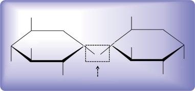
Cellulose is the most abundant polysaccharide available on Earth and consists of chain of glucose monomers. The molecular formula of cellulose is (C6H10O5)n and is an unbranched homopolysaccharide composed of α‐D‐glucopyranose units linked by β‐(1 → 4) glycosidic bonds (Figure I.2). The linear glucan chain forms highly stable regular intra‐ and inter‐molecular hydrogen bonds which stabilize its reticulate structure [85]. It is produced by various sources such as plants, microbial cells (Acetobacter, Rhizobium, Agrobacterium, Aerobacter, Achromobacter, Azotobacter, Salmonella, Escherichia, and Sarcina, etc.) [86], and enzymes (the cell‐free system)
[87]. Compared to microbial cellulose, also known as bacterial cellulose (BC), and bio‐cellulose produced by cell‐free enzymes system [85], which represent the purest forms, the plant cellulose contains several impurities in the form of lignin and hemicellulose, which necessitate its further treatment by various chemical methods. However, BC and bio‐cellulose are directly used in various applications without further pretreatment. Further, these possess unique structural, physico‐chemical, mechanical, and biological features, such as high water‐holding capacity (WHC) (100–200 times its dry weight), slow water release rate (WRR), higher crystallinity (60–90%), high tensile features (with elastic modulus 1 to 15 MPa and elongation at break 10–30%), an ultrafine fiber network, and moldability into 3D structures. which bestow BC with high potential value [85, 88–90] and broaden its applications in different fields, such as biomedicine, opto‐electronics, food technology, and separation processes, etc. [91]. BC is rarely soluble in common solvents such as water and organic and inorganic solvents, owing to its highly extended hydrogen bonding [92, 93]. However, several solvents systems have been developed such as LiCl/ dimethylacetamide (DMAc) [94], N‐methylmorpholine‐N‐oxide (NMMO) [40], ionic liquids (ILs) [95], and alkali/ urea (or thiourea) aqueous, which can dissolve BC [96]. BC has mainly found applications in the biomedical field where it is used as wound dressing material, for burns, artificial skin, vascular grafts, scaffolds for tissue engineering, tissue regeneration, and artificial blood vessels, etc. [89, 97, 98]. Also, it has been used for the preparation of several commercial products such as tires, headphone membranes, high performance speaker diaphragms, high‐grade paper, makeup pads, diet food, and textiles, etc. [98]. Furthermore, it is used as carrier in drug delivery systems, enzyme immobilization, and ion exchange membrane, and as biodegradable and biocompatible sensors and actuators [99, 100].
I.5.2.3 Chitosan
Chitosan is an abundant polysaccharide present in nature. Chemically, it is composed of randomly arranged β‐(1 → 4)‐linked D‐glucosamine and N‐acetyl‐D‐glucosamine, representing deacetylated and acetylated units, respectively. Its chemical structure is similar to that of cellulose except for the replacement of the –NH2 group instead of –OH moieties in the glucose units of cellulose backbone. It is synthesized through deacetylation of chitin shells of crustaceans, such as shrimps, using an alkaline solvent such as sodium hydroxide (NaOH) in excess as a reagent and water as a solvent. The chemical reactions are completed in two steps under first‐order kinetic control where activation energy (48.76 kJ/mol) of the first step is higher than the second and yields about 98% chitosan as the final product.
The degree of deacetylation (% DD) is determined via nuclear magnetic resonance (NMR) spectroscopy, where it ranges from 60–100% for commercial chitosan. The molecular weight of commercial chitosan ranges between 3.8–20 KDa.
Chitosan is highly soluble in dilute acids, highly biocompatible, non‐toxic, biodegradable, does not provoke the immune system, is anti‐cancerous, and environmentally friendly [101]. It biodegrades slowly to harmless products of oligomers and is absorbed slowly in the body. The biodegraded chitosan accelerates wound healing [102]. Owing to these features, it has found immense use in biomedical applications. It is used for drug delivery and wound care, both alone and in the form of composites with other materials such as BC, owing to its antibacterial activity [103]. Also, it is heavily used in agriculture as a seed treatment and biopesticide against fungal invasion. In the wine industry, it is used as a fining agent and for the prevention of spoilage. Further, it is used as self‐healing polyurethane paint coating.
I.5.2.4 Alginate and Other Seaweed‐Derived Polysaccharides
Alginate is an anionic polysaccharide isolated from seaweed. Chemically, it is composed of mannuronic acid and guluronic acid units where mannuronic acid units form β (1 → 4) linkages while guluronic acid units are linked via α‐(1 → 4) linkages . Structurally, mannuronic acid units are distributed linearly and exhibit a flexible conformation while guluronic acid units display a steric hindrance in the vicinity of carboxyl groups. Besides alginate, several other seaweed‐derived polysaccharides, such as carrageenan, fucoidan, laminaran, and ulvan have also been used for various biomedical applications owing to their high biocompatibility, easy availability, and simple isolation strategies [104]. Such polysaccharides can form hydrogels through ionic interaction between carboxylic groups (–COOH) present on their surface with a cationic cross‐linking agent [105]. Besides, divalent cations such as Ca2+, Zn2+, Ba2+, or trivalent cations (e.g. Al3+) may also exist in these hydrogels. Of
the various seaweeds, polysaccharides‐based hydrogels, alginate‐based hydrogels are extensively used as biomaterials for various biomedical applications including as scaffolds in tissue engineering, as carriers in drug delivery, and as model ECMs for biological studies [106]. Similarly, the carrageenan‐based hydrogels are extensively used for the encapsulation of cells, the transformation of growth factors, and the formation of bone and cartilage tissues [107, 108]. The fucoidan and ulvan‐based hydrogels are used in the culturing of cells and improving their activity [109, 110].
I.5.2.5 Hyaluronic Acid
Hyaluronic acid (HA), a non‐sulfated glycosamino‐glycan is constituted of repeated units of D‐glucuronic acid and D‐N‐acetylglucosamine linked together via alternating β‐1,4 and β‐1,3‐glycosidic linkages [111]. In nature, it is present in the form of a long straight chain of anioinc polysaccharide. It is extensively found in the human body where it is dispersed in different tissues. Generally, it has the ability to cross‐link by simple freeze‐thawing in the absence of any cross‐linker or organic solvent [112]. It can be potentially modified into different forms due to the presence of several functional groups in its chemical structure, such as –COOH, –OH, and N‐acetyl functional group. It can form hydrogels through various cross‐linking methods, such as chemical, enzymatic, or photo‐cross‐linking which find different applications in the biomedical and electronics fields. It has been extensively used for biomedical applications, such as cell motility, wound care and healing, cell signaling analysis, fabrication of different matrices, and angiogenesis, since it accounts for a major portion of ECM of skin, cartilage, and vitreous humors [91].
I.5.3 Proteins
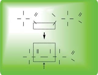
Proteins are biomacromolecules comprised of one or more long chains of amino acids. Each amino acid is comprised of an α‐carbon, amino group (–NH2), carboxyl group (–COOH), and a variable side chain designated as R‐group (–R). The amino acid residues are linked together through a peptide bond between the –COOH and –NH2 groups of two consecutive amino acids (Figure I.3). The peptide bond has two resonance forms which contribute some double‐bond character and inhibit rotation around its own axis which lead to the coplanar conformation of α‐carbon. The protein synthesis is carried out inside the living cells in a two‐stage process: transcription and translation, which take place in the nucleus and the cytoplasm of the cell, respectively. During transcription, the information from the segment of DNA known as the gene encodes the information for the formation of messenger RNA (mRNA). The mRNA
Figure I.2 Chemistry of glycosidic bond formation in carbohydrates.
then forms the respective polypeptide through translation which involves the formation of an assembly of ribosome, ribosomal RNA (rRNA), and transfer RNA (tRNA). The sequence of amino acids in a protein is determined by the sequence of nucleotide in its respective gene designated in the form of a genetic code. A single genetic code is comprised of three nucleotides (triplet codon). These genetic codes encode 20 different amino acids designated as standard amino acids, however, some organisms contain additional non‐standard amino acids, such as selenocysteine and pyrrolysine. Besides biological synthesis, proteins are also synthesized chemically by various methods collectively known as “peptide synthesis” which mainly use organic synthesis methods [113]. These methods include liquid‐phase synthesis, solid‐phases synthesis, and using solid supports, etc. Usually, the structure of protein is determined by X‐ray crystallography and NMR spectroscopy.
A linear chain of amino acid is called a polypeptide, however, polypeptides containing less than 20–30 amino acid residues are generally not designated as proteins, rather they are called peptides or oligopeptides. Besides amino acids, certain proteins contain additional parts termed prothetic groups or cofactors, which impart specialized function to a protein. Proteins exist in four levels of structural organization: the linear sequence of amino acids (i.e. polypeptide) represent the primary structure of the protein. The secondary structure of proteins contains α‐helices and β‐sheets which are stabilized by hydrogen bonds. The spatial distribution of the secondary structure represents the tertiary level of protein organization. It is stabilized by non‐local interaction such as hydrophobic interaction, salt bridges, sulfide bonds, and post‐translational modifications. The quaternary structure of proteins is the complex form which is formed by several polypeptide chains designated as subunits. These subunits either perform individual functions or account for the cumulative function of complex
protein molecule. Similar to other biomacromolecules, proteins are essential components of living organisms and perform different functions. For example, some proteins functions as enzymes where these catalyze biochemical reactions to perform various metabolic activities. Similarly, some proteins perform structural and mechanical functions, such as actin and mysoin proteins in muscles and the cytoskeleton and maintain the cell shape by forming a system of scaffolding. Other classes of proteins serve as cell‐signaling and ligand‐binding components, transcription factors, membrane receptors, antibodies, transport molecules, and vaccines, etc. Other proteins, for example, collagen and gelatin, are extensively used for biomedical application owing to their potential to form hydrogels. Collagen, the main component of ECM, possesses a right‐handed triple helix and is comprised of three left‐handed polypeptide helices that are twisted into a right‐handed coil. It forms hydrogels with other materials through chemical, enzymatic, and photo‐cross‐linking [114–116]. Similarly, gelatin biopolymer derived from collagen through hydrolytic degradation is widely used in pharmaceutical, food, and biomedical applications [117]. However, it has limited thermal stability and possesses weak chemical cross‐linking ability.
I.5.4 Nucleic Acids
Nucleic acids are biomacromolecules essential for all forms of life. Chemically, these are composed of nucleotides which are linked to each other through covalent bonds. Typically, sugar of one nucleotide forms a covalent bond with the phosphate of subsequent nucleotide. Each nucleotide is composed of 5‐carbon sugar (pentose), phosphate (PO43¯) group, and a nitrogenous base. The sugar and phosphates in nucleic acids are linked together in alternate fashion through a phosphodiester bond which is formed through the reaction of –OH groups in phosphoric acid with –OH group of another molecule [118] (Figure I.4). There are two types of nitrogenous bases, purines and pyrimidines, based on their chemical structures (Figure I.5). Two types of nucleic acids are known, DNA and RNA. Strings of nucleotides bind together to form helical backbones. Typically, one and two strings form the backbones of RNA and DNA, respectively. DNA and RNA differ from each other in their chemical composition, DNA contain adenine (A), guanine (G), cytosine (C), and thymine (T), while RNA contains uracil (U) instead of thymine besides other three bases. Besides structural arrangement and chemical composition, DNA and RNA also differ from each other in the sequence of nucleotides. The sequence of nucleotides is of immense importance and contains the information for the synthesis of biological molecules
Figure I.3 Chemistry of peptide bond formation in proteins.
(i.e. protein), molecular assemblies, cellular and sub‐cellular structures, organs, and organisms, etc.
I.5.4.1 DNA
Deoxyribonucleic acid (DNA) carries the genetic information of a cell and is referred to as a “genome.” A genome is the structural and functional organization of a cell. It contains all the information for the growth, shape, size, pattern, development, and functionality, etc. of a cell, tissue, organ, and organism. Typically, the DNA molecule is present in the nucleus of eukaryotes and forms a complex with histone proteins, referred to as a
chromosome. In prokaryotes, the chromosomes are suspended in the cytoplasm. A DNA molecule is typically comprised of two polynucleotide strings which form a double‐helical structure. These strands run in opposite directions to each other. The nucleotides on two separate strands in DNA are bonded to each other through hydrogen bonding. Typically, a double hydrogen bond is formed between A of first strand and T of another strand while a triple hydrogen bond is formed between G of one strand and C of another strand. The DNA backbone is flexible enough to resist any cleavage. Under some specialized conditions, the DNA molecule is twisted like a rope through a process called DNA supercoiling. In a relaxed state, one strand in DNA molecule encircles the axis of the double helix once after every 10.4 base pairs (bp) on average, however, in a twisted state, the strands become tighter or are loosely arranged [119]. Supercoiling can be either positive (twisting in the direction of the strand) or negative (twisting in the opposite direction of the strand). Most DNA molecules in nature exist as negatively supercoiled, which is caused by an enzyme topoisomerase. DNA molecules can exist in different conformations such as A‐DNA, B‐DNA, and Z‐DNA, however, only B‐DNA and Z‐DNA are known to be identified in functional organisms [120]. Both DNA strands contain the same biological information which is replaced when two strands separate from each other during mitotic cell division. The strand of DNA which is the same as the mRNA formed as a result of transcription is termed the sense‐strand, while the complementary strand is called the anti‐sense strand. However, both sense and anti‐sense sequences can co‐exist on the same DNA strand. To date, the function of the anti‐sense sequences is unknown [121]. It has been suggested that anti‐sense RNAs might be involved in regulating gene expression through RNA‐RNA base pairing [122]. It is worth mentioning here that about 98% of the total DNA in humans does not code for any protein and is referred to as non‐coding or junk DNA.
DNA forms temperature, pH, enzymes, and UV light‐responsive hydrogels through physical entanglement or chemical cross‐linking upon extraction from the nucleus (eukaryotes) or pooling out from cytoplasm (prokaryotes). For example, it forms hydrogels with cationic poly‐electrolytes [123] or metal ions, such as silver [124] for their applications in cell culture and differentiation studies [125] and 3D bioprinting [126].
I.5.4.2 RNA
Ribonucleic acid (RNA) is typically a single‐stranded molecule, however, it forms an intra‐strand double helix through complementary base pairing. Similar to DNA, it is also comprised of nucleotides linked via phosphodiester bonds, except it contains uracil (U) nitrogenous base
Uracil (U)
Figure I.5 Illustration of chemical structures of various nitrogenous bases in nucleic acids.
Figure I.4 Chemistry of phosphodiester bond formation in nucleic acids.
instead of thymine (T), which is linked to adenine (A) via a triple hydrogen bond. Unlike DNA, it contains ribose sugar at the C‐2 position, hence it is less stable and prone to hydrolysis. The presence of this functional group leads to A‐type conformation of RNA [127] which results in the formation of a deep and narrow major groove and a shallow and wide minor groove [128]. Further, due to the presence of the –OH group in the conformationally flexible regions of the RNA molecule, it can chemically attack the adjacent phosphodiester linkage and cleave the sugar‐phosphate backbone [129]. It plays several vital roles in living systems, such as coding, decoding, gene expression and regulation, catalyzes biological reactions, and sense and communicates responses to cellular signals, etc.
Typically, there are three types of RNA molecules: messenger RNA (mRNA), ribosomal RNA (rRNA), and transfer RNA (tRNA). Collectively, these three types of RNA molecules direct the protein synthesis in living systems, an important event of life. mRNA represents the coding genetic material which is produced inside the cytoplasm through the transcription of the sense strand of the DNA (serving as a template) molecule using the enzyme RNA polymerase. tRNA delivers amino acids from the “amino acids pool” to the ribosome where tRNA links the amino acids together via peptide bond to form a polypeptide or protein after post‐translational modification. Specifically, each tRNA molecule translocates a specific amino acid from the pool, thus, there are 20 different types of tRNA molecules to bring specific amino acids to the ribosome assembly. Recent research has shown that besides three typical RNAs, there also exist some regulatory RNAs in the cell, such as micro RNA (miRNA), small interfering RNA (siRNA), Piwi‐interacting RNA (piRNA), CRISPR RNAs, antisense RNA, and Xist, etc. These RNA molecule s are generally involved in the regulation and down‐regulation of genes. For example, miRNA, comprised of 21–22 nucleotides, forms a complex with enzymes and cleaves the complementary mRNA and thus blocks its translation into a polypeptide [130]. The siRNAs, comprising 20–25 nucleotides, are produced through viral breakdown and interfere with the translation of mRNA using the same mechanism as miRNA [131]. The piRNA found in germline cells in animals is comprised of 29–30 nucleotides and serves as defense mechanism against the transposons and plays a vital role in gametogenesis [132]. CRISPR RNAs serves as a regulatory system similar to RNA interference [133]. Similarly, the antisense RNAs mostly downregulate the gene expression by binding to a mRNA and form a double‐stranded RNA which is degraded enzymatically, however, a few are known to activate transcription [134, 135]. Besides the above‐mentioned coding RNAs,
a non‐coding RNA also exists such as Xist which inactivates the X chromosome in females by coating it [136].
I.6 Part III Biomaterials
Biomaterials are substances that are engineered to interact with a living system for medical purposes which can be either therapeutic (tissue engineering and regenerative medicines) or diagnostic. These are non‐viable materials and are implanted into the body to replace a missing part or repair a damaged tissue. The study of biomaterials is called “biomaterials science” or “biomaterials engineering,” which is about five decades old. It is a multi‐disciplinary field which encompasses different fields, such as tissue engineering, materials science and engineering, medicine, cell and molecular biology, and biochemistry, etc.
By nature, biomaterials are either derived from nature or synthesized using different strategies from various materials, such as organic and inorganic metals, polymers, ceramics, or clay, etc. The role of biomaterials can be passive, such as their use as a heart valve, or bioactive, where they function interactively, for example, hydroxy‐apatite‐coated hip implants. Biomaterials are routinely used for broad spectrum applications, such as in dental applications, surgery, drug delivery, tissue engineering, wound healing, pharmaceutics, and several others. In general, different types of materials are used for various biomedical and tissue engineering applications which can be broadly classified into four classes [137]:
1) Naturally derived polymer materials, for example, collagen, gelatin, starch, alginate, and chitosan, etc. These materials are better due to their recognition by biological systems [138].
2) Acellular tissue matrices, for example, bladder submucosa and small intestinal submucosa. Similar to naturally derived polymeric materials, these are also recognized by biological systems and are preferred candidates for tissue engineering [138].
3) Synthetic polymers, for example, poly(glycolic acid) (PGA), poly(lactic acid) (PLA), poly(caprolactone) (PCL), and poly(lactide‐co‐glycolide) (PLGA), etc.
4) Ceramics, for example, hydroxyapatite (HA) and β‐tricalcium phosphate (TCP).
I.6.1 Summary of Par t III Biomaterials
In Part III, “Biomaterials,” we discuss different classes of biomaterials, such as thermal‐ and photo‐deformable liquid crystal polymers, intriguing bioinspired movements and their potential applications, dendritic polymer‐based micelles and their function in drug delivery,
bone‐inspired biomaterials and their applications in bone tissue engineering, human dental caries restoration, protein‐based hydrogels and their applications, and various fiber‐reinforced composites as well as damping structures and self‐healing materials, etc.
I.6.2 Features of Biomaterials
I.6.2.1
Biocompatibility
Biocompatibility of biomaterials refers to their ability to remain in direct contact with living tissues without causing a toxic effect or provoking an immunogenic response [139]. Usually, the biocompatibility of biomaterials is the function of their liquid‐absorbing capabilities that support prolonged and sustainable growth of living cells. Biocompatibility is determined for individual materials and composites. Under certain circumstances, the biocompatibility of biomaterials is not up to the desired levels, therefore, their biocompatibility is enhanced by making their composites with more compatible polymers. For example, the biocompatibility of BC has been enhanced by making its composite with several polymers, such as gelatin, hydroxyapatite, and chitosan [103, 117]. Generally, the implantation of an acellular material triggers a series of events similar to a foreign body response, which leads to further acute inflammation response and in some cases developing a chronic response. However, a biomaterial should not provoke such a response, which is highly related to its biological properties, internal and external architecture properties, surface features, porosity, and the source of materials used for the fabrication of biomaterial implant [140]. Any deviation from the normal wound healing process determines the level of biocompatibility of biomaterials [141].
I.6.2.2 Biodegradability
Biomaterials when used as in vivo implants must degrade into non‐toxic products at a controlled degradation rate that closely matches the regeneration rate of the host tissues . When biomaterials are used as scaffolds, these are degraded when cells are deposited, and they secrete their own ECM and eventually form a 3D structure that closely mimics the natural microenvironment of the host tissues. The in vivo degradation process of the scaffold is a function of certain variables, such as the material structure (e.g. the chemical composition, the molecular weight and the molecular weight distribution, crystallinity, and structural and morphological features, etc.), the macroscopic features (e.g. the shape and size of the implant, and porosity, etc.), and the microenvironment (e.g. the pH and temperature of the culture medium, and the presence of enzymes, cells, and tissues, etc.). Optimization of the degradation process and minimiz
ing its effects are crucial, for example, it may result in local drop of pH if the released degradation products contain acidic compounds, which may lead to necrosis or inflammation.
I.6.2.3 Porosity, Pore Size and Shape, and Micro‐ and Macro‐Structure
Porosity of biomaterials is defined as the percentage of void spaces [142]. Tissue engineering applications of biomaterials require a high level of porosity (>90%) to offer a high surface area for the seeding of high cell density, migration and the proliferation of cells, and neo‐vascularization [143]. The porous structure of the biomaterials enhances the biological and mechanical interlocking between the implanted scaffold and the host tissue at the site of implantation and ensures greater stability and avoids fibrous encapsulation. Similarly, appropriate pore size and shape of the pores in biomaterials ensure cell viability and tissue regeneration by accumulating a large number of cells and the absorption of high nutrient concentrations. However, a balance must be maintained between the pore size and the mechanical strength of the biomaterials since an increase in pore size generally leads to lower mechanical strength. Usually, pores of biomaterials, that are too large or too small, result in the failure of cell adhesion, proliferation, and migration into different parts of the biomaterial. Therefore, it is essential to define an appropriate size and shape of the pores of biomaterials for a specific cell type and desired application. In general, small pores favor greater cell adhesion and differentiation in vitro while larger size pores favor greater cell adhesion and proliferation in vivo However, the pore size is not the only parameter that ensures the successful development of regenerated tissue, several other factors, such as pore interconnectivity and distribution are also essential which guarantee the transport of nutrients, diffusion of gases, and removal of wastes from cells.
I.6.2.4 Mechanical Features of Biomaterials
Biomaterials used for tissue engineering applications must demonstrate high mechanical properties to withstand in vitro manipulation and stressed condition in the in vivo microenvironment. High mechanical strength of biomaterials preserves the required space for cell attachment, proliferation, and migration as well as matrix formation. The biomaterial implant must demonstrate mechanical properties closely mimicking those of the host tissues to provide temporary support to the regenerating tissues.
I.6.2.5 Surface Topography
Surface features of the biomaterials play a vital role in their applications in tissue engineering and regenerative
medicines. The surface roughness of biomaterials ensures efficient cell adhesion, proliferation, differentiation, and viability. Also, the scale of topography, such as micro‐, nano‐, and mezo‐scale biomaterials, the shape of the biomaterials, such as ridges, steps, grooves, pillars, and pits, and their orientation have profound effects on the overall cell behavior and phenotypic expression [144]. In general, nano‐scale (100–300 nm) biomaterials offer improved cell adhesion and proliferation for most cell types. It is generally believed that nano‐scale materials modulate the interfacial forces between implants and cells which leads to the regulation of cytoskeleton formation and the organization of the cell membrane receptors and also triggers the complex intra‐cellular molecular signaling [145]. Besides the scale of biomaterials, their surface roughness also determines the cell‐to‐cell interactions, adhesion, and proliferation. For example, highly rough surfaces of biomaterials usually fail to establish interconnections, which leads to compromised adhesion and the proliferation of cells. Thus, surface properties and interactions between cells and ECM are of immense consideration for cell signaling and subsequent cell attachment, proliferation, and formation of phenotypes [146].
I.6.2.6 Simple Manufacturing and Sterilization Strategy
Biomaterials used for various applications should be manufactured by simple, robust, reliable, and cost‐effect strategies. Moreover, the manufactured materials must be easily sterilized by common sterilization strategies, such as heat, chemicals, autoclavation, and UV sterilization, etc.
I.6.3 Current Advances in Biomaterials
Science
Some of the advanced applications of biomaterials are:
● Smart polymers: These are injectable hydrogels that are used for the delivery of growth factors, gene and gene segments, and viable cells to the target site, such as damaged tissues.
● Biomimetic elastomers: These are elastic polymers which mimic the mechanical features of the host tissue and play the vital functions of the proteins found in the host body.
● Engineered skin: This is fabricated from collagen and collagen‐synthetic composites through electrospinning strategies to improve the collagen strength and elasticity for various applications.
● Heart patches and blood vessels: Researchers across the world have evaluated the potential of different biomaterials for the formation of synthetic heart patches and blood vessels. These biodevices have been fabri
cated by various strategies, such as electrospinning and several others.
● Immuno‐isolation systems: Attempts have been made to provide protection to the cells against various factors by using different biomaterials to avoid immune attack. This practice has also resulted in the improved efficacy of cell transplantation.
● Aligned fiber assays: Using electrospinning strategies, highly aligned fibers have been fabricated for their application in diagnosis and treatment of several highly metastatic cancers.
● Islet delivery: Researchers have developed biomaterials for pancreas replacement to preserve the functionality of islets after their harvesting from the donor and to assist their delivery to the recipient. It can result in the long‐term survival and health of the patient.
● Tendon repair: Using biomaterials, tissue‐engineered constructs have been designed to shorten the recovery time for repair of tendons and to improve their function.
I.7 Scope of the Book
The importance of bioinspired materials leads to the highly interdisciplinary character of this field, which brings together natural scientists (biologists, chemists, and physicists) and engineers. Thus, an active interaction across disciplines is the key to the real development of this old research area, which is now attracting many researchers worldwide. However, as underlined by several recent reviews on the subject, this condition is yet to be fully met, due to the rather limited understanding of the building principles of living entities which are numerous and complex, and because the definition of the scope and novel applications remain to be further clarified and exploited. Hence, conveying information in the field and disseminating the bioinspired solutions already discovered are of real importance, and would contribute significantly in propelling this promising research area. Therefore, the aim of this book is to introduce a comprehensive overview of the bioinspired materials science and engineering, discussing biofabrication approaches, and applications of bioinspired materials as they are fed back to nature in the guise of biomaterials. Some biological compounds, such as carbohydrates, proteins, and nucleic acids, etc. have been mentioned with regard to what is learned from them, and how they are useful in the engineering of bioinspired materials.
The book is divided into three main Parts: biofabrication, biomacromolecules, and biomaterials. Illustrating the bioinspiration process from materials design and conception to the application of bioinspired materials, this book presents the multidisciplinary aspect of the concept
Another random document with no related content on Scribd:
But a threatening cloud, the more dangerous because unseen, hung over this gorgeous festival, which seemed to promise a long life of honor and renown to the recipient of a nation’s applause and gratitude. Three days after his triumph Ismail lay a corpse in the palace of the Alhambra.
Among the captives taken in the campaign of Martos was a girl of dazzling beauty. Her possession was disputed by several soldiers, whose hands, bloody with the carnage of a city taken by storm, were about to sacrifice her, when she was rescued by Mohammed-IbnIsmail, a cousin of the Emir. No sooner had the latter learned of the occurrence and of the extraordinary charms of the captive than he despatched the eunuchs to conduct her to the royal harem. The just remonstrances of Mohammed were treated with contempt. Not satisfied with the injury he had inflicted, Ismail ordered his cousin to leave the precincts of the court, and curtly informed him that his proper place was with the enemies of his sovereign. The exasperated prince, who had already signalized his valor in many a campaign, whose blood had been shed in defence of his country, whose birth exalted him to a level with the throne itself, and whose sense of justice revolted at the unprovoked outrage he had endured, had no inconsiderable following among the dignitaries of the palace and the officers of the army.
A few hours sufficed to mature a plot; the support of Othman, commander of the royal guard, was obtained, and the conspirators only awaited a moment favorable to the execution of their design. The Emir, walking with his vizier in the gallery of the Alhambra and wholly unsuspicious of danger, was stabbed by Mohammed; the resolute defence of the minister availed nothing in the face of superior numbers, and he perished by the side of his master, while the assassins, who had previously provided means of concealment, escaped in the general confusion.
The wounded monarch was borne into the palace, where he soon expired. His death was concealed—his injuries were even represented as trifling—until, in anticipation of a fatal result and to secure the succession, allegiance was sworn to his son, a youth of twelve years, who ascended the throne under the name of
Mohammed IV The diligence of the second vizier apprehended a number of the conspirators, whose heads were exposed on the battlements of the castle. The treacherous Othman was foremost in protestations of loyalty and devotion to the new ruler. The chief assassin fled to Malaga, and the ill-concerted and bloody enterprise dictated by wrong and accomplished by cowardice was productive of no other result than a change of rulers and an increased public attachment to the family of the murdered king.
The character of the latter was worthy of the great place he occupied and of the glorious traditions of his dynasty. His personal courage and success in war won for him the affectionate admiration of his people. His intervals of peace were diligently employed in the construction of mosques and palaces, of baths and fountains. He exercised with liberality and discrimination the distribution of alms, as enjoined by the Koran. The hanging gardens planted under his supervision were copied from those of ancient Babylon. The police regulations inaugurated by him established the safety of the streets and suburbs by day and night; for the better apportionment and collection of taxes the cities were divided into different quarters, and the alien tributaries designated by distinctive costumes. He sternly repressed the fanaticism of the theologians, ever a prolific source of public anxiety, disturbance, and confusion. The leisure moments of Ismail were passed in the society of the learned, in the tournament and the chase, in the pleasures of horticulture, in the construction of magnificent edifices.
The youth and inexperience of Mohammed IV. were supplied by the political sagacity of his vizier Al-Mahruk, a man of ability but of inordinate ambition, and absolutely unscrupulous in the means of gratifying it. His policy, solely directed to the centralization of power in himself, was the ultimate cause of his destruction. He lost no opportunity to humiliate the nobles. The brothers of the Emir were, in turn, removed from the court; one, upon some frivolous pretext, was imprisoned in Almeria, where he died; another was exiled to Africa; a third was forced to seek concealment in a remote village on the frontier. The jealous intolerance of the vizier endeavored to remove every possibility of a successful rival. The possession of eminent
talents and virtues was a provocation of oppression; the most able and experienced statesmen and commanders, apprehensive of violence, left the capital, and a feeling of alarm and discontent became general in every quarter of the kingdom.
This state of affairs, so dangerous to the stability of government and the maintenance of peace, lasted until the young prince arrived at the age of sixteen years, when Moslem custom permitted his assumption of the reins of government. One of his first acts was the degradation and imprisonment of the obnoxious minister. In his place was appointed Ibn-Yahya, who enjoyed the respect and confidence of all classes, and whose judicious counsels were well calculated to guide the career of a young, ambitious, and inexperienced sovereign. Othman, the former commander of the royal guard, whom disappointed ambition, a consciousness of guilt, and the fear of detection had driven from Granada, now planned a new enterprise to retrieve his fallen fortunes. At his instigation the peasantry of the rugged district of Andarax rose in rebellion. The insurgents were defeated by the forces of the Emir, but the mountainous region they infested, and whose fastnesses the royal troops were unable to penetrate, prevented their dispersion. The intrigues of Othman obtained at once the interference of the Castilians and of the Emir of Morocco. Again the aid of foreign enemies was invoked to revive the hopes of a defeated faction; the patriotic ardor which might have preserved the declining empire was sacrificed to the gratification of private resentment; and the ablest of the Moors, unwilling to profit by the melancholy lessons taught by the history of the khalifate, renounced every noble impulse for the sake of avenging their private injuries.
In the vicinity of Cordova the army of Mohammed sustained a disastrous defeat; the survivors retreated to Granada; and the Emir, infuriated by the reflection that the imprisoned vizier Al-Mahruk was indirectly responsible for the misfortunes which afflicted the commencement of his reign, commanded his immediate execution. The efforts of Mohammed to counteract the African influence enlisted by the rebels met with no better success. Algeziras was stormed and taken, and the vizier Ibn-Yahya was killed in the
assault. Ronda and Marbella fell into the hands of the rebels, who soon threatened the city of Granada itself. Consternation seized the inhabitants of the capital; and Mohammed, assembling the remains of his defeated army and attended by the principal cavaliers of the court, went forth to meet the enemy. Gallantly seconded by his nobles, at the head of a force insignificant in numbers but remarkable for courage and resolution, he speedily recovered his prestige and his influence. All the cities captured by the Africans were retaken. The stronghold of Baena surrendered. The walls of Casares were destroyed by artillery Within sight of the latter city the Moslems, in a bloody encounter, destroyed a Christian force sent to relieve it. Thus, within the space of a few months, Mohammed counteracted the effect of the recent disasters to his arms, restored to his dominions the territory of which treason had deprived him, and acquired new renown by a victory gained over an army superior in numbers and thoroughly versed in all the stratagems and resources of regular and partisan warfare. These military exploits firmly established his reputation. No prince of the Alhamares, of his years, ever achieved such celebrity. Nature had bestowed upon him every gift of mind and person which could elicit the approbation of the wise or arouse the enthusiasm of the multitude. His face was handsome, his stature above the middle height, his limbs models of athletic symmetry. Skilled in every exercise, he delighted in those passages of arms so popular among the Moorish chivalry. Prominent in a nation of bold and dexterous horsemen, he was universally accounted the finest lance in the Moslem army. Grave in demeanor, pleasing in address, elegant in manners, his attractive exterior only served to enhance the noble traits of a virtuous and enlightened mind. He was a friend of letters; and learned men accompanied him during his expeditions, who could observe the course of events and perpetuate the remembrance of such facts as might contribute to the profit of the country and the glory of its king.
In the ever-changing panorama of the Reconquest there frequently appears the Moslem of Africa, descendant of the Almoravide or the Almohade dynasties; proud of the fame of his ancestors, and perpetual claimant of the legacy of their valor; as ambitious of the regaining of Cordova and the purification of its
desecrated temple as were his contemporaries, the crusaders, of the conquest of Jerusalem and the recovery of the Holy Sepulchre. The Emirs of Fez, of the family of the Merynites, had risen to great distinction among the rulers of Africa. Their dominions included the entire northern portion of that continent. Their capital was the resort of all that was intelligent and accomplished among the Mussulman nations. In the incessant political disorders of the Peninsula, hundreds of merchants, scholars, philosophers, had sought amidst the quiet and security of distant regions that refuge from incessant revolution and Castilian conquest no longer to be enjoyed at Granada. Not a few of these found their way to Fez. Picturesque in its situation, the taste and liberality of its rulers had made it a beautiful city. Its gardens, planted with every variety of tropical plants and flowers, and refreshed by innumerable fountains, offered a vision of Paradise to the tired and thirsty wayfarer who had toiled for many leagues through the stifling heat and drifting sands of the desert. A high degree of civilization had been attained by the people of Morocco, who, with the natural inclination of the Arab mind to scientific investigation and mathematical studies, had made considerable progress in astronomy, chemistry, botany, and medicine. Their efforts were materially aided by the precious manuscripts once part of the great library of the khalifs, and the treasures of private collections rescued from barbarian ignorance and Christian bigotry during the destruction of the Ommeyade empire. To Fez, where it was revered as a priceless relic, and where perhaps to this day it is still preserved, was transported by the Almohades the famous Koran, partly written by the hand of the martyred Othman, which was formerly exposed for the veneration of the faithful in the Kiblah of the Great Mosque of Cordova. Heedless of the change in national conditions, of the decline of religious enthusiasm, of the decay of that martial spirit which renders the fanatical soldier invincible, the Emirs of Morocco never lost sight of their favorite project, the recovery of Spain. Their repeated efforts for that end were rendered inoperative by the religious feuds between their subjects and the Andalusian Moslems; the resources of both kingdoms, which might have been profitably exerted against the infidel, were wasted in the infliction of mutual injuries; and the
Castilian sovereigns, taking advantage alternately of the animosities of either faction as best suited their purposes, directed their arms against each other for their common ruin.
In the year 1333, a body of Moorish cavalry, seven thousand strong, under command of Abd-al-Melik, a prince of the blood, disembarked at Algeziras. Invited by the rebels of Granada, and expected to unite with the Castilian forces against Mohammed, their operations were begun in an unexpected quarter. With a celerity that indicated the previous arrangement of a well-conceived campaign, the invaders and their countrymen of Algeziras invested Gibraltar. Considering the defences of that fortress, greatly improved since the Castilian occupation, the enterprise seemed hopeless. But either through the peculations of the governor or from neglect of the court to provide the garrison with supplies, the place was in a few weeks reduced to the greatest distress. Deprived of all ordinary means of sustenance, the hungry soldiers devoured the leather of their belts and bucklers. When too late, the difficulty of the situation was realized. The selfish and tyrannical policy of Alfonso XI. had alienated the attachment of his subjects. The voluptuous King had lingered too long in the arms of his mistresses, and the pugnacious nobility were engaged in gratifying their hereditary grudges in the congenial occupation of private war. Official incompetence and delay had prepared the way for great disasters. The African fleet commanded the sea. No force available by the Christians could dislodge from its intrenchments the well-appointed army of Abd-alMelik, with success within its grasp. The Castilian princes, as well as the nobles, listened with haughty indifference to the patriotic appeals of their sovereign. So low had the royal authority fallen that some cavaliers of the highest rank disregarded the peremptory messages of the crown; others demanded compensation for contributing to the defence of their country. Alfonso, abandoned by the nobles, found his only reliance in the uncertain and selfish adherence of foreign soldiers of fortune, attracted to Spain by the expectation of plunder; and in the co-operation of the grand-masters of the military orders, who, compelled by their vows, furnished a lukewarm and reluctant support to the throne. Long before the Castilian army could arrive at Gibraltar, messengers brought the unwelcome news of its surrender.
The standards of the Prophet were raised, amid the acclamations of the soldiery, upon the highest point of the citadel, and the African vessels occupied the harbor. The garrison was suffered to retire with the honors of war, and the exultant Moslems, intoxicated by victory, already pictured to themselves the speedy recovery of the seats of their ancestors and the revival of the ancient glories of the khalifate.
The King of Castile, convinced that the Moors were not firmly enough established in their new conquest to maintain a siege, pushed forward to Gibraltar. Harassed by the enemy’s cavalry, his force reached its destination after many conflicts of doubtful issue and unimportant results, and the Africans were, in their turn, besieged. In their exposed situation, with a vigilant enemy in their rear and the sea patrolled by the African fleet, the Castilians endured unspeakable hardships. The day was one incessant conflict, the night a succession of alarms. The convoys were intercepted. Famine, with its attendant horrors, stalked through the wretched camp. The faltering loyalty and insufficient discipline of the troops encouraged desertion, and the numbers of those spared by want and disease began to be sensibly diminished. Many who fled from their standards encountered a more deplorable fate, for none escaped the vigilance of the Moorish scouting parties; and captives were so numerous that in the market of Algeziras the choice of Christian slaves could be obtained for a doubloon of gold. To add to the general distress, the Emir of Granada was approaching at the head of a numerous body of troops with the design of effecting a junction with Abd-al-Melik. At this critical moment, when the Christian cause seemed all but lost, the course of events was changed by one of those unexpected occurrences which, while common in the annals of those times, is apparently inexplicable unless attributed to the influence of the chivalrous instincts that during the prosecution of the Moslem wars often changed ferocity into courtesy and enmity into friendship. The narrow space which separated the hostile camps was, according to the practice of the age, the scene of many knightly encounters. As the result of one of these, proposals of peace were offered and accepted, a truce of four years was signed, and the enemies, who but a few hours before had fiercely contended for each other’s destruction, now mingled together upon terms of
familiar intimacy An interchange of presents took place between the monarchs of Castile and Granada. The different degrees of civilization existing in the two kingdoms, and the marked superiority of the Moors in the knowledge and adaptation of the mechanical arts, are disclosed by the accounts that have descended to us concerning this exhibition of royal courtesy. The articles presented by the King of Castile are scarcely alluded to by the ancient chroniclers. Had they been objects of curiosity, elegance, or value, Castilian pride would not have been silent concerning them. On the other hand, the gifts of the Emir of Granada, their intrinsic worth, the excellence of their workmanship, the number, variety, and setting of the jewels with which they were adorned, are the subject of minute and accurate description. They included splendid arms and armor; among them a helmet enriched with rubies of extraordinary size, and a sword, with a damascened blade of the finest temper, whose scabbard, formed of overlapping plates of gold, was studded with magnificent topazes, sapphires, and emeralds. Not the least remarkable of these articles of elegant luxury were beautiful silks of many colors and pieces of cloth of gold. In Granada, the manufacture of silk had long since reached perfection, a considerable portion of the great bazaar of the capital was reserved for its merchants, and the superior quality of its product had brought it into great demand in every port accessible to commerce. In Castile, distant scarcely a day’s journey, this fabric, not beyond the reach of persons in moderate circumstances under the advanced civilization of the Spanish Arabs, was practically unknown, and so far from being an article of merchandise was an object of curiosity, and worth far more than its weight in gold.
The ceremonies of the treaty ended with a banquet, and Mohammed, forgetful or regardless of the prejudices of religion, accepted the hospitality of the King of Castile. This concession to a Christian misbeliever offended the bigotry of the Africans; the fancied partiality shown to the Emir of Granada by his host was resented as a national affront; and the ill-concealed mutual jealousy of the rival sectaries of Islam was again emphasized by a bloody tragedy On his return to the camp, Mohammed was waylaid and slain by his fierce and treacherous allies, who had so greatly profited by his aid,
and who could neither comprehend nor suffer the generous courtesy which recognized the virtues of good faith and toleration even in an hereditary foe. The Emir had scarcely breathed his last when the vizier Redwan, by a bold stroke characteristic of the crooked methods of Oriental politics, secured at the same time the public tranquillity and the continuance of his own power. Hastening to Granada, he caused Yusuf, the younger brother of Mohammed, to be proclaimed Emir, and conducted him with a magnificent escort to the Moslem camp, where allegiance was at once sworn to him by the army
The year 1339 opened with extensive preparations for another African invasion, whose object was avowedly the conquest of the entire Peninsula. Every means to insure success was taken by the shrewd and active Abul-Hassan, Sultan of Fez, whose talents had raised his empire to the first rank among the Moslem powers of the West. Fanatical and eloquent missionaries were despatched to preach the Holy War among the wild tribes of the Desert. A treaty of alliance was concluded between the Sultan and Jaime III., King of Majorca. The friendly relations interrupted by the murder of an emir were resumed with Granada. For the moment, the instinct of selfpreservation and the interests of a common faith outweighed national jealousy and the bitterness of theological hatred. An innumerable army was raised and equipped. The African navy, already more formidable than that of the enemy, was greatly strengthened. The possession of Gibraltar and Algeziras invited an enterprise which Moslem fanaticism easily persuaded itself was practicable, and, in case of failure, afforded means of security and retreat. Daily, for months, soldiers accompanied by their families crossed the strait. The sagacity of Abul-Hassan convinced him that by the presence of their wives and children the fidelity of the troops would be confirmed, their valor animated, and their confidence in the success of an undertaking, conducted with the sanction of religion, fully assured. Great numbers of these armed colonists entered Granada, where they were received with every evidence of consideration by Yusuf, who saw, with great satisfaction, this important addition to the military strength of his kingdom.
Information of these events, serious enough in themselves, and doubtless much exaggerated by fear and ignorance, spread terror through the realms of Portugal, Aragon, and Castile. The rivalry of the monarchs, the enmity of the nobles, the ambition of the clergy, were, for the time, laid aside in common apprehension of the approaching deluge. The naval forces of the three kingdoms were united under the command of the Castilian admiral, Don Geoffrey Tenorio. His fleet consisted of but thirty-six vessels, ill-manned, imperfectly equipped, and wholly unfitted to cope successfully with the swift and well-appointed galleys of the Sultan of Fez. In the general consternation, and to counteract the religious fervor of the Moslems, the sanction of divine aid was solicited through the Pope, and the Holy Father issued from Avignon a grant of plenary indulgence to all Christians who should participate in the impending contest.
The commencement of hostilities, near the close of the year 1339, was marked by a series of predatory expeditions, undertaken in turn by each nation, and characteristic of the disorderly and indecisive conflicts so popular in those times. After the capture of considerable booty, the African general, Abd-al-Melik, encamped with the bulk of his army in the plain of Pagana. The Christians, hastily assembled at the summons of their lords and commanded by the heads of the noble houses of Guzman and Ponce de Leon, names subsequently famous in the conquest of Granada, by a forced march surprised the enemy’s camp. Attacked in their tents, stupefied by slumber, and confounded by the din of combat, the Moslems, incapable of either resistance or flight, perished by thousands. Abd-al-Melik, pierced with many wounds, died among the reeds of a neighboring stream where he had concealed himself. The loss of the invaders exceeded ten thousand men, and a multitude of captives were led away in chains. A treasure of great value was secured by the victors; and the booty acquired in the last campaign, composed principally of cattle and sheep, was retaken and appropriated for the use of the Castilian army.
The effects of this brilliant exploit, in which were exhibited alike the skill and prowess of the Castilian soldiery, were, as usual,
nullified by aristocratic jealousy and court intrigue. The signal advantage obtained was not pursued; and the Moors, encouraged by the want of spirit displayed by the enemy, were aroused to fresh exertions by the consciousness of power and the mortification of defeat. Bent on revenge, the Sultan, Abul-Hassan, redoubled his preparations for the coming invasion. Every man subject to his authority throughout his vast dominions was enlisted. The ports of Northern Africa resounded with the din incident to the repairs and the equipment of the fleet. The Sultan himself superintended the embarkation of the troops at Ceuta, and assumed command of the expedition. The Moorish armament, comprising two hundred and ten vessels, of which more than seventy were galleys of war, finally sailed from Ceuta; and the Moorish host, nearly two hundred thousand strong, landed without accident at Gibraltar and Algeziras. Every resource at the command of Abul-Hassan had been employed to collect, and to provide with munitions of war, this immense body of men. All the most powerful motives which actuate the human mind had united to further the project of invasion,—royal ambition, private vengeance, the admonition of religious duty, the thirst of empire, the hope of Paradise. For nearly two centuries so formidable a force had not threatened the Christian domination in the Peninsula. It was not without reason that the number of pilgrims to famous shrines was quadrupled; that the intervention of local saints was invoked in every hamlet; that the terrified inhabitants of Andalusia asked themselves if the inheritance of their fathers, won foot by foot from the infidel, was to be wrested from their hands at one blow by the barbarians of Africa; if the cathedral of Cordova, that priceless trophy of conquest, still existing in all its pristine beauty and consecrated to the worship of God, should once more be occupied by the slaves of the Arabian Prophet.
The enemy’s fleet had passed the strait during the night, apparently through the supineness of the Spanish admiral, whose inferior force, however, could not have even delayed its progress; but, from this apparent neglect of duty, a rumor arose that Don Geoffrey Tenorio, one of the most honorable and high-spirited of men, had betrayed his trust for a bribe. Stung to the quick by the unjust imputation, the brave soldier ordered his ships to prepare for
battle. His adversaries outnumbered him three to one, and no courage or dexterity could compensate for the disadvantage of position or the disparity of numbers. The conflict was short and bloody. The Christian admiral, after a desperate struggle on the deck of his ship, fell sword in hand; the majority of the Christian vessels were taken or sunk; and only five succeeded in reaching the harbor of Tarifa. Abul-Hassan, from the loftiest minaret of Ceuta, witnessed the victory which established his maritime supremacy in the Western Mediterranean, and destroyed for nearly a generation the naval power of the kingdom of Castile.
In September, 1340, the Sultan of Fez formally assumed command of his troops at Algeziras. Not long afterwards the army of Granada, commanded by Yusuf, arrived, and the long-expected campaign began in earnest. The chronicles of the time differ greatly in their numerical estimates of the allied host. That it was very large, however, does not admit of doubt, and, even after due allowance for priestly exaggeration and Castilian gasconade, it would seem to have exceeded two hundred and fifty thousand men. Of these, nearly a hundred thousand were Mauritanian and Granadan horsemen, the finest light-armed cavalry in the world. The military skill which disposed of this great force corresponded in no degree with the irresistible power it was capable of exerting if intelligently directed. The most obvious course would have been to advance rapidly into the country of the enemy, already paralyzed with fear at its approach, and bring about an engagement before the Christians were fully prepared. The capture of Tarifa was, however, in the eyes of the two sovereigns, a more certain advantage than the precarious issue of a pitched battle on their own ground with the redoubtable chivalry of Castile, and, in consequence of their determination, a line of intrenchments was drawn around that city. It is uncertain whether any incentive other than mere caprice influenced the Moslem commanders in their decision. Tarifa was a place of comparatively small strategic value. So long as the more important fortresses of Gibraltar and Algeziras remained in the hands of the Moslems, affording ready communication with the shores of Africa, no material advantage could result from its possession. Its harbor was neither extraordinarily safe nor commodious. While the country in its vicinity
was rich and fertile, its extent was not great enough to justify the expenditure of any large amount of blood and treasure for its subjection. And finally, the formidable character of the defences of Tarifa, which had, upon more than one occasion, demonstrated that a garrison insignificant in numbers could readily maintain its position against an immense army, should have convinced the Moorish princes of the difficulties to be encountered in its reduction, in addition to the probabilities of ultimate disaster. The siege of the city, once decided upon, was pushed with the utmost energy. Quantities of munitions of war, provisions, the ponderous engines used in military operations, and a few pieces of rude artillery were transported from Ceuta. The Castilian galleys which endeavored to intercept these supplies were wrecked by a tempest, and the sailors were killed by the enemy or captured and compelled to renounce their religion. The apathy of the Christians, partly the result of constitutional indifference, but largely due to royal oppression, seemed about to abandon their country to ruin. Alfonso XI., reduced to despair, convoked an assembly of the grand masters of the military orders, the most eminent prelates, and such of the principal nobles as he had not degraded, exiled, or put to death. After a pathetic appeal to their patriotism, he deposited upon a table his sword and crown, and, leaving with them these insignia of royal dignity as mute representatives of his honor and distress, he retired from the room. In the deliberations which followed, it was resolved to at once attempt the relief of Tarifa, an undertaking which, if successful, would avert a national misfortune. Every effort was exerted to assemble an army. The nobles summoned their retainers and vassals. The clergy proclaimed a crusade, raised the holy banner blessed by the Pope, and inflamed the religious zeal of their audiences by all the artifices of bigotry and all the powers of eloquence of which they were masters. Appeals were made to the Kings of Aragon and Portugal. A fleet of galleys was obtained from the Republic of Genoa, a proceeding which utterly exhausted the already bankrupt treasury of the kingdom, while the well-known duplicity of these mercenaries caused no little apprehension lest their power might yet be turned against their allies through the machinations of a rich and unscrupulous adversary.
After an investment of several weeks, after numerous assaults and many stratagems, all of which were repulsed or foiled, the undaunted garrison of Tarifa still maintained unimpaired the honor and reputation of the Castilian arms. To this handful of heroic soldiers the prospect was indeed discouraging. From the battlements of the castle, as far as the eye could reach, could be descried the countless tents of the besieging army. Amidst the coarse brown shelters of camel’s hair, the home of the migratory Arab, appeared the more pretentious quarters of the various division commanders, indicated by pennons of gaudy colors and by patrols of heavily armed sentinels. On a slight eminence, in the centre of the vast encampment, stood side by side the royal pavilions of the Sultan, Abul-Hassan, and Yusuf, Emir of Granada. Embellished with every adornment procurable by the boundless resources of wealth and power, they were conspicuous from afar. Their material was blue and crimson silk, profusely and elaborately embroidered. Globes of silver surmounted the stakes which sustained their folds. Before the tent of the Sultan of Fez waved the great standard of the Holy War,—of green silk inscribed with passages from the Koran, and with the name of Allah repeated hundreds of times in characters of gold. With armor and weapons glittering with jewels, a numerous guard watched over the safety of their sovereigns,—fierce warriors of the Zenetah, and of the Beni-Saraj, those “Sons of the Saddler,” destined in after-years to play a prominent part in the history of Granada, and whose valor, amidst the sinking fortunes of the Moslem empire, sustained in many sanguinary battles the reputation of their ancestry and the terror of their name. Outside the harbor was ranged the hostile fleet, covering the sea with its snowy sails; its gay ensigns emblazoned with the devices of the commanders or with the mottoes of the Koran assumed as their peculiar insignia by the princes of the House of Fez.
Well might the hearts of the little band of Christians in the beleaguered city of Tarifa sink within them. To all appearances, they were abandoned by their sovereign. No tidings of approaching relief could reach their ears. The din of incessant battle resounded through the streets. The walls were crumbling under the blows inflicted by formidable engines of war. More than once had scaling-parties
obtained a foothold on the ramparts, only to be repulsed by superhuman efforts. Even in capitulation there was little prospect of safety, for such of their comrades as had fallen into the enemy’s hands were tendered the alternative of apostasy or death. For in this Holy War, waged more earnestly for the extension of religious faith than for the acquisition of territory or the spoils of conquest, the humane rules which ordinarily governed the surrender of enemies or the treatment of prisoners were either suspended or abrogated. Their stubborn and prolonged resistance had exasperated the besiegers, and there was slender hope of quarter in the event of submission or capture. The Moslem army was largely composed of barbarians, ignorant of the laws and usages of civilized nations; and, whether taken by storm or surrendered, the city would inevitably be sacked and would probably be destroyed. In the last moment of extremity and despair the Christian banners were discovered from the battlements. In the organization of the approaching army the efforts of the Spaniards had been absolutely exhausted. Castile, in the fourteenth century, could not command the men, the funds, the military resources, which had been under the control of the grim old warrior Ferdinand III. The population was diminished by wars, sedition, famine, disease. Agriculture was impossible where life and property were constantly unsafe. Articles which in the contiguous kingdom of Granada were in the hands of every peasant were unknown to most Castilians. The flocks, once the chief source of Estremadura’s and Andalusia’s wealth, had been swept away by the alternate incursions of friend and foe. The extravagance of mistresses, the peculation of officials, the exactions of the clergy, the rapacity of the nobles, had drained the public treasury. The administration of the finances was mainly in the hands of Jews and churchmen, who thwarted each other whenever possible, and always at the expense of the state; who, not unjustly, regarded each other with suspicion; and who, in turn, were cordially hated by those on whom were imposed the onerous burdens of tribute and taxation. Successive regencies organized for plunder; the flagrant abuse of power, and the prostitution of justice to the gratification of personal revenge; the insolence of royal concubines; the sanguinary tyranny which disgraced the throne; the invasion of private rights and the
insecurity of private property; the impunity of crime; the omnipresent evidences of distress and penury; the degradation of labor, and the distinction attending the prosecution of successful rapine, had embittered public sentiment, and alienated the allegiance of a gallant and romantic people who formerly held the royal dignity of Castile as second only to the omnipotent authority of God. It was a momentous crisis in the history of Spain. The existence of an extensive monarchy, the integrity of a religion hoary with the venerable traditions of thirteen hundred years, were at stake. The numerical superiority of the Moslems was overwhelming. In the Christian ranks, on the very eve of battle, dissension still reigned, and princes of the royal blood were suspected of treasonable correspondence with the enemy. Under such circumstances, when every hand was needed, the publicity of such rumors, giving rise to mutual distrust, greatly impaired the efficiency of the army. The importance of the contest was evinced by the rank of those who followed in the train of the sovereigns of Castile and Portugal. The Primate of Spain; the Archbishops of Santiago, of Seville, of Braga; the bishops of Palencia and Mondoñedo; the grand masters of every martial brotherhood in both kingdoms; the representatives of every noble house from the Pyrenees to the Mediterranean, from the frontiers of France to the shores of the Atlantic, rode in the train of the monarch. The duties of these belligerent prelates were not restricted to the celebration of masses, to the invocation of saints, to the shriving of sinners. Nearly all of them had laid aside the sacred habiliments of their profession, and appeared sheathed in mail at the head of companies of well-appointed retainers. No contemptible adversaries were they, these sturdy champions of the Cross, equally at home before the quiet altars of magnificent cathedrals or surrounded by tumult and carnage in the very front of the line of battle. Such had been the custom of members of the Christian hierarchy even before the battle of the Guadalete. The crusading character of the Moorish wars, undertaken for the spread of religion, indorsed by the infallible authority of the Pope, assisted by the generous piety of foreign princes, had imparted a martial cast to every ecclesiastical organization in the Peninsula. There were more military orders in Spain than in any other country in the world. Not only were the
Templars and Hospitallers represented there by wealthy priories and commanderies, but no less than four powerful bodies of monastic knights owed their origin to the wars of the Reconquest. The influence of these military monks in politics and war was extensive and formidable. They appointed regencies. They made and unmade sovereigns. Their counsels directed the measures of great principalities and kingdoms. Under the cloak of religious austerity they concealed many odious vices, ambition, venality, licentiousness, cruelty, avarice. In conjunction with the Church, they absolutely controlled the policy of the monarchy of Castile. To no class of its subjects was that monarchy so greatly indebted for its origin, its extension, its glory, the consolidation of its power, the formation of its manners. Ecclesiastical domination, established during a crusade of seven hundred years, made possible the atrocities of the Inquisition. It placed its seal upon the national character, noticeable in the grave and haughty demeanor, the taciturn disposition, the suspicious nature of the modern Spaniard. It was not without far-reaching results that the iron grasp of episcopal despotism was placed upon a people at its formation, and continued through long and eventful centuries of alternate success and disaster. As no caste contributed so much to the greatness of Spain as the clergy, none profited so much by its opportunities. Theirs was the most opulent branch of the Catholic hierarchy in Christendom. Their primate, first in precedence among prelates of corresponding dignity, ranked next to the Pope. No other country could boast such rich benefices, such vast domains, such princely revenues; religious houses like palaces in their variegated marbles and mosaics; cathedrals which even after ages of neglect are still matchless specimens of grandeur and beauty, filled with works of art of unapproachable excellence, furnished with sacred vessels of massy gold and sparkling gems, lighted by windows whose gorgeous tints, mellowed by age, offer to the admiring and awe-stricken worshipper a veritable glimpse of Paradise. Notwithstanding all these evidences of opulence and splendor, with its boundless possibilities for human happiness and human progress, the country did not advance. Its subsequent acquisitions cursed instead of benefiting it. The present degeneracy and weakness of the Spanish monarchy afford a
melancholy example of a country founded upon, sustained by, and destroyed through the influence of superstition.
On the morning of the thirtieth day of October, 1340, upon the banks of the Salado, an insignificant stream, but one destined to immortality in the annals of the Spanish Reconquest, the two great armies prepared for battle. The first intelligence of the enemy’s approach was the signal for the abandonment of the siege. The outposts were recalled. The lines of circumvallation which for so long had enclosed the suffering and famished city were deserted. The cannon and catapults whose projectiles had opened many breaches in the walls were broken up or burnt. With the first light of dawn the King of Castile and his entire army received the communion administered by the Archbishop of Toledo, whose sacerdotal robes were thrown over his armor. The decimated garrison of Tarifa, leaving its defences, took up a position in the rear of the Moslems. The left wing of the latter was commanded by Abul-Hassan; in front of him was the great standard of the Faith; in his hands the open Koran. At the right was posted Yusuf with the chivalry of Granada. In the mighty host of the invaders there was little knowledge of tactics and still less of discipline, each tribe fighting independently under the banner of its chieftain, and relying on the impetuosity of the first attack; in case of repulse equally unable to rally or by skilful evolutions to take advantage of the errors or the momentary disorder of an enemy. At this distance of time, it is impossible to even correctly approximate to the numbers of the opposing forces, as each was interested in magnifying the strength of the other, either to increase the credit of victory or to diminish the ignominy of defeat. The numerical preponderance of the Moors was, however, unquestionable. Their superiority in this respect was largely modified by the character of their adversaries. The Christian knight, sheathed with his horse in steel, was more than a match for a score of illarmed, half-naked barbarians. A few resolute cavaliers, acting in concert, could rally many thousands of fugitives; to an undisciplined mob, once stricken with a panic, numbers were only an impediment.
Thus, upon the opposite banks of the Salado were ranged the hostile armies whose respective success or misfortune was to
decide the fate of the Peninsula. After a few skirmishes the Christians succeeded in crossing the stream. A body of nobles, by a flank movement, entered the enemy’s camp and destroyed it. Alfonso XI., advancing with the main body, encountered AbulHassan and was at once enveloped by the entire left wing of the Moslem army. The Castilians were almost overpowered; the royal guards were struck down by a hail of missiles; and the King, in despair, was with difficulty restrained from rushing almost alone upon the lances of the enemy. At this moment the flanking party and the garrison of Tarifa fell suddenly upon the rear of the Africans. Taken by surprise, the ranks of the latter were thrown into disorder, and, the confusion spreading on all sides, they broke into flight. In another part of the field the King of Portugal was engaged with the Emir of Granada. The troops of the latter, now forced to sustain the onslaught of the entire Christian army and dispirited by the retreat of their allies, abandoned their position. Their retirement became a rout, and the immense multitude, defenceless, and crowded together in an unwieldy mass, fell an easy prey to their merciless pursuers.
Nothing is so remarkable in this decisive battle as the short time it took to gain it. But a few hours sufficed to destroy that gigantic armament which required the combined efforts of two powerful kingdoms many months to organize and bring into the field. Frightful slaughter ensued. The plain, slippery with blood, was strewed with tens of thousands of corpses. A crimson torrent rushed through the narrow and precipitous channel of the Salado. The dead far exceeded the prisoners in numbers, but the greater part of the defeated army escaped to Granada. Both the Emir and Abul-Hassan regained their capitals by sea. The harem of the Sultan of Fez, and several of his sons, who, in the vain confidence of victory, had accompanied him, fell into the hands of the Christians. The implacable character of the struggle is shown by the treatment of these helpless unfortunates, whom the savage Castilians butchered in their tents. Since the memorable day of Las Navas de Tolosa, no such a display of booty had regaled the eyes of a victorious soldiery. The quantity of gold and silver bullion was so great that the commercial value of those metals was, in consequence, decreased one-sixth throughout the kingdoms of Spain and France. The wealth
represented by bracelets and necklaces, by jewelled scimetars and enamelled daggers, by spurs whose material of massy gold was entirely concealed by their sparkling settings, by heaps of gems of unusual size and dazzling brilliancy, by precious ingots, requiring the united efforts of many men to lift them, was beyond all computation. To these attractive objects were added others of less interest, perhaps, but of more utility,—magnificent saddles and housings set with sapphires and topazes; pavilions of silk brocade; garments curiously embroidered with texts from the Koran; robes stiff with cloth of gold and silver; thousands of Arabian horses renowned for swiftness, gentleness, beauty, and endurance. The money subsequently obtained from the ransom of illustrious captives formed no inconsiderable amount of the spoils of this great victory. One hundred of the finest chargers, fully caparisoned, each led by a Moor of rank in splendid apparel, the royal standard of Castile, and the captured arms and armor of the Sultan of Fez were sent to the Pope at Avignon, as evidences of the power of papal intercession and as trophies of Christian triumph.
With the defeat of the Salado disappeared the active interference of the Sultans of Africa in the affairs of the Peninsula. They no longer seemed to possess either the capacity or energy to conduct great military enterprises to a successful issue. Henceforth defensive warfare alone exercised the talents and wasted the resources of the kings of Granada, the sole representations of Saracen power in Europe, in a conflict which, protracted for nearly two centuries longer by the suicidal feuds of Spanish princes, was destined to exhibit features that seem to belong rather to the fabulous realms of romance than to the rugged domain of history; while the royal line of the Alhamares, preserving from destruction the remnants of Moorish civilization transmitted from the Western Khalifate, by the protection of the arts and the encouragement of letters, for a time seemed about to restore the glories of Cordova and to render instinct with life and vigor the fast-vanishing phantom of Moslem greatness.
Abul-Hassan justly imputed the calamity which had overtaken him to the cowardice of his allies, who fled almost before they had crossed swords with their adversaries; and, henceforth, the


