15 minute read
The Underpinnings of Motor Learning


The primary motor cortex (M1) along with the supplementary motor area (SMA) is known to be critical for motor skill learning. Recent research showed that repetitive training of a particular motor skill could mediate changes in motor map organization (Hosp, et al., 2011). For instance, sequential skill training interventions expand movement representations that refer to trained limb areas (i.e. execution of a particular pattern of movement becomes possible) in the motor cortex of rodents, primates, and humans. These changes are associated with functional and structural synaptic plasticity which occurs in the motor cortex. Animal research studies demonstrate a significant increase in synaptic density in layers II/III or pyramidal neurons in layer 5 as the new skill is acquired, however, there are no plastic changes observed in the process of skill learning. Synaptic plasticity is the key element in the acquisition of new motor skills as its molecular mechanisms help to enhance (LTP) the neural circuits associated with the newly acquired skill or break (LTD) the neural circuits that are no more relevant (the circuits degrade as there is no repetitive skill training and reinforcement).
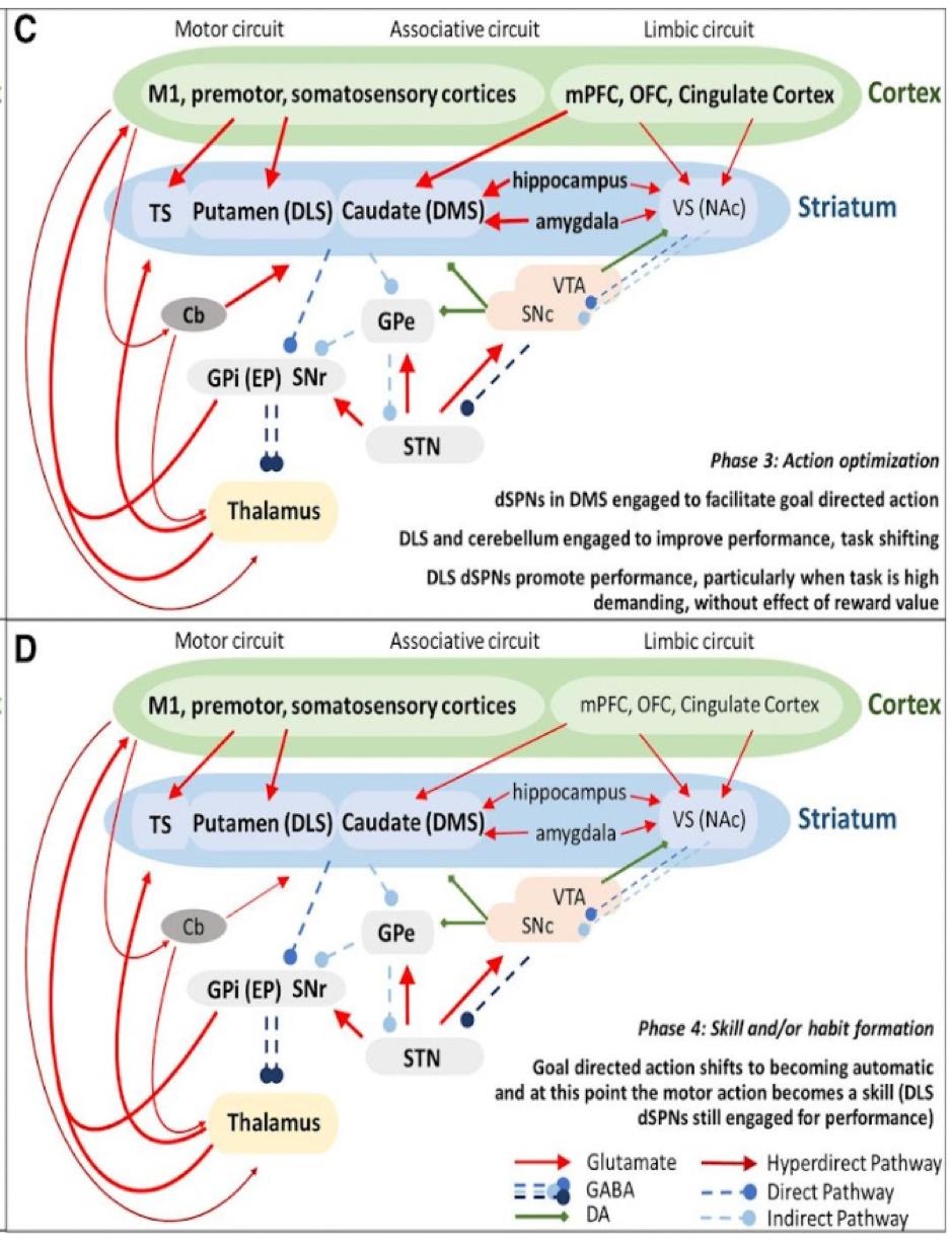
As discussed earlier, motor skill learning and performance are highly dependent on the basal ganglia pathways along with the nigrostriatal pathway that connects the VTA and dorsal striatum. According to the research, the dorsal striatum can be mostly involved in “automatized fine skills and micromovements embedded in an action”. (Cataldi, et al., 2021) More clearly, the role of the dorsal striatum in motor learning can be identified from the distinct steps of the learning process (Figure 1).
Advertisement
The New York University Integrative Psychology Review wp.nyu.edu/nyuipr
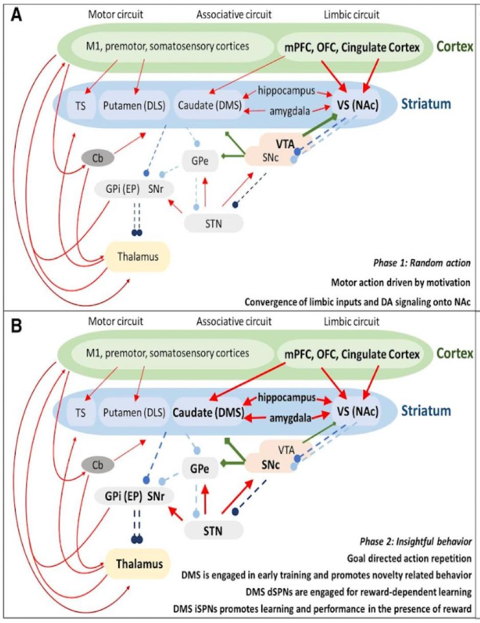
The researchers found that the first step in learning is based on random actions driven by motivation followed by the 2nd step which depends on “insightful behavior” (a person forms an association between a motor action and a specific goal, deciding on the most appropriate ways of achieving the goal to begin repetitive training of the skill). During the 3rd step the goal outcome is optimized with the adjustment of motor activity to reinforce the learned skill until it becomes a stable skill or habit in the 4th step. The nigrostriatal pathway may be particularly important in steps 3 and 4, since DA is involved in motivation and reward mechanisms (tested through pharmacological toxins that block the activation of DA1 and DA2 receptors) necessary for skill reinforcement and strengthening of the formed association between a motor action and action goal (e. g. for a planned movement to reach an object with a hand) (Cataldi, et al., 2021).
Additionally, various lesions to the basal ganglia pathways result in motor impairments. For instance, the lesions in a monkey's caudate and putamen are known to be associated with slow movements and decreased amplitude of executed movements.
Equally important is the fact that motor learning depends on procedural memory according to the case of H. M.; a patient subjected to a lesion of ⅔ of his hippocampi. As the result of the lesion, H. M. had impaired long-term memory which resulted in the inability to form new memories (anterograde amnesia), as well as to recollect old memories (retrograde amnesia). However, contrary to declarative memory, procedural memory remained intact in H. M., so he was able to show improvement in motor learning tasks consistently in the mirror-tracing task. The case of H. M. suggests the importance of the hippocampus for the 2nd step in motor learning (Scoville and Milner, 2000).
Overall, the evidence from behavioral studies, lesions, and pharmacological manipulation with DA receptors demonstrate the importance of basal ganglia in the motor learning process which is also supported by procedural memory.
Dopamine effects on synaptic plasticity in motor learning:
As was mentioned above, the dorsal striatum receives dopaminergic axonal projections from the substantia nigra (SN) and VTA. The role of the dopaminergic pathways in motor learning refers to motivation-reward mediation as well as the reinforcement of relevant motor actions (have positive outcomes) or degradation of irrelevant motor actions (have aversive outcomes) within steps 3-4 in motor learning discussed earlier. Aside from motivation-reward mediation, dopamine can indirectly regulate the velocity, accuracy, and execution of planned motor actions in motor learning (e.g. the inhibition of SN activity prior to the executed motor action can result in failed attempts to execute a movement and reduction of the following motor performance) (Molina-Luna, et al., 2009).
The striatum is preliminarily composed of spiny neuron cells which are targeted by the dopaminergic neurons that project from the VTA areas to the dorsal striatum. The spines receive the dopaminergic input symmetrically (at their “necks” and “heads”). The striatum also receives excitatory inputs from the M1 and thalamus. The DA neurons form synaptic connections with the medium spiny neurons (MSNs) that express DA receptors of different types (D1, D2, D3, etc.).
There are 2 main pathways in the striatum which convey direct spiny neuron projections (dSPNs) and indirect piny neuron projections (iSPNs). dSPNs express D1 excitatory receptors, whereas iSPNs express D2 inhibitory receptors. The difference in the expressed dopaminergic receptors for 2 distinct pathways accounts for the different effects of DA release in the striatum. On the striatal pathway level, the DA release allows either inhibition or promotion of movements through the preceding suppression/activation of thalamic activity upon DA release. By taking into account this regulatory mechanism and Hebbian homosynaptic plasticity
The New York University Integrative Psychology Review wp.nyu.edu/nyuipr theory it is possible to link the nigrostriatal pathway with the molecular mechanisms of LTP and LTD which can be involved in the process of motor learning as a person acquires a new motor skill (e.g. a post-stroke patient during the training exercise in the recovery therapy).
The latest research findings (Kida and Mitsushima, 2018) suggest that dopaminergic neurons within VTA can convey a representation of movement. This idea is supported by the fact that DA neurons are involved in the interaction of cognitive and motor systems which can be explained through the exploration of SPNs on the molecular level. Moreover, it was demonstrated that the changes within the nigrostriatal pathway related to the aging process can lead to slower cognitive activity as well as reduced DA release, especially prominent in impaired muscular tonic responses and smoothness of movement associated with Parkinson’s disease (PD).
On the molecular level, the shaping of motor behavior patterns and actions can depend on the dopaminergic signaling that mediates LTD and LTP. The experimental data showed that the inhibition of K+ voltage-gated channels in the dorsal striatum (K+ conductance is inhibited with a pharmacological drug) reduces the probability of LTD and verifies the existence of distinct molecular mechanisms for LTD and LTP regulation within the nigrostriatal pathway connected to M1 and, therefore, involved in motor learning. The levels of DA release are correlated with the effectiveness of skill learning in the situation where a person is involved in repetitive training of a new motor skill. In such situations, the dopamine precursor (Levodopa) distributed as a medication is associated with the improvement of motor activity in stroke patients, as they learn new motor skills. The acquisition of new motor skills requires subsequent plastic changes within the synapses of the existing neural circuits which can be modulated by DA, as D1 and D2 receptors are also present in M1. In accordance with the molecular mechanisms of LTP and LTD discussed earlier, M1 neurons can either express LTP or LTD, upon activation of
D1 or D2 receptors respectively. The expression of LTP in M1 will enhance the neural circuit associated with a newly trained skill, whereas the expression of LTD will result in the degradation of the irrelevant circuits which underlie the motor skills which are no longer trained for a long time (Trusel, et al., 2015).
As the research showed, motor skill learning may enhance synaptic strength, claiming that LTP actually strengthens the synapses as the process of learning takes place (Kida and Mitsushima, 2018).
The collective research data (Galvan, et al., 2001; Molina-Luna, et al., 2009; Rioult-Pedotti, et al., 2015) is consistent with the idea that DAergic neurotransmission in the primary motor cortex promotes the learning of new motor skills through the modulation of M1 synaptic plasticity modulating. Animal experiments (conducted with rats) with the eliminated DA terminals within M1 (intracortical injection of 6-hydroxydopamine (6OHDA) was made) demonstrate the impairments in the motor learning process. After the injection, the rats performed significantly worse in the “reaching task”. In this case, it would be reasonable to think if the impairments in the learning process are linked with disturbances of LTP in M1. Available data shows that inhibiting dopaminergic receptors in M1 decreases the probability of expressing LTPs.
Similarly, it was found that enhancing new motor skill learning via M1 LTPs may be associated with activating phospholipase C upon activation of D1 and D2 receptors in rats. (The inhibition of phospholipase C was linked with a decreased probability of LTP expression) Finally, the research that systematizes the chain of processes related to motor learning described above tested a hypothesis of the role of the dopaminergic VTA projection to M1 in the mediation of motor skills learning as the result of induced synaptic plasticity events within M1. The researchers applied the Retrograde Tracing from M1 and tyrosine hydroxylase immunohistochemistry in rats in order to confirm the presence of DA cells in VTA. Additionally, the
The New York University Integrative Psychology Review wp.nyu.edu/nyuipr inhibition of VTA dopaminergic neurons with the antagonists of D1 and D2 resulted in impaired motor learning in rats subjected to the reaching task as opposed to controls. Whereas the learning potential was restored upon the injection of a DA precursor (Levodopa) into the primary motor cortex of the antagonist-exposed rats (Kida and Mitsushima, 2018).
After the review of the scientific findings that investigate the links between the key structural components and processes associated with DA and motor learning, the theoretical mechanism of modulation of synaptic plasticity in motor learning by DA can be proposed, which includes the following chain of events: Dorsal striatum receives the DAergic input from the SN via direct and indirect pathways conveying the excitatory or inhibitory effect of DA1 and D2 receptors on the LTPs and LTDs. The effects of LTPs and LTDs determine the function of the spiny motor neurons (SPNs) and modulate the changes in synaptic plasticity within M1 indirectly, as the new motor skill is acquired and reinforced later.
Dopamine effects on synaptic plasticity in motor learning in Parkinson’s disease:
The logic described in previous sections of this paper is largely based on animal research. Due to the ethical concerns and specific constraints of human-subject research, the exact molecular and cellular mechanisms of dopaminergic impact on the synaptic plasticity in motor learning can be assessed in the research with Parkinson’s disease (PD) patients.
It is known that the disruptions of DA neuromodulation in humans is associated with a set of neurological disorders such as PD, attention deficit hyperactive disorder (ADHD), schizophrenia, and cocaine addiction (Pisani, et al., 2005).
For Parkinson’s disease the disturbances of dopaminergic pathways (convey excitatory inputs which are involved in the control of movement execution) along with the extrapyramidal pathway are thought to contribute to motor dysfunctions. Such disturbances conventionally are thought to be associated with the lowered activity of the striatonigral pathway (Whone, et al., 2003; Obeso, et al., 2008; etc.).
The studies featuring the role of basal ganglia and dorsal striatum in PD patients (Whone, et al., 2003) usually identify the impairment of medial spiny neurons and low DA levels as the main causes of the inhibition of thalamocortical and brainstem motor systems. The lack of DA within the striatum in PD patients diminishes motor control, making it hard for them to learn new motor skills and favoring the development of PD symptoms such as tremors, slowness, and stiffness of movement. Instead, excessive DA concentrations often result in the development of hyperkinetic disorders, such as chorea (induced jerky involuntary, uncontrollable movements, etc.) or neurological tics.
Following the logic of animal studies described above in which the scientists manipulated the DA receptors activity with the implication of injected pharmacology (regulates LTP/LTD), it is reasonable to expect that changes in synaptic plasticity of the striatum as the result of a disrupted activity of DA receptors may underlie the impairment of the spiny neurons (DA imbalance leads to the disrupted bidirectional input in SPNs) in PD as well as the observed neurological symptoms (indirect pathway expressing D2 receptors dominates in the pathological PD condition). In addition to motor performance, memory systems involved in motor learning may also be impaired. The evidence derived from the PD research in humans (Obeso, et al., 2008), which is centered on the plastic reorganizations of the motor cortex regions that receive DAergic inputs, suggests consistent findings with the animal research (inferred from electrophysiology data and behavioral observations). In the first place, there was an identified association between the synaptic plasticity in M1 (contributed to changes in motor neural circuits in PD) and striatonigral dopaminergic projection. Another confirmed association was the different roles of D1 and D2 receptors in the regulation of synaptic plasticity within the primary motor cortex (M1). Finally, the dysfunction of SPNs as the result of DA deprivation within the striatum was correlated with the observed behavioral deficits in the process of motor learning along with alterations in memory functioning in PD.
These observations in human subjects with PD explain the potential influence of DA on synaptic plasticity in motor learning on the neural pathway-level and provide similar findings in rat studies with a degree of validity. However, due to the limitations of the technology available to be used in research with human subjects, it is hard to assess the plastic changes in motor learning mediated by DA on the cellular and molecular levels.
The research with mice subjected to artificially modeled PD-like conditions showed that the elimination of D1 receptors or the injection of D1 antagonistic drugs induced the lack of long-term potentiation (LTP), disrupting the stable activation of SPNs (or their complete degradation) in the striatum (Xu, et al., 2017). The inhibition of LTD also yielded similar results. Therefore, intact LTP cellular mechanisms may play an important role in the stable functioning of SPNs in PD patients, however, this consideration requires more profound future research.
Equally important is that the study with modeled PD physiological conditions justifies the antagonistic role of D1 and D2 receptors in the mediation of D1-dependent LTP and D2dependent LTD, providing a scientific account for the maintained balance of the 2 pathways (direct/indirect) within the striatum which authorizes “dynamic adaptations” in synaptic plasticity, as the new motor skills are acquired and retained. Thus, PD-modeled research proposes an inversely parallel explanation for the cellular mechanisms of synaptic plasticity in motor learning, linking D1 and D2 receptors functioning with the functioning of SPNs (degradation or enhancement) (Xu, et al., 2017).
Overall, the context of Parkinson’s disease demonstrates a set of research works that form a solid scientific base for the logical sequence of structures and processes involved in synaptic plasticity in motor learning through the integration of molecular, cellular, organ-level, and behavioral data discussed in the earlier parts of this paper.
Critique of the proposed theory and future research perspectives
Despite the fact that there are multiple sources of scientific evidence of the involvement of the dopaminergic pathways (mainly nigrostriatal) and basal ganglia structures in the mediation of synaptic plasticity in M1 relevant for motor learning, the correlational nature of findings still makes it hard to separate the pathways and structures that are directly involved in the mediation of synaptic changes related to motor learning and reward pathways which are mainly involved in the reinforcement of the learned skills and behaviors.
Ethical concerns and limited technology (mostly electrophysiology and pharmacology, sometimes fMRI) in research with human subjects also make it significantly harder to accurately infer the exact role of the expressed cellular processes such as LTP and LTD in the motor learning process, as they were mostly researched in the context of memory formation and reorganization within the hippocampus. Moreover, further research on the plastic changes in the PD condition that constitute motor deficits tend to question the role of basal ganglia structures in the mediation of motor learning. Taking into account the complexity of the human brain, it may turn out that synaptic plasticity in M1 depends on the combination of activated brain structures and underlying alternative pathways (e.g. the role of mesolimbic DA pathway in motor learning is still not precisely clear despite its role in the reinforcement of learned skills) which go beyond the scope of the proposed DA-M1 mechanism of plastic changes in motor learning. Therefore, more profound research is needed to address this theory.
For a more holistic understanding of the role of DA in the synaptic plasticity in motor learning its impact and mediational effects can be studied in conjunction with the cholinergic pathways that release acetylcholine (ACh) since it is largely involved in muscle contraction and movement execution (Kida and Mitsushima, 2018).
Conclusion
As a result of analysis of relevant research literature, it is possible to observe the molecular and cellular mechanisms of synaptic plasticity in motor learning within the primary motor cortex which refer to the DAergic mediation of LTP and LTD through the expression of distinct receptors D1 and D2 (supported by the implication of the Hebbian monosynaptic plasticity theory to experimental settings with animals). A wide range of animal studies and studies in the context of PD with human subjects link the DAergic neuromodulation with the function of the nigrostriatal pathway (SN → dorsal striatum), relying on the analysis of the excitatory and inhibitory impacts of LTP and LTD on the normal functioning of SPNs in the striatum which are believed to be involved in the mediation of changes in synaptic plasticity within M1 in the process of motor learning. The research in the context of PD also allows us to generalize key findings of the animal studies (which explore the dysfunction of the striatum, the effects of DA precursor and pharmacological agonists on the functioning of SPNs) in order to prove the consistency of the proposed mechanism of synaptic plasticity in motor learning.
References:
Albanese, A., Altavista, M. C., & Rossi, P. (1986). Organization of central nervous system dopaminergic pathways. Journal of neural transmission.Supplementum , 22, 3-17. Beeler, J. A., Cao, Z. F. H., Kheirbek, M. A., Ding, Y., Koranda, J., Murakami, M., ... & Zhuang, X.
(2010). Dopamine-dependent motor learning: insight into levodopa's long-duration response. Annalsofneurology , 67(5), 639- 647.
Bear, M., Connors, B., & Paradiso, M. A. (2020). Neuroscience: Exploring the Brain, EnhancedEdition:ExploringtheBrain . Jones & Bartlett Learning.
Cataldi, S., Stanley, A. T., Miniaci, M. C., & Sulzer, D. (2021). Interpreting the role of the striatum during multiple phases of motor learning.
TheFEBSJournal
Filion, M. (1979). Effects of interruption of the nigrostriatal pathway and of dopaminergic agents on the spontaneous activity of globus pallidus neurons in the awake monkey. Brain research , 178(2-3), 425-441.
Galvan, A., Floran, B., Erlij, D., & Aceves, J. (2001). Intrapallidal dopamine restores motor deficits induced by 6- hydroxydopamine in the rat. Journalof neuraltransmission , 108(2), 153-166.
Hosp, J. A., Pekanovic, A., Rioult-Pedotti, M. S., & Luft, A. R. (2011). Dopaminergic projections from midbrain to primary motor cortex mediate motor skill learning. Journalof Neuroscience , 31(7), 2481-2487.
Kida, H., & Mitsushima, D. (2018). Mechanisms of motor learning mediated by synaptic plasticity in rat primary motor cortex. Neuroscienceresearch , 128 , 14–18. Molina- Luna, K., Pekanovic, A., Röhrich, S., Hertler, B., Schubring-Giese, M., Rioult- Pedotti, M. S., & Luft, A. R. (2009). Dopamine in the motor cortex is necessary for skill learning and synaptic plasticity. PloSone , 4(9), e7082.
Missale, C., Nash, S. R., Robinson, S. W., Jaber, M., & Caron, M. G. (1998). Dopamine receptors: from structure to function. Physiological reviews , 78(1), 189-225. Obeso, J. A., Marin, C., Rodriguez-Oroz, C., Blesa, J., Benitez-Temiño, B., Mena-Segovia, J., ... & Olanow, C. W. (2008). The basal ganglia in Parkinson’s disease: current concepts and unexplained observations. Annals of Neurology: Official Journal of the American Neurological Association and the Child NeurologySociety , 64(S2), S30-S46.
Pisani, A., Centonze, D., Bernardi, G., & Calabresi, P. (2005). Striatal synaptic plasticity: implications for motor learning and Parkinson's disease. Movement Disorders , 20(4), 395-402. RioultPedotti, M. S., Pekanovic, A., Atiemo, C. O., Marshall, J., & Luft, A. R. (2015). Dopamine promotes motor cortex plasticity and motor skill learning via PLC activation. PloS one , 10(5), e0124986.
The New York University Integrative Psychology Review wp.nyu.edu/nyuipr
Sejnowski, T. J., & Tesauro, G. (1989). The Hebb rule for synaptic plasticity: algorithms and implementations. In Neural models of plasticity (pp. 94-103). Academic Press. Sheynikhovich, D., Otani, S., & Arleo, A. (2013). Dopaminergic control of long-term depression/long-term potentiation threshold in prefrontal cortex. The Journalofneuroscience:theofficialjournalofthe Society for Neuroscience , 33(34), 13914–13926.
Speranza, L., di Porzio, U., Viggiano, D., de Donato, A., & Volpicelli, F. (2021). Dopamine: the neuromodulator of long-term synaptic plasticity, reward and movement control. Cells , 10(4), 735.
Scoville, W. B., & Milner, B. (2000). Loss of recent memory after bilateral hippocampal lesions. 1957. The Journal of neuropsychiatry and clinical neurosciences, 12(1), 103–113. Trusel, M., Cavaccini, A., Gritti, M., Greco, B., Saintot, P. P., Nazzaro, C., ... & Tonini, R. (2015). Coordinated regulation of synaptic plasticity at striatopallidal and striatonigral neurons orchestrates motor control. Cellreports , 13(7), 1353-1365.
Wickens, J. R., Reynolds, J. N., & Hyland, B. I. (2003). Neural mechanisms of reward-related motor learning. Current opinion in neurobiology , 13(6), 685-690.
Whone, A. L., Moore, R. Y., Piccini, P. P., & Brooks, D. J. (2003). Plasticity of the nigropallidal pathway in Parkinson's disease. AnnalsofNeurology:Official JournaloftheAmericanNeurologicalAssociation andtheChildNeurologySociety , 53(2), 206-213.
Xu, T., Wang, S., Lalchandani, R. R., & Ding, J. B. (2017). Motor learning in animal models of Parkinson’s disease: aberrant synaptic plasticity in the motor cortex. MovementDisorders , 32(4), 487-497.
The New York University Integrative Psychology Review wp.nyu.edu/nyuipr
