
15 minute read
Socket Graft Management


SOCKET GRAFT MANAGEMENT: WHAT YOU NEED TO KNOW
By: David H. Wong, DDS | Diplomate, American Board of Periodontology
Advertisement
Think for a moment about how common tooth extractions are in dentistry. Now think about bone loss, how it’s virtually irreversible, and how it’s associated with disease and inflammation. How are these two related and what can be done about it?
As we explore this question, we need to ask more questions: What happens to the bone after a tooth is extracted? How long does it last? Is it preventable? How?
INTRODUCTION
There are many reasons why teeth are extracted: severe periodontal disease, non-restorable caries, root fractures, orthodontics, pathology, etc. What the literature says about extractions and bone loss may be surprising. For example, tooth loss creates 40% to 60% alveolar bone loss in the first 2-3 years and then a resorption rate of 0.5% to 1% every year for the rest of the patient’s life.3 Bone loss also occurs both horizontally and vertically. While the extraction socket does fill with bone, the socket is also often occupied with connective tissue.2
What is the cause of the quantity and rapidity of bone loss? The reason is best explained by the bundle bone, which occupies the facial plate of a tooth. This bundle bone is very avascular and receives its blood supply from the periodontal ligament, the periosteum, and the alveolar marrow spaces.1 Once a tooth is removed, the loss of bundle bone is rapid and is responsible for the majority of the bone loss that occurs early on.
This bone loss occurs nearly universally in all individuals as well as with all teeth to varying degrees. The most common treatment to minimize (not eliminate) bone loss is to place a bone replacement graft into the socket at the time of extraction. There are several notable benefits to grafting an extraction socket:5
• Enable the placement and stability of implants • Reduce the loss of bone volume • Reduce the need for additional bone journal | Nov/Dec 2020grafting
• Provide quality bone for implant osseointegration • Improve the esthetics of the final prosthesis • Regenerate bone faster • Protect the adjacent teeth
SURGICAL CONSIDERATIONS TO SOCKET SITE MANAGEMENT
When it comes to extracting a tooth, care must always be taken to preserve the facial and lingual cortical plates. Without these valuable structures, a simple socket graft may turn into a more complex procedure. For the purposes of this article, socket graft management will be discussed assuming a four-walled socket. Situations where a bony wall is missing (advanced periodontal disease, root fractures, endodontic lesions, etc.) often require additional planning and different execution. To simplify socket grafting procedures, every effort should be made to minimize trauma. Utilizing an “atraumatic” surgical technique is of utmost importance.
GRAFT MATERIAL SELECTION
When it comes to choosing bone replacement graft materials, the options are endless. Fortunately, for basic socket grafting needs several materials will suffice. Bone graft materials are generally categorized into four groups: autografts, allografts, xenografts, and alloplasts.
• Autograft - Donor and recipient sites are in the same individual.
• Allograft - Donor and recipient sites are from two different individuals of the same species, i.e. cadaver bone. These graft materials may also be further subcategorized into mineralized or demineralized products.
• Xenograft - Donor and recipient sites are from two different individuals of different species, i.e. animal bone.
• Alloplast - A group of synthetic bone grafts such as ceramics and glasses and a multitude of other materials. It is important to note several features about grafted bone compared to “native” bone. Grafted bone yields similar bone-to-implant contact as with native bone (40%-65%) and offers good primary stability at the time of implant insertion. It also does not impair early osseointegration. Grafted bone is able to sustain loading conditions long-term.
THE SCIENCE OF BONE GRAFTING
There are several mechanisms at work when it comes to how bone graft materials are able to aid in the regeneration of bone in an extraction socket. Osteogenic materials are able to directly form bone. This is an ideal property. Meanwhile, osteoinductive graft materials stimulate bone growth by influencing undifferentiated mesenchymal cells. Finally, osteoconductive bone grafts lead to bone formation by serving as a scaffold for bone growth.
Despite the properties that bone graft materials possess, several other factors must be present to allow for proper and predictable bone growth. According to Wang and Boyapati6 four critical factors must be present:
• Primary closure/Passivity of the flap
• Angiogenesis
• Space Maintenance of the graft
• Stability of the graft
When performing bone grafting procedures, all of these factors should be considered to allow for maximum predictability and success.
CASE STUDY
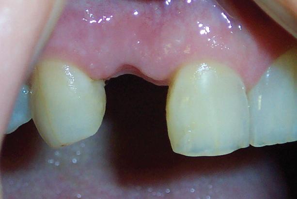
A 65-year-old healthy female presented with a hopeless lateral incisor (#7). The tooth had fractured at the gingival margin and was determined to be non-restorable (Fig. 1). She presented with the crown of the tooth bonded to the adjacent teeth. Her ultimate goal was to have the tooth replaced with a dental implant. This case series focuses on site preparation with extraction of the tooth and placement of a socket graft.
Following informed consent, local anesthesia was achieved. As with every extraction, the first priority was to remove the tooth as atraumatically as possible (Fig. 2). The ultimate goal was to protect the facial plate and avoid fracturing the bone. Once the tooth was successfully removed, the extraction socket was thoroughly evaluated.
To evaluate the socket, a periodontal probe or explorer is used to examine the socket walls. In cases where a fenestration or dehiscence is present in either the facial or lingual cortical plates, various utilizations of flap designs are available as well as the addition of additional materials such as barrier membranes or blood-derived growth factors. In this situation, a fourwalled socket was present, so the following steps were taken to graft the socket:
1. 0.5 cc of mineralized cortical bone was hydrated in sterile water for 15-20 minutes (Fig. 3). Many other bone substitutes are available which would suffice, and sterile saline is often utilized to hydrate the bone graft material instead of sterile water.
2. The socket was then thoroughly debrided of any inflammatory tissue using scalers and hand instruments Figure 1 Figure 4 REFERENCES 3. Once hemostasis subsided, the mineralized cortical bone was introduced into the socket and gently condensed utilizing a cotton tip applicator. Care was taken to not pack the particles too tightly so as not to interfere with proper fluid exchange and clotting. Particular attention was also paid to ensuring that the bone graft material was placed at the level of the adjacent bone, not the level of the gingival margin.
4. The soft tissue was partially closed by tying a figure 8 suture over the socket with 4-0 chromic gut suture (Fig. 4). An Essix retainer was then utilized as the temporary prosthesis (Fig. 5).
5. The healing at 12 weeks shows a mature socket that is now prepared for either a dental implant or fixed partial denture (Fig. 6).
FINAL THOUGHTS
Once extraction of the tooth and grafting of the socket is complete, it is generally recommended to allow 12 weeks before considering permanent tooth replacement with options such as a fixed partial denture or a dental implant.4
ABOUT THE AUTHOR:
Dr. David Wong is a board-certified periodontist in private practice in Tulsa, Oklahoma.
Dr. Wong received his undergraduate education and dental training at the University of Oklahoma. He then went on to complete a three-year residency in periodontics at the University of MissouriKansas City. He is a Diplomate of the American Board of Periodontology as well as an Adjunct Associate Professor at the University of Pennsylvania School of Dental Medicine and an Associate Clinical Professor at the University of Oklahoma College of Dentistry. He is a published author in several peer-reviewed dental journals but has also reached a mainstream audience in media such as Fox News and the Wall Street Journal. Dr. Wong presently resides in Tulsa with his wife and three children where he maintains
Figure 2


Figure 5


a full-time private practice. Figure 3
1. Araujo MG et al. Dimensional ridge alterations following tooth extraction. An experimental study in the dog. J Clin Periodontol, 2005; 32(2): 212-8. 3. Christensen GJ. Ridge Preservation:Why Not? J Am Dent Assoc. 1996; 127: 669-670
4. Neiva R et al. Analysis of tissue neogenesis in extraction sockets treated with guided bone regeneration: clinical, histologic, and micro-CT results. IJPRD. 2011; 31(5): 457-469 Figure 6
5. Pagni G, et al. Postextraction alveolar ridge preservation: Biological basis and treatments. Int J Dent. 2012: 151030.
Collect What You Produce: INSURANCE MANAGEMENT
By: Cathy Jameson, PhD | Part 7 of a ten-part series
“Be insurance-aware but not insurance-
driven.” (Cathy Jameson)
Dental insurance is an asset to both you and your patients. You can increase the amount of care you provide and your patients can receive the dental care they need. While insurance is beneficial, there are frustrations. Your patients would like to have better coverage for needed and desired care, and you would like to be paid more equitably for the care you provide.
Insurance-related challenges that you may face include: (1) little to no increases in yearly maximums resulting in an insurance plan that covers less and less care; (2) insurance companies dictating your fees and/or types of treatment; (3) inaccurate communication to your patients about your fees; and (4) a major impact of managed care on the profession.
Approximately 85% of all dental insurance is PPO (Preferred Provider Organization), which is a major change in insurance coverage. Four out of every five dentists accept at least one PPO. There are fewer and fewer total fee-for-service (private pay) providers. According to the ADA, 50% or more of your income will flow through your practice in the form of insurance reimbursement. It is imperative that you manage this system correctly. The purpose of this article is to explore how best to manage insurance. It is not a comprehensive treatise on insurance management. If you have specific questions related to insurance, I refer you to Dr. Charles Blair’s books on managing insurance and on coding (Coding with Confidence and Administration with Confidence), or you can contact our consulting division at Jameson Management (info@jamesonmanagement. com or www.jamesonmanagement.com).
THE PERSONAL NATURE OF INSURANCE People feel very close to their insurance plans. Thus, the way you handle your patient’s insurance is important. No matter how good or poor the plan, do not be critical of it in front of the patient. It’s a benefit they’ve paid for or received journal | Nov/Dec 2020 from their employer. If you’ve chosen to participate in a managed care program (and adjust your fees accordingly) it’s your choice. It’s not the patient’s fault if the insurance company is difficult to deal with or doesn’t pay well. Handling insurance is a process that needs and deserves special attention.
INSURANCE VERIFICATION It is important that you verify a patient’s insurance program before treatment is rendered. Gather relevant information about the benefit package. Is the patient eligible? What are the benefits?
PRE-DETERMINATION OF BENEFITS Unless an insurance company requires a pre-determination of benefits, don’t provide one. Most patients who ask for a pre-determination do so because dental practices have taught them to do so. Some practices are so apprehensive about asking for money that they defer it by saying, “Let’s see what your insurance is going to pay, then we can see what we need to do— or see how much you will owe.” That’s a stall tactic – one that insurance companies love!. Approximately 50% of people who file a pre-determination of benefits will never proceed with treatment.
According to insurance experts, a predetermination of benefits should be done only if (1) the insurance company requires it, (2) the patient requests it, or (3) the patient refuses to accept accountability for the strengths and weaknesses of their dental plan.
Just because a patient receives a predetermination of benefits doesn’t mean the insurance company will pay. If a patient has an extensive treatment plan and insists on a pre-determination, consider filing the pre-determination but start only the restorative care. By the time a response is received, you would then be ready for more extensive or complicated treatment. If you are going to accept assignment of benefits, consider the following:
1. Quote the entire fee and let the patient know that they are ultimately responsible for the entire fee, regardless of what insurance reimburses.
2. File insurance claims as a service to the patient. Take the assignment of benefits.
3. Collect the estimated patient portion at the time of the service. Do not under any circumstances file the insurance and wait to collect the patient portion after insurance has paid. This is financial suicide.
4. Let the patient know up front that they are responsible for the account; the insurance contract is between them and the insurance company. Put into writing that if for any reason insurance hasn’t paid in 45 days (or whatever time frame you choose) the patient will need to pay the balance in full. And if the insurance company doesn’t pay at all, the patient will be responsible for the total amount.
5. Be sure you have a written financial agreement on file. (Many people never remember that they ever agreed to anything.)
6. Keep track of this entire process. If insurance hasn’t paid within the agreedupon time frame or if payment leaves a balance remaining, call the patient immediately and collect the balance with a credit card or patient financing program or send a statement with a self-addressed stamped envelope.
7. Prepare a pre-authorization form that is secured and firewalled in your records.
8. Place any post-insurance balance (or the full balance if insurance doesn’t pay at all) onto the patient’s healthcare financing program or on a bankcard.
MORE ABOUT INSURANCE MANAGEMENT Your insurance system must be managed/ monitored with extreme dedication. What do you collect per year? What is 50% of that? Your management of this much money is a critical factor of the business of your practice and must be handled accordingly. All management systems in the dental practice need to be both time- and cost-efficient, including insurance management.
About 77% of the population has some dental insurance, leaving 23% without coverage. The average maximum annual insurance coverage remains $1,000 to $1,500. The 77% with insurance may need assistance with the part not covered by their annual maximum. It goes without saying that the 23% without coverage will need assistance for a greater portion of their dental costs.
If you are filing insurance, develop a controlled system for filing and following up on all claims. You should consider contacting a management expert who can help you set up that system, learn to administer it effectively, and make sure that you get great results (1) if you do not have a 7-10 working-day turnaround on all claims, (2) if you have 30-day past due claims, and/or (3) if you don’t really know the status of your insurance. Too much money is at stake.
SUMMARY Be insurance-aware, not insurancedriven. Don’t let what an insurance company will or will not cover determine the type of treatment you recommend. Remember that insurance companies are not interested in whether or not the treatment you recommend is best for the patient. They are only interested in whether or not something is a covered benefit, and they will pay accordingly. Consider the following “Rule of 10 Insurance Musts”:
1. Decide whether or not you are going to accept assignment of benefits and manage accordingly.
2. Practice verbal skills and learn to 3. Get insurance verifications quickly -- before the patient arrives, if possible.
4. Move away from unnecessary insurance pre-determinations.
5. Collect the patient’s estimated portion at the time of the service.
6. Make sure your patients understand that they are ultimately responsible for the full treatment fee.
7. Develop a foolproof system for filing and collecting insurance claims.
8. Make sure you are receiving insurance payments within 7-10 working days and that you have no past due claims.
9. Collect any remaining post-insurance balance from the patient immediately. Do not let any such outstanding balances sit on your books and lose value every month.
10. Insurance collections are a major part of your entire income. Honor this with good management, time, attention, care and respect.
Make it easy for people to pay. Convenience is important to busy people in today’s world. Insurance has been a wonderful supplement to people’s dental care and to the growth of dental practices. Now more people can use insurance benefits because they have a way to finance the difference after insurance pays with patient financing programs.
ABOUT THE AUTHOR:
Cathy Jameson, PhD, is the founder of Jameson Management, Inc., an international management, hygiene, and marketing firm which offers proven management and marketing systems for helping organizations improve in a positive, forward-thinking culture. Jameson holds a doctorate in management from Walden University where she focused her research on transformational leadership. She has been inducted into the College of Education Hall of Fame and is a Distinguished Alumna of Oklahoma State University. She serves on the Board of Governors there. Jameson has been named one of the top 25 Women in Dentistry and has received Lifetime Achievement Awards from the Excellence in Dentistry Organization and from the Academy of Dental Office Managers. She was a finalist for the Stevie Award for outstanding entrepreneurial women. She is a member of the American Association of Female Executives, National Speaker’s Association, Academy of Dental Management Consultants, National Society of Leaders and Success and Chi Omega Women’s Fraternity. Jameson has lectured in all US states and in 31 countries. She has had over 1,500 articles published throughout the US and the world. She is the author of eight books, including the 3rd Edition of her bestseller, Collect What You Produce and Creating a Healthy Work Environment. These can be purchased from Amazon.
For more information on Dr. Jameson’s lecture or personal consulting services, contact her at Cathy@ jamesonmanagement.com. For more information on the consulting services of The Jameson Management Group, contact www.info@jamesonmanagement.
com or www.jamesonmanagement.com


