
2 minute read
AEGD Case Studies


Case #1
By: Aziz Pradhan, DMD
Advertisement
A 67-year-old male presented to the University of Oklahoma Advanced Education in General Dentistry Program in December 2020 for a comprehensive dental exam and treatment plan. The patient’s chief concern was that he would like to take care of his oral health after not seeing a dentist for several years. He is taking medications to manage high blood pressure, high cholesterol levels, and an enlarged prostate. A carotid artery stent was placed in December 2022.
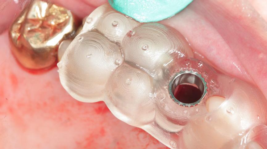
The patient presented with missing teeth #1, 3, 16, 17, 19, an FPD 2-x-4 with recurrent caries on #2, coronal fracture of #12, recurrent buccal caries on #15 under an existing amalgam restoration, and a vertical root fracture on #19. The periodontal diagnosis for the patient was localized Stage I Grade A periodontitis. Initial treatment consisted of localized SRP, sectioning of FPD 2-x-4, extraction of #2, and extraction of #19. Implants were placed and restored to replace #2 and #3. Tooth #15 was restored with a gold crown. Due to the extent of the coronal fracture on #12, the tooth was deemed non-restorable (Figure 1). A CBCT scan showed adequate palatal bone for an immediate implant.

The implant on #12 was placed using a fully guided protocol. Intraoral scans were taken and uploaded as STL files to Dentslpy Sirona’s inLab CAD software. Tooth #12 was digitally removed, and the edentulous space was replaced with a digital wax up of #12 (Figure 2).
REALGUIDE ™ was used to fabricate the surgical guide. STL files of the maxilla with #12 digitally removed and of the digital wax up were uploaded into the REALGUIDE ™. Additionally, the CBCT file was uploaded into the planning software. An Astra 3.6X9mm implant was planned to ensure adequate engagement of palatal bone and acceptable prosthetic positioning (Figure 3). After planning was completed the guide design was uploaded to the InLab CAM Software and printed using Dentsply Sirona’s Primeprint and guide cylinder was added.
Tooth #12 was atraumatically extracted with an elevator and forceps technique. The surgical guide was seated (Figure 4) and the Astra guided implant sugery kit was used to prepare the osteotomy. Symbios demineralized corticated and cancellous bone was placed into the osteotomy followed by placement of the implant to depth achieving a primary stability of 15 Ncm. A cover screw was placed, and the site covered with additional graft. A collagen plug was used to cover the site followed by placement of Figure-8 and simple interrupted chromic sutures. A post-operative periapical radiograph was taken (Figure 5) and the patient was dismissed with post-operative instructions.
About The Author
Dr. Aziz Pradhan is originally from Boston, MA. He attended Tufts University and earned his B.S. in Biology. He attended Tufts School of Dental Medicine where he received his D.M.D. After completion of the AEGD residency, he plans to practice in Dallas, TX.


