1 minute read
Lymphatic drainage


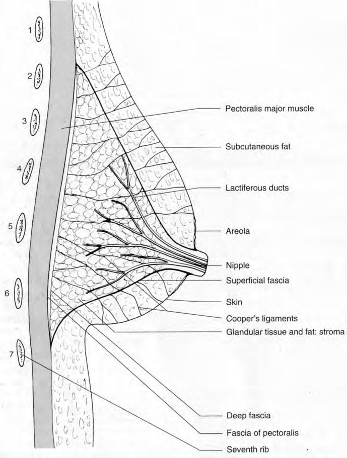
Fig. 9.1 Breast: sagittal section.
• The lateral thoracic branch of the axillary artery supplies 30%, mainly upper outer quadrant; and • Perforating branches of the anterior intercostal arteries.
Advertisement
Venous drainage accompanies the arteries to the axillary and subclavian veins and the azygos system.
LYMPHATIC DRAINAGE (see Fig. 9.2) There are superficial lymphatics under the skin of the breast and a particular concentration in the subareolar plexus, beneath the nipple. Lymph flows unidirectionally from superficial to deep in the breast to the perilobular and deep subcutaneous plexus. Lymph in the deep plexus then drains centrifugally from the nipple to the axillary and internal mammary chains. However, the majority of drainage is to the axillary chain, w i th less than 5% draining to the internal mammary chain.
The axillary lymph nodes are arranged in groups, arbitrarily referred to as levels. Level I nodes lie lateral to the lateral border of pectoralis minor. Level II nodes lie behind pectoralis minor. Level I II nodes lie medial to the

Fig. 9.2 Breast: anterior view demonstrating relationship to chest wall, blood supply and lymph drainage.
medial border of pectoralis minor. Nodes may also lie in the breast tissue. The most common location is in the upper outer quadrant and axillary tail. The significance of identifying the nodal groups is that breast cancer is thought to spread in a sequential fashion, initially to the level I nodes. If the level I nodes are not involved, then it is unlikely that other 'higher level' nodes w i ll be involved. Thus finding negative level I nodes may save the patient further axillary surgery in the case of breast cancer. The status of the level I nodes is also of major prognostic significance. This forms the basis for sentinel node mapping techniques (see below).
The internal mammary nodes lie in the intercostal spaces in the parasternal location, adjacent to the internal mammary vessels in the extrapleural fat.


