4 minute read
FIT TO PRINT


FROM DIAGNOSIS TO TREATMENT, TAMPA GENERAL HOSPITAL AND USF HEALTH ARE LEADERS IN 3D PRINTING TECHNOLOGY
BY SETH SOFFIAN
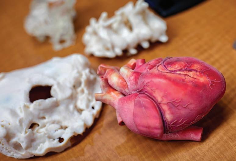
Summer Decker, PhD, was describing the look and feel of a human liver—tough and dense, with complex vasculature and distinct segments—when she darted away for something better than words. She returned to her offi ce in the radiology department holding an exact replica, created on a 3D printer, of a cancer patient’s actual liver.
Immediately clear on the multicolored, multi-textured object was the precise location of the mass that needed to be removed and the intricate blood vessels that surgeons would need to navigate in order to do so.
“We’ve been able to give [surgeons] a clear version, so they know exactly where that tumor is and exactly where the vessels are,” said Decker, a key fi gure leading the internationally renowned 3D printing efforts at Tampa General Hospital and director of 3D clinical applications, vice chair for research and innovation in radiology, and associate professor at the USF Health Morsani College of Medicine at the University of South Florida.
Sophisticated models like the one Decker displayed provide doctors in cardiology, oncology, trauma, and other disciplines with more than just an all-angles look at complex problems. They also allow physicians to test different devices and techniques for optimum fi t and suitability before performing surgical procedures. Printing capabilities are so good— down to the micron level, equal to one thousandth of a millimeter—that they surpass what X-rays, MRIs, CT scans, and other images can provide.
“Even the fi nest scan cannot be as good as what the printers can print,” Decker said.
Having seven of the most state-of-the-art 3D printers available enables the TGH and USF Health team to create models in rapid fashion, revolutionizing how physicians envision and execute treatment plans.
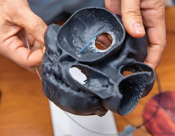
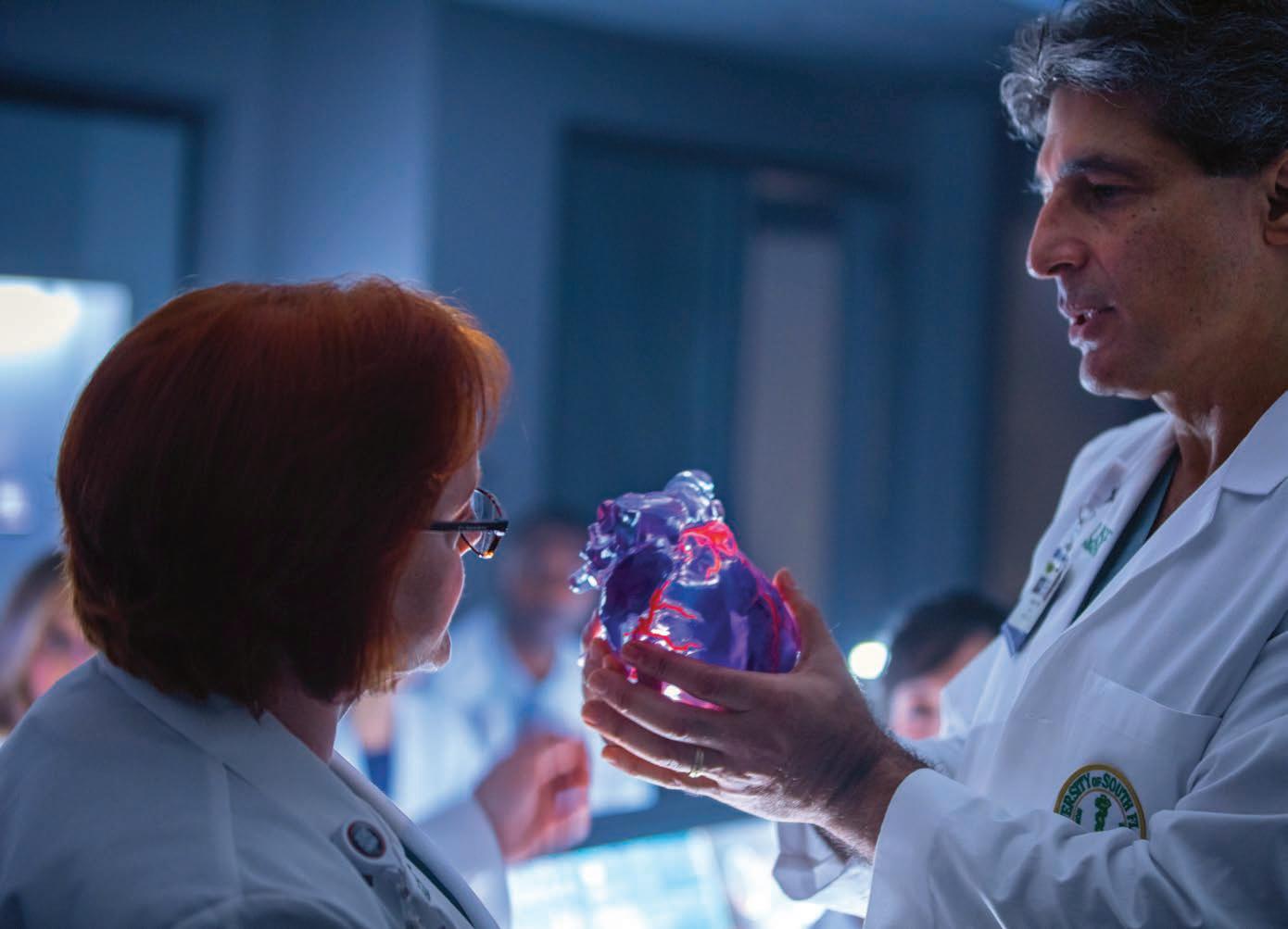
Advances in 3D printing currently in use at TGH include creating models in a biomimetric material that mimics human tissue (right) and using reconstructions to guide surgeries (below). Opposite page: Summer Decker, PhD, and Dr. Fadi Matar examine a 3D-printed heart.
Dr. Krishna Nallamshetty
One of the foremost benefi ts is the ability to forecast possible ramifi cations of a procedure. “It’s been helping tremendously,” said Dr. Fadi Matar, an interventional cardiologist at TGH and associate professor of internal medicine at the USF Health Morsani College of Medicine, who now won’t touch a complex case without a patient’s 3D model. “A new heart valve can compromise other structures. We look at the impact of the device on the model. A lot of times our fears are not substantiated, and then we can proceed.”
Another improved outcome from such advancements are shorter surgeries, which can mean less time under anesthesia for patients. One case at TGH saw the shattered half of a patient’s face—damaged by a gunshot—reconstructed using a mirrored 3D print of the undamaged side. This model helped to cut surgical time for the procedure from 11 hours to three.
The use of 3D printing in medical research dates back well beyond a decade. But TGH and USF—early adopters of their own research program that fed clinical breakthroughs as well—continue to benefi t from and contribute to groundbreaking work in the fi eld.
“We get referrals from all over the country for high-complexity patients,” said Dr. Krishna Nallamshetty, chief of staff and comedical director of radiology at TGH and an associate professor of radiology and cardiology in the USF Health Morsani College of Medicine. “The people who have used the technology are amazed by how useful it can be and what the impact is on patient care.”
In fact, fi rms in aerospace, automotive, fi lmmaking, and other industries turn to TGH when adapting new technology to their medical divisions. “We actually advise industry,” said Decker, who trains radiologists, residents, medical students, technologists, and other medical professionals in 3D printing at facilities around the world. “We sit on the committees to say, ‘This material works. This doesn’t. This one works for hearts. This one doesn’t.’ We can really guide the industry to give us what we need medically.”
The 3D-printed liver Decker displayed, for example, uses what is known as biomimetic material, which mimics human tissue. It is superior to anything available even a couple of years ago, she explained.
Matar is such a proponent of 3D printing that he has begun researching heart replica models that can actually pump, unlike existing static models. It’s the kind of continued collaborative work the USF Health and TGH team hopes to see more of as the imaging team moves from the USF Health South Tampa Center for Advanced Healthcare to the main hospital.
“Every doctor who comes into radiology will be able to see what we can offer,” Nallamshetty said. “Then we can suggest to them how to best apply some of these really innovative techniques to help take care of our patients better.”
Such achievements need not be limited to life-like tissues or the most complex cases. In 2020, for instance, USF Health earned international acclaim for their rapid development of a widely deployed 3D-printed nasal swab that helped alleviate global supply shortages at the outset of the COVID-19 pandemic. The USF-patented design, which was tested at TGH and other health care facilities, has now produced more than 80 million swabs in more than 57 countries around the world.
From materials to surgical prep and application, the promise of 3D printing knows no bounds. “I think we’re still just at the tip of the iceberg,” Nallamshetty concluded. “The best is yet to come.”