Cardiac Care
SPRING 2024 An Update for Physicians from the Heart, Vascular and Thoracic Institute The Hypertrophic Cardiomyopathy Program: A Center of Excellence Leading the Way in Care p. 10 Feature Article Same-Day Discharge After TAVR p. 4 TCAR: Innovations in the Treatment of Carotid Artery Stenosis p. 5 Multimodality Cardiac Imaging
Hypertrophic Cardiomyopathy p. 12
for
José L. Navia, MD, FACC

Dear Colleagues,
Vice
Chief,
Cleveland Clinic in Florida Regional Heart, Vascular and Thoracic Institute
Director, Heart and Vascular Center, Weston Hospital Chairman, Cardiothoracic Surgery, Cleveland Clinic in Florida
S.
Donald Sussman Distinguished Chair in Heart and Vascular Research
Teamwork is essential to the success of any organization, and it is what makes Cleveland Clinic in Florida thrive. We have multidisciplinary teams in every specialty working together to provide the best in patient care, and we regularly collaborate with experts at our main campus in Cleveland, Ohio, and with other healthcare organizations in Florida. We know that the best outcomes happen when knowledge, expertise and best practices are shared.
Our hypertrophic cardiomyopathy (HCM) program is a notable example of this. As one of Florida’s leading referral centers for HCM care, it is recognized by the Hypertrophic Cardiomyopathy Association as one of only 2 centers of excellence for the condition in the State of Florida. You will find an overview of the program, which is our feature story for this issue, on page 10.
Imaging, of course, is of the greatest importance for the correct diagnosis of HCM. Our HCM physicians and imaging specialists are experts in multimodality cardiac imaging and they work closely together as a team to care for our high number of HCM patients (see page 12).
Cleveland Clinic surgeons in Cleveland and Florida are among a small group who have pioneered myectomy for obstructive HCM. We perform high volumes of the procedure and, through a unique collaboration with Cleveland Clinic in Ohio, we are able to offer this surgical option for patients in South Florida. To learn more, read the article on page 14.
Also in this issue, we discuss the feasibility of same-day discharge after the TAVR procedure (page 4), and the latest innovation in the treatment of carotid artery stenosis (page 5).
We hope you enjoy all of the articles in this issue of Cardiac Care, our first of 2024. We welcome the opportunity to partner with you on specialized care for your patients, should you need the assistance.
Respectfully,
José L. Navia, MD, FACC
Medical
José L. Navia, MD, FACC naviaj@ccf.org
Nicolas Brozzi, MD, FACC brozzin@ccf.org
Managing
Table of Contents Same-Day Discharge After TAVR ................................ 4 TCAR: Innovations in the Treatment of Carotid Artery Stenosis .......................................... 5 A Message from Cleveland Clinic Weston Hospital’s Vice President and Chief Medical Officer .................... 6 Patient Spotlight: Woman Finds Lifesaving Care for Hypertrophic Cardiomyopathy at Cleveland Clinic in Florida ......................................... 8 Feature Article: The Hypertrophic Cardiomyopathy Program: A Center of Excellence Leading the Way in Care 10 Multimodality Cardiac Imaging Guides the Diagnosis and Management of Hypertrophic Cardiomyopathy .................................. 12 Surgical Treatment of Hypertrophic Cardiomyopathy 14 Transforming Cardiovascular Care: Three of the Latest Innovations 16 Mark K. Grove, MD, Retires After Long Career at Cleveland Clinc 18 New Staff .............................................................. 19 Cardiac Care is produced by The Heart, Vascular and Thoracic Institute in Florida.
Editors
Editor Christine Harrell
Director Suzette Lopez Marketing Evelyn Arias, Senior Director Christina Garcia, Regional Service Line Manager To reach a staff member or to inquire about our services, please call: clevelandclinicflorida.org/heart Cleveland Clinic Weston Hospital Cardiology 954.659.5290 Cardiothoracic Surgery 954.659.5320 Cleveland Clinic Indian River Hospital Cardiology 772.778.8687 Cardiothoracic Surgery 772.563.4580 Cleveland Clinic Martin Health Cardiology and Cardiothoracic Surgery 772.419.2137
Art
Same-Day Discharge After TAVR: An Important Innovation for Improved Patient Experience
Mauricio G. Cohen, MD, FACC, FSCAI Director, Structural Heart Interventions
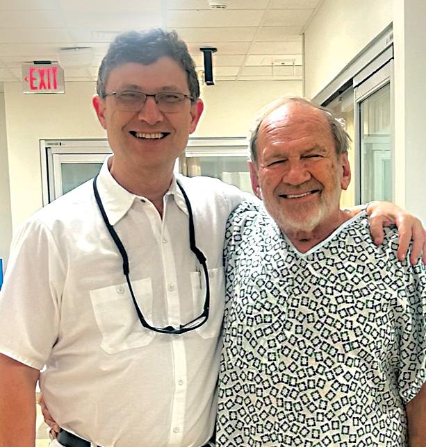
Transcatheter aortic valve replacement (TAVR), a minimally invasive procedure for severe aortic stenosis, has traditionally involved a hospital stay for post-procedural monitoring. However, advances in technique have enabled a paradigm shift towards same-day discharge, offering an opportunity for patients to enjoy an expedited recovery in the familiar comforts of home.
Cleveland Clinic in Florida is committed to innovation in cardiac care including embracing sameday discharge protocols for eligible TAVR patients, which offers several notable benefits to both patients and healthcare systems. Patients experience reduced exposure to hospital-acquired infections and enjoy a faster return to their routine activities. This approach also optimizes hospital resources, allowing for efficient utilization of beds and a smoother patient flow within the healthcare system.
Procedural factors determine eligibility for same-day discharge
The feasibility of same-day discharge relies mostly on procedural factors, as neither patient age nor the Society of Thoracic Surgeons (STS) risk score are predictors of readmission. Results of the multicenter PROTECT TAVR study in 2022 showed a 2.8% rate of 30-day cardiovascular readmission among 124 patients selected for same-day discharge after TAVR. The mean age of the patients was 78 years and median STS score was 2.4%.
Procedural factors include a minimalist approach under moderate sedation, primary percutaneous femoral access, secondary transradial access, completion of a femoral angiogram to confirm hemostasis, and absence of complications.
Post-procedural care includes a 6-hour observation period with telemetry monitoring for delayed rhythm disturbances, hemodynamic stability, and comfortable ambulation. Patients with new (such as LBBB) or pre-existing conduction deficits require longer observation. Transthoracic echocardiography is performed before discharge.
The results of an additional study confirmed that when the abovementioned procedural factors and
post-procedural care were in place, same-day discharge after TAVR was feasible without risk to patient safety post-discharge.
Our multidisciplinary team, comprising experienced interventional cardiologists, cardiac surgeons, anesthesiologists, and specialized nursing staff, meticulously evaluates each patient to ensure suitability for same-day discharge, considering additional factors such as social support and proximity to a medical center, should a problem arise. Patients discharged on the same day are provided a direct phone number for questions and are expected to return for a follow-up visit that includes electrocardiogram within 24 to 48 hours.
As part of our ongoing commitment to optimizing patient care, Cleveland Clinic in Florida remains dedicated to advancing patient management strategies. Our relentless pursuit of excellence, coupled with a patientcentric approach, continues to shape the future of cardiovascular care, allowing us to offer innovative solutions like same-day discharge after structural interventions to enhance patient outcomes and experiences.
Dr. Cohen cohenm10@ccf.org
CARDIAC CARE - SPRING 2024 4
TCAR: Innovations in the Treatment of Carotid Artery Stenosis
Morris Sasson MD, RPVI
Associate Staff, Department of Vascular Surgery
Until recent years, open carotid endarterectomy (CEA) has been the standard of care for many patients with severe or symptomatic carotid stenosis. However, minimally invasive procedures such as transfemoral carotid stenting and now TransCarotid Artery Revascularization (TCAR) have emerged as safe and effective minimally invasive alternatives for patients with carotid disease. The
Department of Vascular Surgery at Cleveland Clinic in Florida provides multispecialty care and access to the industry’s most advanced technology and procedures, which now includes the use of TCAR for many patients in need of carotid interventions.
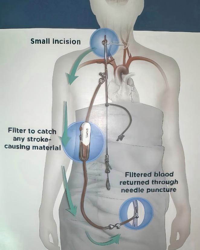
TCAR is a less invasive, clinically proven, approved treatment option for carotid artery disease. The procedure begins with a small incision just
above the clavicle. A temporary sheath is placed directly into the carotid artery in an area away from the disease. The sheath connects to a filter and flow reversal system outside of the body and then to another small sheath that is placed directly into the femoral vein. The difference in pressure causes the reversal of blood flow away from the brain, decreasing the risk of a stroke. A stent is then inserted through the sheath to open the stenotic and diseased carotid artery. Because of the blood flow reversal, any debris that might break loose during stent placement won’t travel up to the brain, rather it travels downward and gets trapped in the filter, and the filtered blood returns into the vein.
The postoperative course is very important for faster recovery after carotid surgery. Close coordination of care with the ICU team focusing on adequate hemodynamic support including strict blood pressure management, makes a substantial difference in the patient’s recovery. Patients can expect to go home within 24 hours of the procedure.
When compared to traditional open CEA, studies have shown that patients undergoing TCAR usually recover quickly with less pain and smaller scars. TCAR patients also have a lower risk of cardiac complications, myocardial infarction and cranial nerve injury, and have a shorter procedure time when compared to open CEA.


TCAR procedure showing severe carotid stenosis on initial angiogram (top) followed by stent deployment and final angiogram with resolution of the stenosis (bottom).
Proper selection and identification of patients who are candidates for TCAR is critical. TCAR is now available for patients of all risk levels, including those with symptomatic and severe asymptomatic carotid stenosis as well as high anatomical or medical risk for open surgery.
Since 2022, Cleveland Clinic Weston Hospital has performed TCAR routinely using a minimally invasive approach. Our center performs the highest volume of TCARs in South Florida. Outcomes have been excellent, with 100% technical success rate and no reported strokes for our patients. Our expertise in open vascular surgery and CEA combined with newer, advanced endovascular techniques such as TCAR, has translated into the best possible patient outcomes.
Dr. Sasson sassonm3@ccf.org
ENROUTE Transcarotid Stent System with blood flow reversal from the carotid artery to the femoral vein.
5 CLEVELAND CLINIC FLORIDA
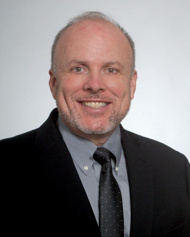
A Message From Cleveland Clinic Weston Hospital’s Vice President and Chief Medical Officer
F. Scott Ross, MD
Vice President and Chief Medical Officer Cleveland Clinic Weston Hospital
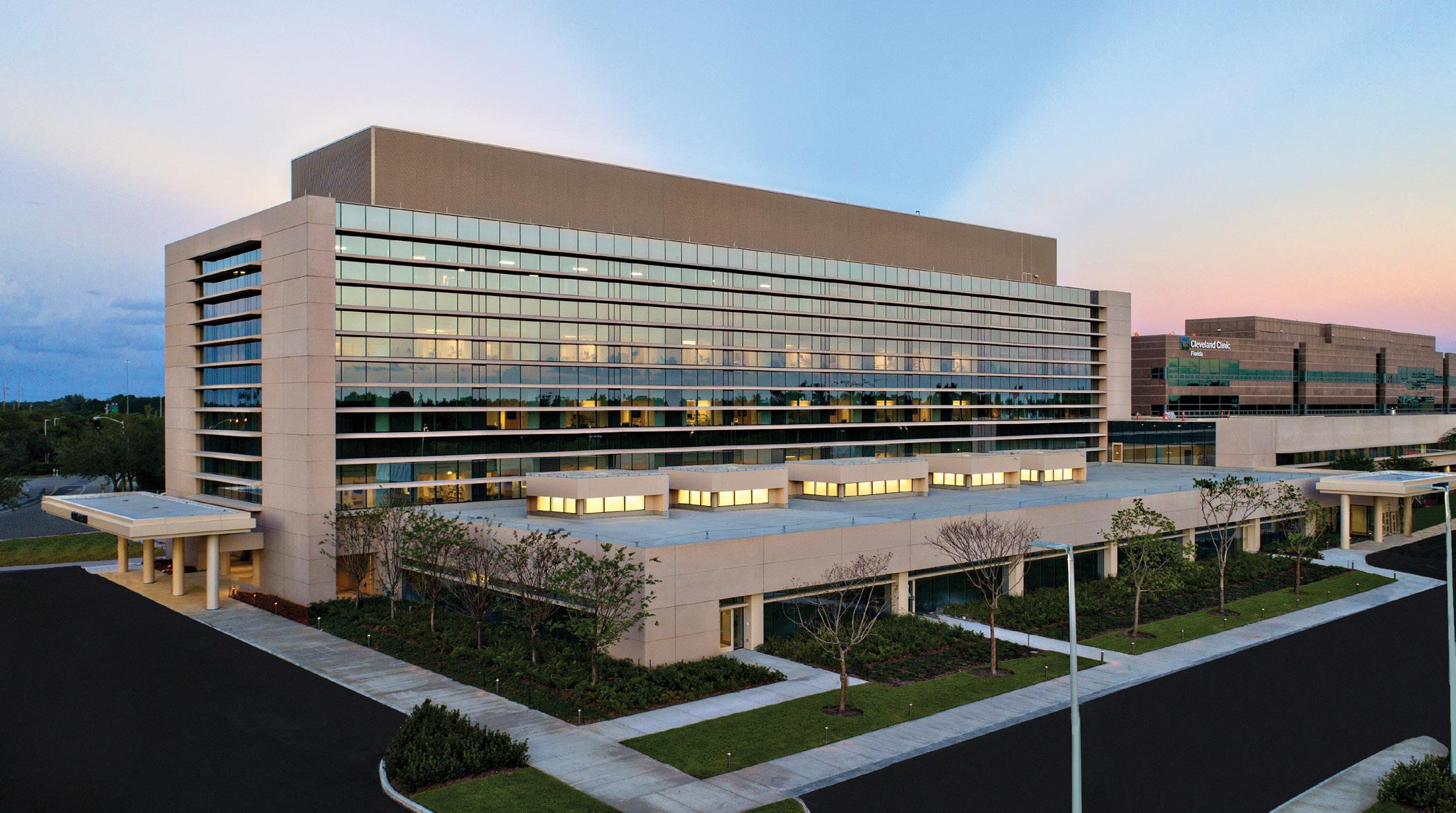
In the realm of cardiovascular care in South Florida, the Heart, Vascular and Thoracic Institute (HVTI) at Cleveland Clinic in Florida is a name that resonates profoundly. A beacon of medical excellence, this institute has not only redefined the standards of cardiac care, it has etched a remarkable trajectory of innovation and patient-centered service.
A Pioneering Legacy
Since its inception in 2001, the HVTI has dedicated its efforts to advancing care for cardiovascular diseases including diagnostic and interventional cardiology, cardiac and vascular surgery and vascular medicine. In 2014, with the establishment of a heart transplant program, Cleveland Clinic Weston Hospital became a a true referral center for the most
critically ill patients in South Florida. Despite its young age, the HVTI has achieved a monumental milestone, celebrating its 200th heart transplant within its first seven years. This achievement not only underscores the surgical expertise of the institute but also highlights its commitment to fulfilling a dire need for the most critically ill patients in South Florida, the Caribbean, and Latin America. Our transplant program is now ranked among the top 5 in the country by the Scientific Registry of Transplant Recipients.
The HVTI’s growth has not been limited to the heart transplant program; advancements in other areas of heart and vascular care have propelled Cleveland Clinic in Florida into its position as a leading healthcare center. The HVTI is at the forefront of vascular medicine, and has the largest department of its kind in the Southeast United States. Offering a comprehensive array of services for all vascular conditions, including rare types such as fibromuscular dysplasia, our vascular medicine department serves as a referral center for hereditary thromboembolic diseases and rare pathologies related to vasospastic, aneurysmal, inflammatory, and noninflammatory vascular diseases. The collaborative model of care, a hallmark of Cleveland Clinic, has been seamlessly integrated into the fabric
of the institute. The multidisciplinary teams, comprising interventional and noninterventional specialists, work cohesively under one roof, ensuring effective communication and the highest quality of care.
Innovations Driving Patient-Centric Care
The HVTI is committed to innovation. A testament to this commitment is the state-of-the-art cardiac electrophysiology labs at Cleveland Clinic Martin Health in Stuart as well as Cleveland Clinic Weston Hospital. These facilities allow for diagnostic, therapeutic, and interventional procedures, including the implantation of pacemakers and treatment of abnormal heart rhythms, and they exemplify the institute’s dedication to providing comprehensive cardiovascular care under one roof. Our center also has been an enrollment site for multiple randomized clinical trials investigating new drug targets and innovative procedures.
A Collaborative Model for Expanding the Frontiers of Cardiovascular Care
With a legacy firmly rooted in cardiovascular medicine since the 1930s, the Cleveland Clinic Foundation in Ohio paved the way for Cleveland Clinic in Florida to emerge as a leader in the field. This leadership
CARDIAC CARE - SPRING 2024 6
is further solidified by the HVTI’s commitment to expand the frontiers of cardiovascular care in the state of Florida. A recent alliance with Lee Health, the largest public health system in the state, creates a platform for sharing best practices, exploring new treatments, and accelerating advances in cardiovascular care. This alliance not only signifies a collaboration between two esteemed healthcare providers but also aligns with Cleveland Clinic’s unique collaborative model of care.
A Commitment to Excellence
Cleveland Clinic in Florida has consistently earned high rankings for heart care and many other specialties, and the HVTI’s trajectory is poised for even greater heights.
In the realm of cardiovascular care in South Florida, the Heart, Vascular and Thoracic Institute at Cleveland Clinic in Florida is a name that resonates profoundly. A beacon of medical excellence, this institute has not only redefined the standards of cardiac care but has etched a remarkable trajectory of innovation and patient-centered service.
but as a beacon guiding the way forward. From pioneering in heart transplants and cardiac surgery, leading the way in the diagnosis and treatment of vascular disorders and offering state-of-the-art electrophysiology labs, the institute’s journey has been one of unwavering commitment to innovation and patient-centric care.
As referring physicians and patients navigate the complex landscape of cardiovascular health, Cleveland Clinic in Florida is here as a trusted partner—a center where the past meets the future, and excellence is not just a standard but a heartfelt commitment to the well-being of every individual under its care.
Research, a fundamental tenet of the Cleveland Clinic mission, continues to drive advancements in cardiac care, including transcatheter valve procedures and heart failure treatment.
Our HVTI stands not only as a testament to the triumphs of the past
7 CLEVELAND CLINIC FLORIDA
PATIENT SPOTLIGHT

Marianela Betancourt remembers that even as a kid she was often short of breath when she played soccer or basketball or ran track. She always chalked it up to being a little overweight.
But as time went on the breathlessness became more noticeable and seemed to happen more often.
About 15 years ago Marianela, who is now 60 years old, woke up in the middle of the night feeling her heart beating “out of control.” Scared and worried she was having a heart attack, she took another dose of her blood pressure medication and called 9-1-1. She was taken to an emergency room near her home in Miami and admitted. There she was administered intravenous medication to bring down her heart rate, which had risen to 200 bpm.
She spent four days in the hospital, and when her heart rate slowed to normal she was discharged. A heart attack had been ruled out, she was
“There is something wrong with my heart.” – A woman’s search for answers leads to lifesaving help at Cleveland Clinic in Florida
referred to a cardiologist and advised to get tested for sleep apnea.

Things got better before they got worse
Marianela continued to have palpitations occasionally but always shrugged it off as the result of maybe too much coffee or the stress from
her administrative job for a fire department. Because healthcare providers often suggested her weight played a role in her health issues, she managed to lose 40 pounds. She was scared to push herself in exercise, however, because without a diagnosis she didn’t know what would trigger the rapid heartbeat again.
In 2015 Marianela went to an annual work conference in Kansas City and said she was “so happy” because she actually felt good enough to be able to socialize and walk around the city with colleagues without getting winded as she had in the past.
However, on the first night of the conference she woke up when her heart started beating rapidly.
“While I was sleeping, again I felt my heart,” Marianela says. “I said to my roommate, who was a very close friend, ‘There is something wrong with my heart.’”
She knew this incident was worse than the last because she had a
CARDIAC CARE - SPRING 2024 8
new symptom along with the rapid heartbeat – wheezing. Her friend called 9-1-1 and Marianela was taken to the hospital again. During the three days she spent there she was diagnosed with pneumonia and put on a higher dose of blood pressure medication and a blood thinner.
Three years later, she was celebrating her birthday at a hotel resort with family when she fell forward coming out of the shower and hit the sink facefirst. She had passed out from what she would find out later was lack of oxygen from her heart condition.
Her health started to decline, she became even more short of breath and weak and passed out and fell more often.
In 2019, still without answers, she asked her son-in-law to drive her to a regularly scheduled appointment with her cardiologist.
“I couldn’t even hold my purse. That’s how weak I was,” Marianela says.
After reading the results of her EKG her cardiologist told her she needed to go to the ER right away. Once there, she passed out in the bathroom and had to have a blood transfusion because of low blood counts.
She told the doctor there “I want answers. I need to know what is happening.”
Finally, answers and relief
The ER doctor who took care of Marianela told her he had been a student of Craig Asher, MD, a cardiologist at Cleveland Clinic Weston Hospital, and that he believed he could help her. He connected Marianela with Dr. Asher’s office, and she soon got an appointment with him. Marianela said Dr. Asher asked her during her first visit if she had a history of falling. “When he asked me that, I knew I was in the right place,” she says. “I knew that he knew what was wrong with me.”
Dr. Asher adjusted her medication, which she said helped her feel a little better. After extensive testing and consultation with Nicholas Smedira, MD, a cardiothoracic surgeon at Cleveland Clinic’s main campus in Ohio, Dr. Asher determined Marianela would need open-heart surgery to repair her heart. She had hypertrophic cardiomyopathy, a disease that causes thickening of the heart muscle, left ventricular stiffness, mitral valve changes and cellular changes.
In December of 2019, Marianela underwent mitral valve repair and a clipping of the atrial appendage, and myectomy (removal of some of the hardening of the heart muscle) due to a stiffening of her heart’s left ventricle. The surgery was performed through a unique collaboration between Cleveland Clinic physicians in Ohio and Florida, including Dr. Smedira, whose specialties include hypertrophic cardiomyopathy and mitral valve repair.
“I’m doing fantastic. I have not had one episode since my surgery,” Marianela says. “On the weekends I can go to four Disney parks with my grandkids. Before this I couldn’t even think of doing that.”
She has regular follow-up visits with Dr. Asher and says she recommends him and Cleveland Clinic to everyone she can.
“The care and attention I received at Cleveland Clinic was beyond,” she says. “And I’m talking about everyone from the person who checks you in and everyone after. They want to be there. They want to help you. It’s amazing. No one can touch Cleveland Clinic when it comes to their service.”

I’m doing fantastic. I have not had one episode since my surgery. On the weekends I can go to four Disney parks with my grandkids. Before this I couldn’t even think of doing that.
- Marianela Betancourt
9 CLEVELAND CLINIC FLORIDA
The Hypertrophic Cardiomyopathy Program: A Center of Excellence Leading the Way in Care
Craig R. Asher, MD Staff, Department of Cardiology Director of the Hypertrophic Cardiomyopathy Clinic

Hypertrophic cardiomyopathy (HCM), one of the most common forms of cardiomyopathy, is a condition in which the heart thickens inappropriately. Historically, the prevalence of HCM has been cited as 1 in 500 individuals. More recently, however, based on gene tests, family screening and early detection with cardiac imaging, it is now estimated to occur more frequently, in 1 in 200 individuals. HCM occurs equally in males and females, is found in all racial and ethnic groups, and has been detected at all ages from children to the elderly. It is found in most parts of the world.
While most patients with HCM have a good quality of life and life expectancy, a wide spectrum of clinical presentations may occur. Because of increased thickness of the heart muscle, heart failure symptoms may develop due to obstruction of blood exiting the heart, or from resistance to blood flow entering the heart. In addition, there may be electrical disturbances with HCM that lead to heart rhythm abnormalities including atrial and ventricular arrhythmias and, less commonly, sudden cardiac death.
Best practices in HCM care
The four pillars of care for patients with HCM are to 1) treat or prevent symptoms or cardiac events; 2) facilitate and ensure family screening; 3) stratify for risk of sudden cardiac death; and 4) provide education, support and resources regarding their condition.
Although many patients with HCM will require no medical therapy, symptomatic patients are treated with medications like beta-blockers, which may provide
relief. Since April 2022, a new disease-specific class of medication (mavacamten, a cardiac myosin inhibitor) was approved for use for the obstructive form of HCM. One of the two landmark trials that studied mavacamten, the VALOR-HCM trial, was led by investigators at Cleveland Clinic’s main campus. With this medication, approximately 80% of patients who were eligible for HCM heart surgery (myectomy), no longer required an operation. Surgical myectomy still remains a timetested definitive option for managing obstructive HCM, and Cleveland Clinic surgeons in Cleveland and Florida are among a small group of surgeons who have pioneered this operation and perform high volumes.
For all patients with HCM, even for those without symptoms, it is important to have a comprehensive evaluation. This usually requires a complete personal and family history, physical examination, electrocardiogram (EKG), echocardiogram and stress echocardiogram, rhythm monitor and, sometimes, genetic testing and a cardiac MRI. Family screening
FEATURE ARTICLE
CARDIAC CARE - SPRING 2024 10
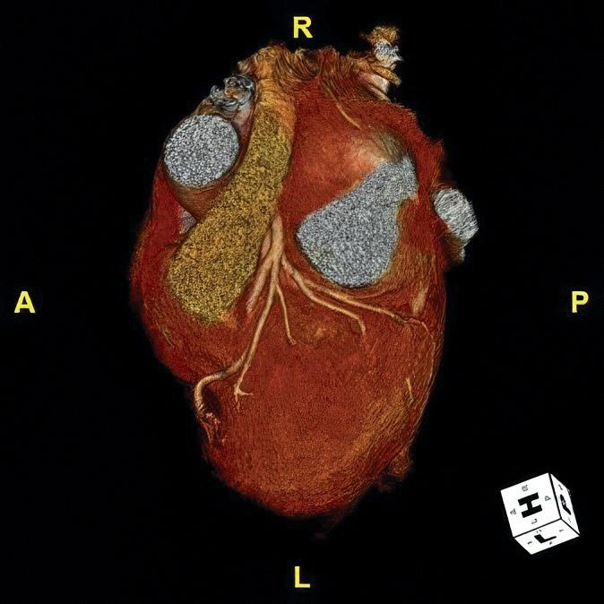
Cardiac computed tomography showing HCM with myocardial bridge.
can be done with interval EKGs and echocardiograms or with gene testing guided by a genetic counselor. Discussions about lifestyle, exercise and the do’s and don’ts of living with HCM are a key aspect of education and patient well-being. Several organizations, including the Hypertrophic Cardiomyopathy Association (HCMA), are excellent resources committed to providing education and advocacy to HCM patients and family members.
A final critical aspect of HCM care involves the assessment of risk for dangerous heart rhythm problems including sudden cardiac death. A complete battery of tests is often required to make this
assessment. There are algorithms and calculators developed by the American Heart Association/ American College of Cardiology and European Society of Cardiology to aid in this determination. However, shared decision-making between patients and physicians is essential to determine which patients may ultimately benefit from prophylactic therapy with an implantable cardiac defibrillator.
Cleveland Clinic Weston Hospital is one of Florida’s leading referral centers for HCM care. We offer a multidisciplinary specialty group with experience in the diagnosis, management and surgery for HCM. Our team utilizes advanced
imaging technologies, genetic testing, traditional and clinical research treatment options and cardiac surgeons specialized in HCM surgery. Weston Hospital’s HCM program is recognized as one of 2 Centers of Excellence in the state of Florida, as designated by the HCMA, with approximately 50 designated centers in the United States.
Dr. Asher asherc@ccf.org
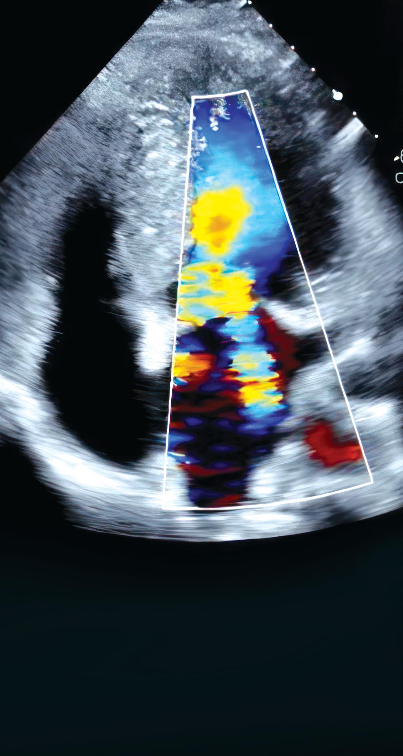
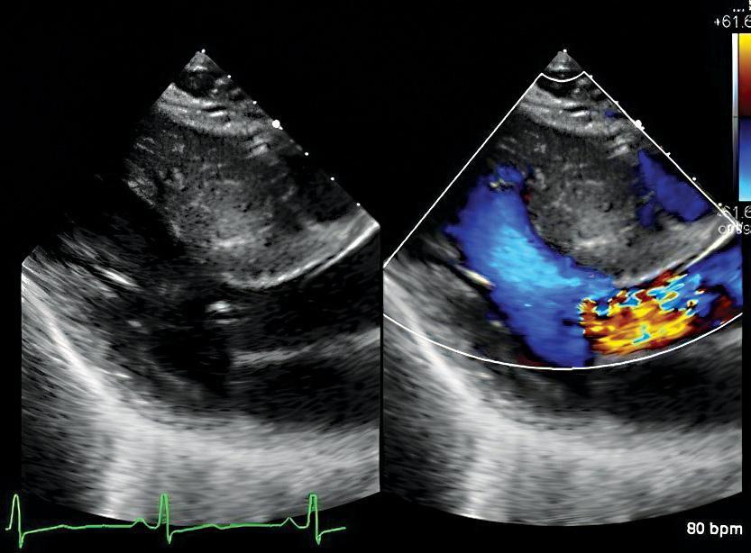
 Transthoracic echocardiography showing systolic anterior motion of the mitral valve due to HCM.
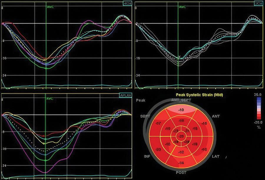
Global longitudinal strain echocardiography of early stage HCM.
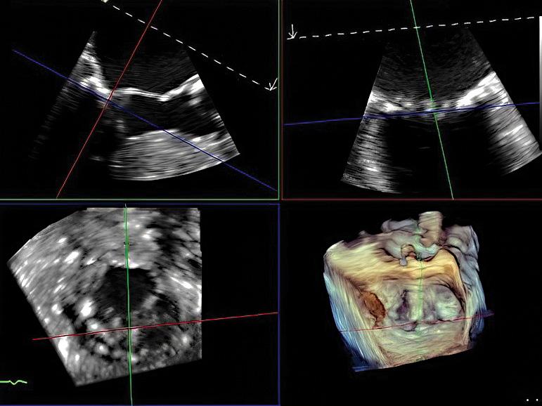
Transesophageal echocardiography showing 3-dimensional multiplane reconstruction of the mitral valve with HCM.
11 CLEVELAND CLINIC FLORIDA
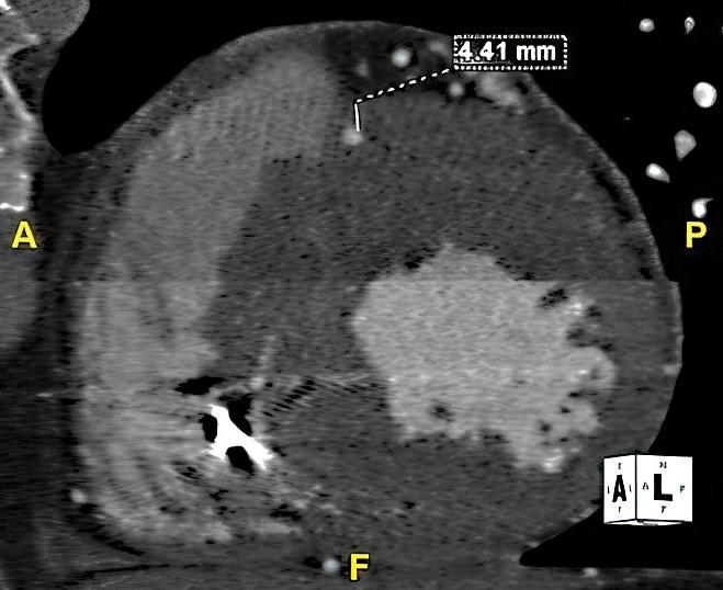
Cardiac computed tomography showing HCM with severe hypertrophy.
Multimodality Cardiac Imaging Guides the Diagnosis and Management of Hypertrophic Cardiomyopathy
David Lopez, MD Associate Staff, Department of Cardiovascular Medicine
Many anatomical and functional abnormalities occur as a result of hypertrophic cardiomyopathy (HCM). Thick walls with disarray of muscle fibers and scar tissue are the primary substrates for common symptoms including shortness of breath, chest pain and palpitations or cardiac events including heart failure, syncope, and even sudden cardiac death (SCD).
Not all HCM results in thick walls and not all thick walls indicate HCM
Multimodality cardiac imaging is the foundation for characterizing, guiding management and determining prognosis of HCM. Most patients with HCM will have an echocardiogram, stress echocardiogram, and cardiac MRI (CMR) at presentation. These imaging studies are coupled with additional tests such as rhythm monitors, gene testing and, sometimes, cardiac catheterization to complete each patient’s evaluation.
CMR is best suited to distinguish mimics or phenocopies of HCM that include physiologic conditions like athlete’s heart and pathologic conditions like cardiac amyloidosis (CA) and Fabry disease (FD). This is an essential first step to verify a patient has HCM. The management of these conditions is vastly different.
Evaluating an HCM diagnosis
Transthoracic echocardiography (TTE) is the first-line diagnostic test in the evaluation of HCM. TTE is not specific for the diagnosis of HCM but it helps exclude other structural abnormalities that cause increased LV wall thickness, including a subaortic membrane and aortic valve stenosis. TTE findings typical of HCM include increased LV wall thickness, systolic anterior motion of the mitral valve, LV outflow tract obstruction (LVOTO), abnormal diastolic function and abnormal myocardial deformation or strain. CMR is usually performed following an abnormal TTE indicative of HCM.
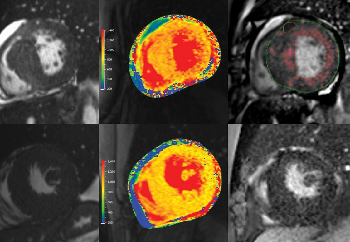
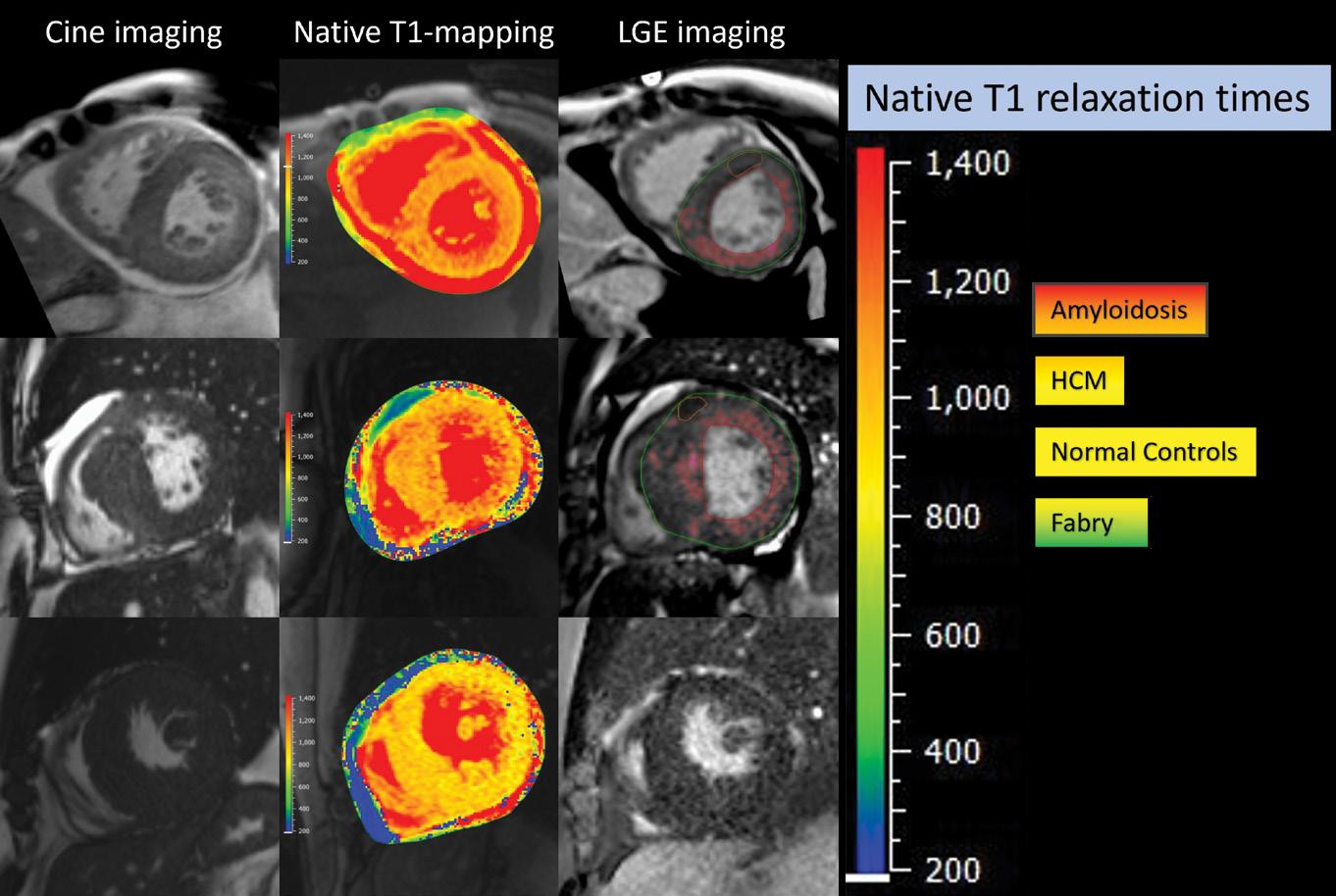
A comprehensive multi-parametric CMR examination allows for confirmation of HCM, characterization of morphology and severity of hypertrophy, assessment of LV function, exclusion of phenocopies, and information for SCD risk stratification. The protocol must include imaging with gadolinium contrast media. CMR cine imaging improves detection of hypertrophied segments and allows accurate measurement of wall thickness. T1-mapping has emerged as a valuable tool for myocardial tissue characterization which helps differentiate HCM, CA and FD (Figure 1). Late gadolinium enhancement (LGE) imaging allows detection of interstitial fibrosis (Also
CARDIAC CARE - SPRING 2024 12
Figure 1: Representative end-diastolic cine (left column), native-T1 mapping (middle column) and LGE images (right column) of cardiac amyloidosis (CA) (top row), HCM (middle row) and FD (bottom row), CA and HCM LGE images illustrate how LGE burden is calculated as a percentage of the myocardial mass. Native T1 relaxation times derived from T1 maps help differentiate these entities. In FD, the T1 relaxation time is shortened, whereas in CA, relaxation times are exceptionally long relative to normal myocardium.
BASELINE
10-YEAR FOLLOW-UP
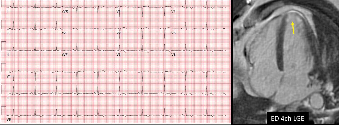
Figure 1). CMR markers associated with increased risk of SCD that inform recommendation for a primary prevention implantable cardiac defibrillator (ICD) include myocardial wall thickness ≥ 30 mm, LV ejection fraction <50%, an apical aneurysm and/or LGE burden ≥15% of the left ventricular mass. Assessment of flow helps characterize location of LVOTO by differentiating flow acceleration due to SAM from nonHCM mechanisms.
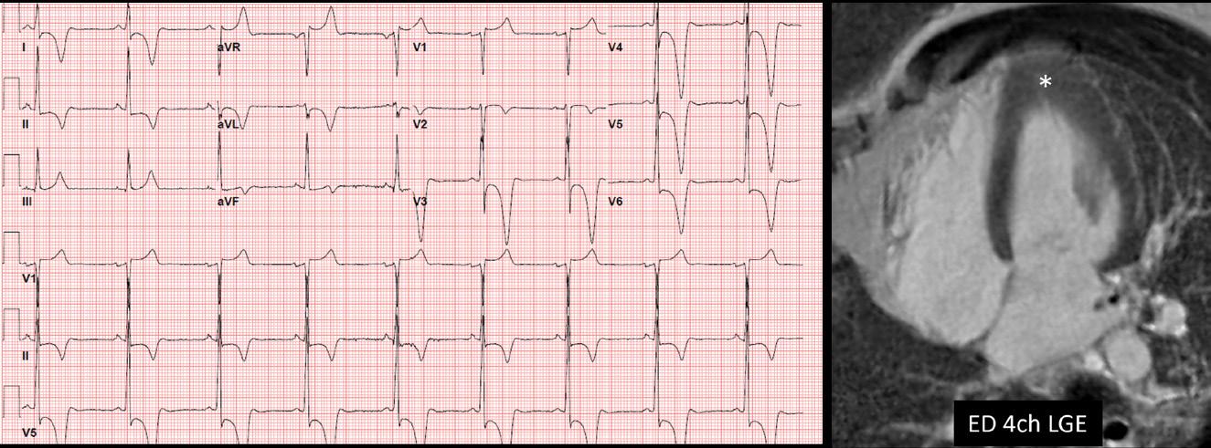
Surveillance CMR is recommended every 3-5 years in those who initially do not meet criteria for primary prevention ICD but have potential to progress and may warrant ICD in the future. Figure 2 illustrates the progression of apical hypertrophy to apical aneurysm in a 10-year followup period. Surveillance ECG may provide clues that myocardial changes
are taking place. Note the loss of QRS and T-wave voltage on ECG across the same monitoring period.
A crucial step in the characterization and management of HCM is to establish the presence or absence of LVOTO, which is not always seen on examinations at rest. Exercise stress echocardiography (SE) is performed to show presence of provokable LVOTO, assess hemodynamic response to exercise and objectively define a person’s exercise capacity. In individuals with obstructive HCM, SE guides treatment recommendations and allows monitoring of therapeutic effects.
Gathering additional
information
Other imaging modalities may be used when echocardiography or CMR do
not provide the necessary information or there is a contraindication. Cardiac computer tomography (CT) can define cardiac anatomy (including detecting a myocardial bridge often present with HCM) but provides limited myocardial tissue characterization. Coronary CT angiography is often used in preoperative planning of young HCM patients undergoing surgical septal reduction therapy. Transesophageal echocardiography is utilized intraoperatively to guide septal myectomy.
Otherwise, it is occasionally also used when there are unresolved concerns for alternative mechanisms of LVOTO or to better characterize the mechanism of mitral regurgitation.
Experience and technological capabilities are key
The road to HCM diagnosis and treatment is often tortuous, even among experienced clinicians. Appropriate use of multimodality cardiac imaging helps to promptly and safely arrive at the correct diagnosis in order to provide the indicated treatment for those with suspected HCM. At Cleveland Clinic Weston Hospital, our HCM physicians and imaging specialists work closely together as a multidisciplinary team to care for high volumes of HCM patients and have expertise in all of these important imaging modalities.
Dr. Lopez lopezd@ccf.org
13 CLEVELAND CLINIC FLORIDA
Figure 2. HCM changes over time. This example illustrates apical variant HCM that progressed to apical aneurysm over a 10-year follow-up period. Note that surveillance ECG can provide clues to myocardial changes. In this case a significant decrease in QRS and T-wave voltage was observed across all ECG leads.
Surgical Treatment of Hypertrophic Cardiomyopathy
Nicholas Smedira, MD
Staff, Department of Cardiothoracic Surgery
Surgical Director Hypertrophic Cardiomyopathy Center
Nicolas Brozzi, MD
Staff, Department of Cardiothoracic Surgery
Surgical Director AMCS Program
Surgical myectomy is one of the oldest open-heart procedures. Introduced in the 1960s by Andrew Glenn Morrow, MD, to remove a limited segment of myocardium from the proximal anterior ventricular septum. The procedure has evolved to allow for a more extended distal resection of hypertrophic myocardium with low operative risks and excellent clinical outcomes.
Surgery for hypertrophic obstructive cardiomyopathy (HOCM) reliably reverses disabling heart failure by permanently abolishing mechanical outflow impedance and mitral regurgitation, resulting in normalization of left ventricular pressures and preserved systolic function.
Perioperative mortality for myectomy has declined to 0.6% in experienced institutions, making it one of the safest open-heart procedures currently performed. Extended myectomy relieves symptoms in >90% of patients by ≥ 1 NYHA functional class, returning most to normal daily activity.
Patients with HOCM are considered for surgical intervention when they have poor exercise capacity for their age (usually NYHA class III/IV), when they develop symptoms not responsive to medical treatment or cannot tolerate medical treatment, and present LV outflow gradient ≥ 50 mmHg by echocardiography at rest or with physiologic exercise provocation.
Technical aspects of the procedure
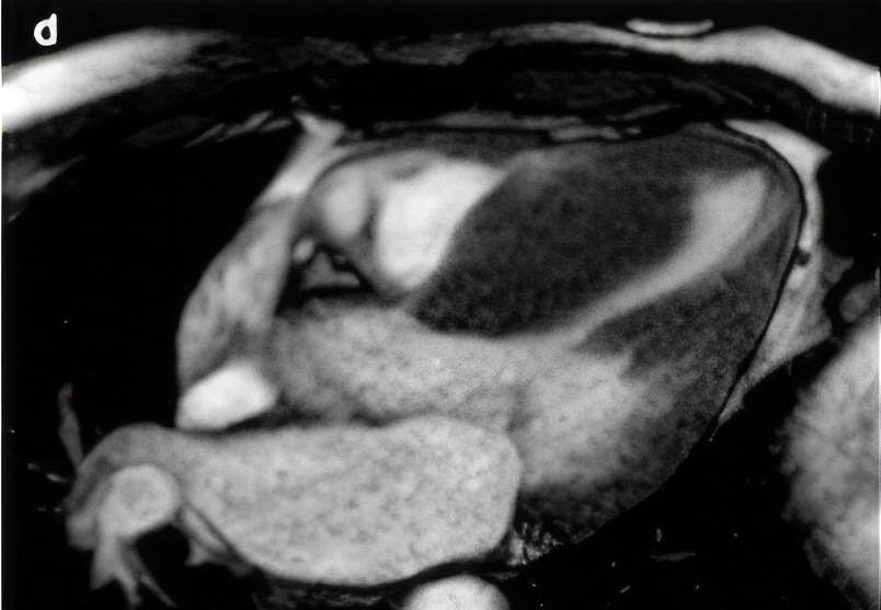
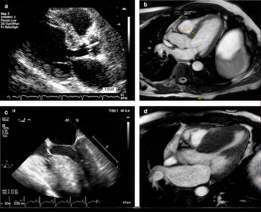
There are infinite configurations of the septum (Figure 1).
Cleveland Clinic surgeons review all preoperative imaging studies, including magnetic resonance imaging and echocardiograms, and occasionally can print a 3-dimensional model of the patient’s heart to understand the anatomy.
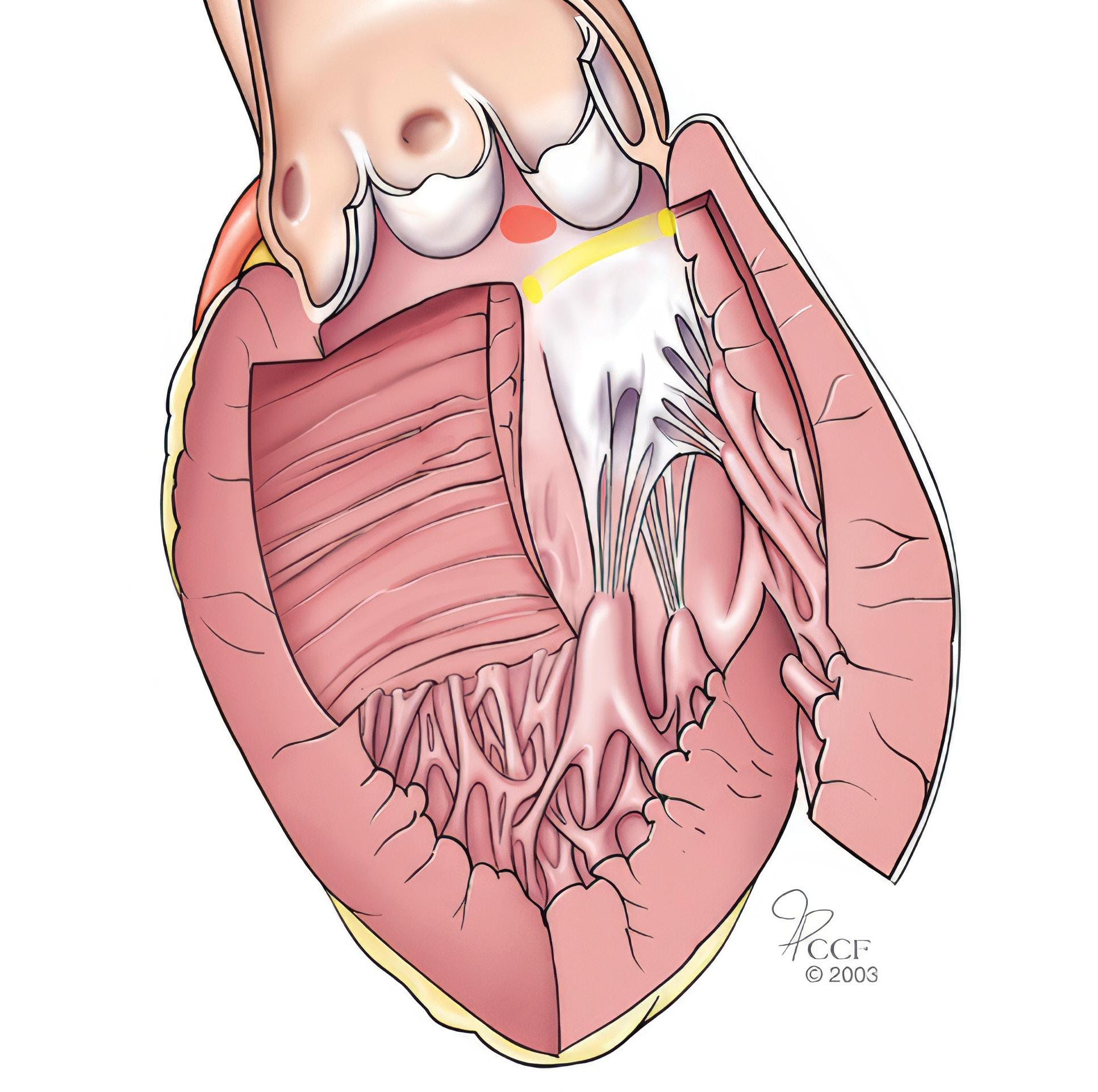
The operation is carried on through an incision of the initial portion of the aorta that allows to retract the aortic valve leaflets and provide exposure of the complex inner anatomy of the left ventricle including the interventricular septum, the papillary muscles with their chordae, and the mitral valve. Long-toothed forceps and scalpel are used to perform a tailored resection of septal myocardial muscle
CARDIAC CARE - SPRING 2024 14
Figure 1: Septal hypertrophy can present on a wide spectrum including (a) limited basal, (b) minimal, (c) generalized, (d) mid-cavitar y.
that starts about 15 mm below the right coronary cusp, away from the membranous septum and the bundle of His, extending longitudinally towards the apex and tapering down laterally as the resection ends in the regions of normal myocardial thickness (Figure 2).
room to discuss the results of the operation, and a functional test is performed at the end of the operation by administration of isoproterenol to produce a transient hyperdynamic state that would produce obstruction and reveal any residual hypertrophy.
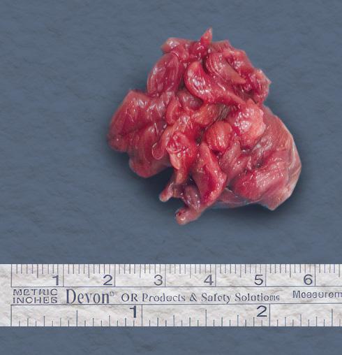
The resected myocardial muscle is placed over a towel, in exactly the location it was removed, to allow the surgeon to estimate the residual septal thickness by subtracting the thickness and length of the resected muscle from the mental 3D image formed during baseline assessment. A standard myectomy excises approximately 8 g of muscle (Figure 3), although some cases will require resection of up to 20 g of muscle.
We will frequently have our dedicated hypertrophic obstructive cardiomyopathy cardiologist in the operating
Additional procedures, such as realignment of the papillary muscles, might be required in selected patients with mid-cavitary obstruction. These procedures are further tailored to specific disposition of the papillary muscles and mitral sub-valvular apparatus.
Safety, outcomes and experience at Cleveland Clinic
Surgery for HOCM is currently one of the safest open-heart procedures with mortality risk <1% in experienced
centers. Resolution of heart failure symptoms is evident with improvement by ≥1 NYHA functional class in >90% of patients with 75% becoming asymptomatic (i.e., from NYHA functional class III/IV to class I). In addition to improved quality of life, surgery reduces frequency of atrial arrhythmias and improves longterm survival to mirror that of the general population.
Cleveland Clinic has performed more than 3,000 myectomy procedures, with mortality 0.7% and <4.5% of patients requiring pacemakers. Due to the strong collaboration between Cleveland Clinic cardiac surgeons in both Ohio and Florida, this surgery is available to patients in Florida.
Dr. Smedira smedirn@ccf.org
Dr. Brozzi brozzin@ccf.org
Figure 2: Illustration of extensive myectomy relieving obstruction of the left ventricular outflow tract.
15 CLEVELAND CLINIC FLORIDA
Figure 3: Image of a standard myectomy specimen. The length of the basal portion of the myectomy measures about 4 cm in width and tapers toward the apex.
Transforming cardiovascular care: Three of the latest innovations
Bernardo Perez-Villa, MD, MSc Senior Engagement Partner, Innovations
It is vital to keep up with cutting-edge developments in the dynamic field of cardiovascular medicine, and many startups armed with pre-market technologies are ready to transform cardiovascular healthcare. Here, we present three promising technologies that may reshape how we address cardiovascular diseases.
SirPluxTM Duo Drug-Coated Balloon
Advanced NanoTherapies Inc. (ANT) has entered the spotlight with its proprietary nanotechnology-driven technology – SirPlux Duo Drug-Coated Balloon – which utilizes 100% biodegradable nanoparticles for precise drug delivery to the target site. This breakthrough technology extends beyond cardiovascular applications to offer noninvasive solutions for various medical conditions in specialties such as orthopaedics and neurology.
One of the hallmarks of ANT’s nanotechnology is its ability to provide sustained drug release and tissue retention, akin to stent-like patency, effectively thwarting restenosis. This is achieved with lower doses of sirolimus and paclitaxel, which harness their combined antiproliferative power. The SirPlux Duo DCB leaves no permanent implant behind, safeguarding long-term arterial health without the risk of late stent thrombosis and offers potential positive economic implications by reducing treatment costs compared to traditional stent-based interventions. In August 2023, ANT announced a $4M Series A extension from a prominent undisclosed strategic medical device company and the company plans to share angiographic data from its ADVANCE-DCB FIH trial in early 2024.
Cardiac Pulmonary Nerve Stimulation
Cardionomic Inc. has created the Cardiac Pulmonary Nerve Stimulation (CPNS) system that holds promise as a potential game-changer in the treatment of acute decompensated heart failure (ADHF). CPNS is a catheterbased investigational device designed to provide acute (≤ 5 days) endovascular stimulation of the cardiac autonomic nerves in the right pulmonary artery, improving inotropy and lusitropy by increasing systemic perfusion and enhancing decongestion in ADHF. Early research has demonstrated

that this system effectively increases cardiac contractility in patients with chronic heart failure while maintaining heart rate stability. Cardionomic Inc. is currently enrolling patients in the United States, the European Union, and Panama for the STOP-ADHF Study, which investigates the safety and performance of the CPNS System in patients hospitalized for ADHF.
Subvalvular SpacerTM
Mitria Medical’s Subvalvular Spacer is a percutaneously delivered nitinol braid designed to treat mitral regurgitation with a focus on preservation rather than resection. The device is placed by puncturing the mitral valve annulus at the desired implant location, and then is delivered through known transcatheter techniques. This novel device sits at the hinge of the posterior leaflet, with the top (smaller) aspect sitting on the atrial side for anchoring, and the bottom (larger) aspect sitting under the leaflet to provide broad support to the displaced leaflet and associated subvalvular apparatus moving them anteriorly to restore coaptation and reduce mitral regurgitation. Mitria completed the first-in-human cases via a surgical approach, treating two patients and resulting in an acute reduction of mitral regurgitation (2-grade reduction from baseline) while maintaining a normal mean gradient after follow-up at 6 and 12 months.
Dr. Perez-Villa perezvb@ccf.org
CARDIAC CARE - SPRING 2024 16
Newsweek
Statista World’s Best Specialized Hospitals 17 CLEVELAND CLINIC FLORIDA
We are honored to be included in
and
Mark K. Grove, MD, Retires After Long Career at Cleveland Clinic
Terry King, MD Retired Vascular Surgeon
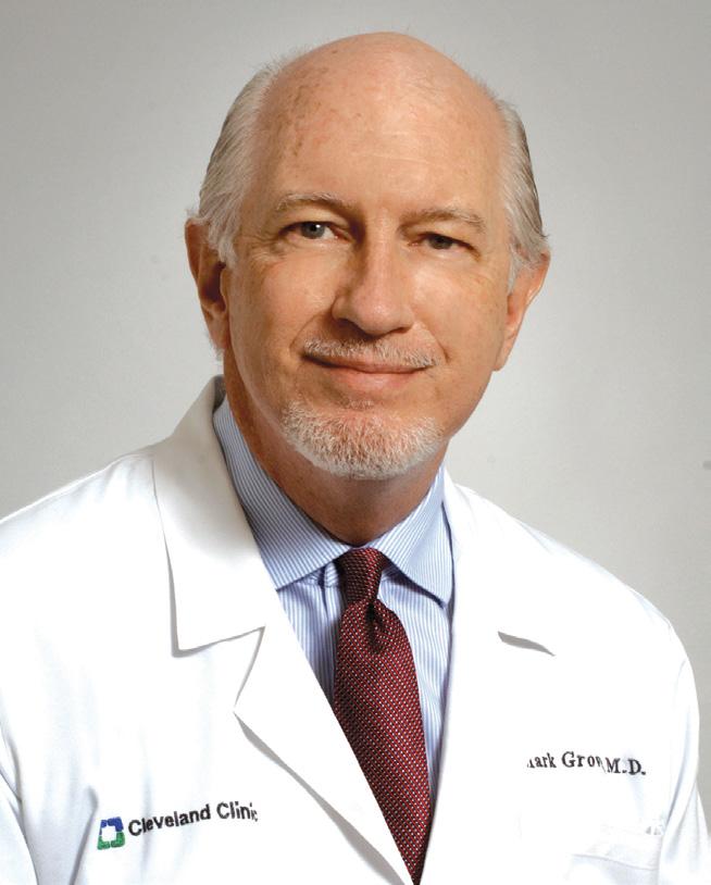
Mark Grove, MD, who retired at the end of 2023, is considered by his colleagues to be a pillar of Cleveland Clinic in Florida. After training in general surgery at Cleveland Clinic’s main campus in Cleveland, Ohio, he went on to complete a fellowship in vascular surgery there as well.
One of his most significant contributions was working with a team of physicians to start a residency program in general surgery in 2012.
In 1995 he joined Mark Sesto, MD, at Cleveland Clinic in Florida. Dr. Grove and Dr. Sesto were the first physicians to practice general
and vascular surgery together at Cleveland Clinic in Florida.
Dr. Grove spent his entire career at Cleveland Clinic in Florida, through its growth and evolution to the Weston campus in 2001. In 2009, after practicing as a vascular surgeon at Cleveland Clinic’s main campus and Naples, Florida, campus, I joined Dr. Grove at the Weston campus.
In 2010, Dr. Grove began to focus solely on the care of vascular patients. He was named Chairman of the Department of Vascular Surgery in 2012. Despite his busy clinical practice, he has contributed to the growth and development of Cleveland Clinic in Florida with his participation in many administrative duties and committees. One of his most significant contributions was working with a team of physicians to start a residency program in general surgery in 2012.
Dr. Grove and I thought alike clinically, and I knew my patients were in the best of hands when I was off call. He was the best of partners in vascular surgery, and we wish him well in his retirement.
Dr. Grove received his medical degree from the Drexel University College of Medicine/Hahnemann University in Philadelphia in 1985. He completed a surgery residency at Cleveland Clinic in Cleveland, Ohio, in 1990. In 1991 he completed a fellowship in vascular surgery, also at Cleveland Clinic. He is board certified in surgery and vascular surgery.
Dr. Grove is a member of the Society for Vascular Surgery and the American College of Surgeons.
CARDIAC CARE - SPRING 2024 18
New Staff New Staff
Welcome New Staff Members
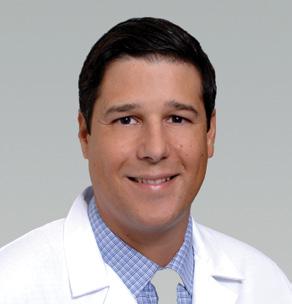

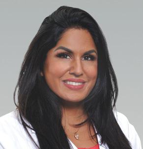
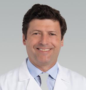
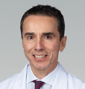
To refer a patient to one of our Heart, Vascular and Thoracic Institute specialists, please call 833.733.3710.
Mauricio Cohen, MD, FACC, FSCAI
Interventional Cardiologist
Director of Structural Heart Interventions
Weston Hospital
Specialty interests: Structural heart disease interventions: left atrial appendage closure, patent foramen ovale closure, atrial septal defect closure, paravalvular leak closure, alcohol septal ablation; Valvular heart disease interventions: transcatheter aortic valve replacement (TAVR), transcatheter mitral valve repair (MitraClip), balloon mitral valvuloplasty, balloon aortic valvuloplasty
Ricardo J. Hernandez, MD, FHRS
Cardiac Electrophysiologist
Weston Hospital
Specialty interests: Cardiac arrhythmia management, syncope, sudden cardiac death prevention, bradycardia, cardiac device implantation (pacemakers and defibrillators), cardiac resynchronization therapy, lead extraction, subcutaneous loop recorder implantation, radiofrequency ablation of arrhythmias, cardioembolic stroke prevention, left atrial appendage occlusion device implantation
Michael W. Howard, MD
Cardiovascular Surgeon
Martin Health
Specialty interests: Minimally invasive coronary and valve techniques, complex aortic arch surgeries, percutaneous valve implantation, ECMO, heart and lung transplants
Nina Thakkar Rivera, DO, PhD
Advanced Heart Failure and Transplant Cardiologist
Weston Hospital
Specialty interests: Heart failure recovery, heart transplant, cardiogenic shock and hemodynamics, critical care cardiology, cardio-oncology, cardiomyopathies
Juan Pablo Umaña, MD
Cardiothoracic Surgeon
Chair, Division of Thoracic and Cardiovascular Surgery
Weston Hospital
Specialty interests: Aortic valve repair and replacement, including minimally invasive and percutaneous treatments, surgery for patients with Marfan syndrome, aortic root replacement with aortic valve reimplantation, Ross procedure, mitral valve repair and replacement and surgery for atrial fibrillation and hypertrophic cardiomyopathy
James Wudel, MD
Cardiothoracic Surgeon
Director, Heart, Vascular and Thoracic Institute
Indian River Hospital
Specialty interests: Multi-arterial coronary artery bypass grafting, surgical treatment of atrial fibrillation, surgical treatment of valvular heart disease including transcatheter therapies
19 CLEVELAND CLINIC FLORIDA
Patients from across the United States, Latin America and the Caribbean turn to the Heart, Vascular and Thoracic Institute at Cleveland Clinic in Florida for life-saving treatment options. Physicians are subspecialty trained in a number of areas and provide compassionate heart care that is second to none.
Departments
& Centers
Cardiology
Cardiac Amyloidosis
Cardiac and Thoracic Surgery
Cardiac Electrophysiology and Pacing
Cardiac Imaging
Cardio-Oncology
Heart Transplant and Mechanical Circulatory Support
Hypertrophic Cardiomyopathy
Structural and Interventional Cardiology
Vascular Medicine
Vascular Surgery
About Cleveland Clinic in Florida
Cleveland Clinic in Florida is a nonprofit, multispecialty healthcare provider that integrates clinical and hospital care with research and education. The Florida region now includes Cleveland Clinic Indian River Hospital, Cleveland Clinic Martin North Hospital, Cleveland Clinic Martin South Hospital, Cleveland Clinic Tradition Hospital, and Cleveland Clinic Weston Hospital, along with numerous outpatient centers in Broward, Palm Beach, Martin, St. Lucie and Indian River counties. The Florida region is an integral part of Cleveland Clinic in Ohio, where providing outstanding patient care is based upon the principles of cooperation, compassion and innovation. Physicians at Cleveland Clinic are experts in the treatment of complex conditions that are difficult to diagnose.
For more information about Cleveland Clinic in Florida, visit clevelandclinicflorida.org
For
Patient Appointments
877.463.2010
Stay Connected
consultqd.clevelandclinic.org/cardiovascular clevelandclinic.org/cardiacconsult clevelandclinic.org/cardiacconsultpodcast
clevelandclinicflorida.org/heart
© 2024 Cleveland Clinic Florida. All Rights Reserved.
REGIONAL HOSPITAL CLEVELAND CLINIC FLORIDA LOCATIONS INDIAN RIVER HOSPITAL TRADITION HOSPITAL MARTIN NORTH HOSPITAL MARTIN SOUTH HOSPITAL WESTON HOSPITAL


 Transthoracic echocardiography showing systolic anterior motion of the mitral valve due to HCM.
Global longitudinal strain echocardiography of early stage HCM.
Transesophageal echocardiography showing 3-dimensional multiplane reconstruction of the mitral valve with HCM.


