4 minute read
Vet Insight: Standing Equine Leg CT

by Edit

Hallmarq was founded 20 years ago by Dr Nick Bolas, who wanted to develop a diagnostic imaging system that would assist vets to accurately diagnose a horse’s injury. Nick and his team of imaging experts worked with local vets in the South-East of the UK, to develop the world’s first Standing MRI system for horses. Since then, Hallmarq has continued to improve equine MRI technology and has also gone on to develop a vet-specific, small animal MRI system, using expertise from across the company
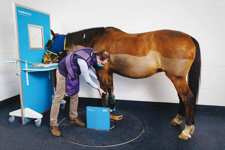
Hallmarq Veterinary Imaging pioneered the development of standing MRI for horses and the new Standing Equine Leg CT (slCT) scanner uses a simple and safe design, with a unique low, flat platform for quick and easy entry and exit of the standing sedated horse to quickly and safely diagnose the cause of equine lameness. The slCT increases access to advanced imaging for equine vet practices and Hallmarq has had success imaging standing sedated horses in collaboration with their UK-based clinical trial sites.
Advertisement
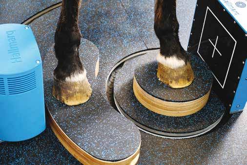
The system uses a dual-concentric ring design which enables the detector plate to remain very close to the region of interest, thereby improving image quality. Using slCT ensures equine practices have access to 3D
Imaging of the distal limb with Hallmarq’s new Standing Equine Leg CT
Photography by Hallmarq Veterinary Imaging
Standing Equine Leg CT
Improving lameness diagnosis
imaging in the evaluation of their lameness cases. Hallmarq is one of the few companies to incorporate motion correction technology to better ensure high quality, clear images in the standing patient.
This technology is often considered out of reach for many clinics and horse owners. However, Hallmarq considers the total cost of ownership in its design decisions, providing an affordable solution to most clinics. For any horse owner, the treat, rest, repeat loop of lameness diagnosis is both costly and time consuming. Early use of advanced imaging to pinpoint the exact cause of the problem minimises the overall cost associated with getting horses back on track.
Hallmarq has capitalised on over 20 years of experience with their unique Standing Equine MRI (sMRI) to create this additional tool for equine vets to fully evaluate and diagnose bony disease in the equine lower limb, including 3D visualisation of fractures. CT combines hundreds of X-rays to create a 3D digital image, which can then be viewed as thin slices from any direction, eliminating overlapping structures and revealing minute detail in bone and cartilage. slCT complements their sMRI which highlights soft tissue and metabolic changes and together they provide an even more powerful diagnostic aid. The value of using both modalities together is one that Dr Alison Fairburn, Specialist in Diagnostic Imaging at Bell Equine Vets also recognises,
Standing Equine Leg CT (slCT) Features
• Fast, 3D imaging with 60 second scan times Susie Godwin & Barrie Upton • Exclusive motion correction technology Joss Ridley, Jayne Darling-Parks & Claire Allen • Simple user interface and easy-to-use system A delicious lunch • Small footprint with an open design for patient safety • • No anaesthesia risk for horse or handlers No additional staffing requirements CPPC Christmas Party; Cowley Manor, Cheltenham; Friday 13 December • • Accessible to most equine clinics Fully supported by Hallmarq’s Ciren Does Christmas Q-care program, providing training, maintenance and system supportWith Christmas on the horizon, Cirencester Park Polo Club Christine Williams & Duncan Branch
Peter O’Rorke, Matt Evetts & Tony Haynes members and friends gathered to celebrate the festive season at the iconic Cowley Manor near Cheltenham. Alongside a “It really complemented what the delicious lunch, served to perfection by the lovely Cowley
MRI had told us about the content and staff, there was laughter and festive cheer aplenty, as guests signal intensity and, in this case, helped exchanged Christmas tidings of great joy! With all eyes turned with surgical planning, providing useful A dual-concentric ring design allows for safe and easy accessto the 2020 season, gossip flew around the room – who will information on the bone margin.” the next big Ciren star be? Which pros will play for which The technology is ideal for lower limb Adding slCT to their state-of-the-art teams next year? Who will take the most falls? Suffice to say, offer our customers a world first.” imaging where it can detect non-displaced diagnostics suite was an obvious next step a wonderful lunch was had by all; the perfect way to begin a Horse owners should contact their fractures, subtle changes in bone density forward for the practice, as Dr. Jonathon thrilling festive season. veterinary surgeon to discuss how advanced and small lesions without the expense, Dixon, member of the referral team and imaging can help in the diagnosis of radiation risks and power requirements of European Specialist in Veterinary Diagnostic Photography by John Hankin lameness. other systems. As installations continue, the new Hallmarq system is now up and running at Rainbow Equine Hospital, North Yorkshire. Yanna Mudie, Ginny Williams & Natalie Meredith Imaging explains, “We are really excited to put into practice the development so far to help facilitate the move into second phase clinical trials and Web: hallmarq.net Tel: 01483 877812 Maureen Moseley, Georgie Seddon-Brown & Isabel Branch