4 minute read
05.3 Kenneth Pearce

from Provost & President's Retrospective Review 2011-21
by Public Affairs and Communications (PAC) - Trinity College Dublin

05
Harnessing mechanics to understand biology and treat disease David Hoey
We all know physical exercise is good for your body. Whether going on a run or lifting weights, your body reacts to physical stimuli with increased muscle mass and strength. Although a little harder to see when looking in the mirror, the same thing is happening to your bones. Your skeleton is exquisitely sensitive to physical or mechanical stimuli. This is first evident in the womb, where muscle twitches in the developing embryo generate forces that contribute to bone and joint shape. The impact of mechanics on the skeleton continues throughout life as bones adapt and remodel in response to changes in physical activity. This is why astronauts who spend long durations in zero gravity need to exercise daily to ensure they do not lose significant bone mass. My lab in the SFI Research Centre, AMBER, is studying mechanics as a potent mediator of bone formation with the aim of harnessing this new knowledge to develop novel therapies and materials to treat devastating bone loss diseases such as osteoporosis, and the fractures that ensue. Osteoporosis is a debilitating bone loss disease associated with an increased risk of fracture. Remarkably, 1 in 3 women and 1 in 5 men over 50 will develop an osteoporotic fracture, which corresponds to a fracture every 3 seconds globally. Moreover, due to the ageing global population, this problem is escalating, with treatment costs predicted to rise to €105 billion by 2050. Using mechanical stimuli to drive bone forming cells – My research over the last number of years has explored how mechanical stimuli, such as fluid shear and pressure, can directly drive production of bone forming cells from stem cells resident within the marrow of long bones. Importantly, through an ERC starting grant, we have taken large strides in determining how these stem cells sense these mechanical stimuli (a process called mechanotransduction) and have pioneered research on antenna-like cellular extensions called primary cilia, which we believe are an important tool utilised by cells to sense mechanics. By identifying these processes, we can develop novel therapies, or mechanotherapeutics, that can activate the mechanisms mimicking the beneficial effects of exercise and so promote bone formation to treat osteoporosis. More recently, through a SFI Frontiers grant, my team is exploring the role of small cargoes released by bone cells called extracellular vesicles which contain regenerative factors that we believe are important in coordinating bone formation. We have shown that mechanical loading of bone cells can enhance the production and potency of these vesicles and we are currently investigating their use as a novel therapy to enhance bone regeneration.
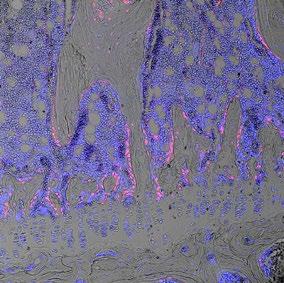
FIG 1
FIG 2
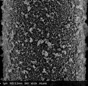
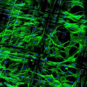
FIG 3
David Hoey is an Associate Professor in Biomedical Engineering in the School of Engineering and Investigator within the SFI Centre AMBER and Trinity Centre for Biomedical Engineering. A Trinity graduate, he was a awarded a B.A.I in Mechanical and Manufacturing Engineering in 2005, PhD in 2009, and elected a Fellow of Trinity College Dublin in 2021. Recipient of a prestigious European Research Council (ERC) Starting Grant and SFI Frontiers for the Future grant, his research focuses on musculoskeletal mechanobiology, mechanotransduction, and materials for tissue regeneration. Contact: dahoey@tcd.ie
FIG 1 – A histological section of a tibia bone from a 12-week old transgenic mouse in which the bone marrow stem cell population is labelled with a red fluorescent protein. Blue stain identifies cell nuclei. Image width: 160µm FIG 2 – A single polycaprolactone fibre, 1/10th the width of a human hair, within a scaffold fabricated using melt electrowriting and coated with a novel bone mimetic nano needle hydroxyapatite layer to enhance bone regeneration. FIG 3 – A melt electrowritten fibrous scaffold, with fibres oriented at 90 degree angles (white lines), seeded with human bone marrow stem cells (green – cytoskeleton; blue – nucleus). Image width: 1mm
3D printing aiding bone repair – Inspired by the native architecture of bone, we have also utilised advanced 3D printing technologies such as Melt Electrowriting to produce mineral coated fibres similar to that seen in bone. These fibres, which are a tenth the size of a human hair, can be accurately deposited to produce scaffolds that can be implanted to significantly aid bone repair. Combined with the mechano-therapies and vesicles identified above, these bioactive materials represent an exciting new strategy to treat osteoporotic fractures which are currently undergoing pre-clinical evaluation.
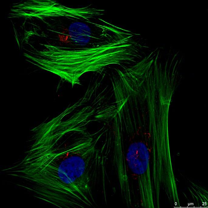
FIG 4 – Human bone marrow stem cells extending a single primary cilium (red linear structure) which we have shown is an important tool used by cells to sense mechanical stimuli. (green – cytoskeleton; blue – nucleus; red – primary cilium)
FIG 4