3 minute read
05.5 Jeremy (Jay) Piggott
from Provost & President's Retrospective Review 2011-21
by Public Affairs and Communications (PAC) - Trinity College Dublin
05
Biomechanics ‘at the heart’ of advances in heart valves Tríona Lally
Heart valve disease involves one or more of the valves in the heart becoming narrowed or leaking and can result in the heart having to work harder to pump blood effectively. There are nearly three million people across Europe aged 65 and over with heart valve disease, and this is set to rise to 20 million within the next two decades due to our ageing population profile. When aortic heart disease is left untreated, about half of sufferers die within two years of developing symptoms. A common treatment for heart valve disease is to replace the patients’ own valve with a bioprosthetic heart valve. These valves are formed from biological tissue derived from animals, where the tissue is treated with chemicals to preserve its structure. The tissue is then cut into leaflets and attached to a metal support, to mimic the function of a native leaflet. Whilst these bioprosthetic valves mimic the function of native heart valves, their success is often limited by the lifespan of the animal tissue used for the leaflets, which can become damaged through prolonged use.
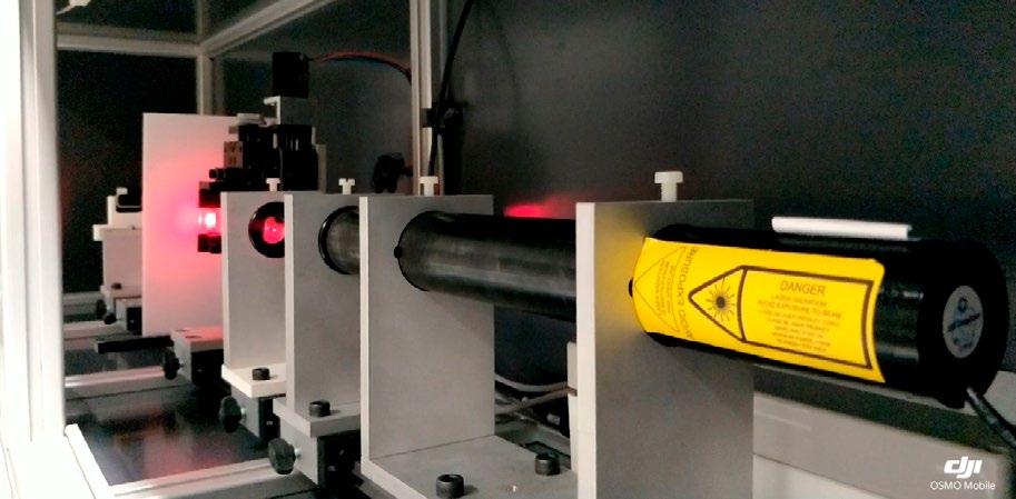
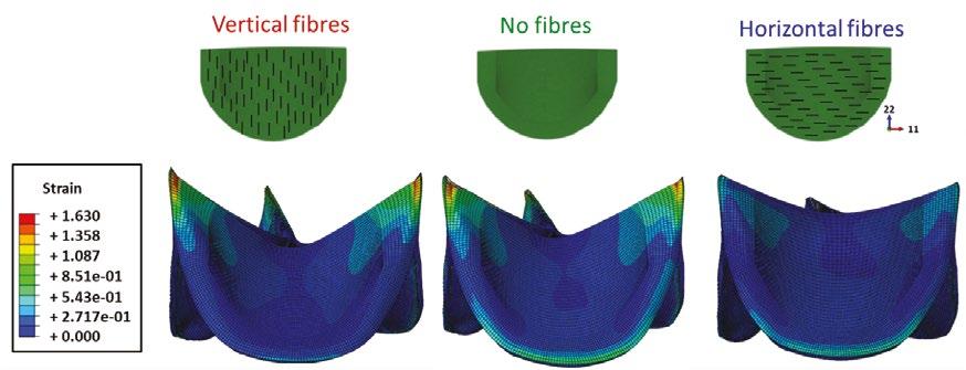
Improving bioprosthetic valves – Our research group in the Trinity Centre for Biomedical Engineering has been working closely with leading medical device manufacturer Boston Scientific, co-funded by the Irish Research Council and the SFI Advanced Materials and Bioengineering Research Centre (AMBER) to improve the lifespan of these valves. This is being achieved in two ways: (i) by understanding the underlying structure of the biological tissue from which the leaflets are currently manufactured and screening these materials to ensure that the leaflets are mounted on the valves in the optimum way, and (ii) developing new polymers inspired by nature which mimic the natural fibre patterns of native heart valve leaflets. The animal-derived leaflet tissue has a naturally fibrous structure, where these fibres are responsible for the strength and integrity of the tissue. Within our group, researchers have developed an in-house laser system to non-destructively ascertain the underlying fibre structure of these leaflet tissues (see Figure 1). This information is then used in computational models to investigate the performance of different leaflet fibre patterns, and ultimately identify optimum fibre orientations of the leaflet material, to increase device longevity (see Figure 2).
3D printing – Whilst this laser screening strategy has enabled improvements in existing bioprosthetic valves, we are now advancing these computational models to better inform the development of 3D printed biomimetic leaflet materials, with a view to creating synthetic materials that mimic the underlying structure and strength of native heart valve leaflets. In the future, these materials are likely to replace the use of animal tissues in bioprosthetic heart valves, and increase the lifespan of replacement heart valve devices.
It is anticipated that by 3D printing novel biocompatible polymers, fibre reinforced valves can be designed and manufactured which are better matched to individual patient’s needs and improve outcomes for those suffering from heart valve disease.
Tríona Lally received her BEng and MEng from University of Limerick and her PhD from Trinity College. She began her academic career as a lecturer in Dublin City University in 2004 and joined the School of Engineering in Trinity in 2015 where she is currently a Professor in Biomedical Engineering and Director of the Trinity Centre for Biomedical Engineering. Tríona was elected Fellow of Trinity College Dublin in 2021. She is a recipient of an ERC Starting Grant for her research, focused predominantly on understanding the role of mechanics in cardiovascular disease progression. Contact: lallyca@tcd.ie
FIG 1 – Small Angle Light Scattering (SALS) system built in-house for ascertaining the underlying fibre architecture of animal tissue from which bioprosthetic heart valve leaflets are made. By passing a laser beam through the tissue, the resulting scattered light pattern can used to identify the underlying fibre pattern in that region of the tissue. FIG 2 – Computational models of leaflets in bioprosthetic heart valves enabling the influence of leaflet fibre orientation on the valve longevity to be fully ascertained based on the strain in the tissue.
FIG 1
FIG 2
Vertical fibres No fibres Horizontal fibres
Strain +1.630 +1.358 +1.087 +8.51e-01 +5.43e-01 +2.717e-01 +0.000


