ISSN 2348-313X (Print)
International Journal of Life Sciences Research ISSN 2348-3148 (online)
Vol. 10, Issue 4, pp: (8-14), Month: October - December 2022, Available at: www.researchpublish.com

ISSN 2348-313X (Print)
International Journal of Life Sciences Research ISSN 2348-3148 (online)
Vol. 10, Issue 4, pp: (8-14), Month: October - December 2022, Available at: www.researchpublish.com
Yasir Awad Ahmed1 , Abdulrahman Abdulkhalig Alshamrani 2 ,Abdullah Hussain saeed alomari3 , Ali Ahmed Yahya Motmy4 , Abdulrahman Saleh Suliman5
1 Corresponding Author, Senior Lab Tech, PCLMA ,King Fahad Medical City ,Riyadh .KSA
2 Medical technologist, King Fahad Medical City, Riyadh .KSA
3 Medical technologist, King Fahad Medical City ,Riyadh .KSA
4 Technician Laboratory,King Fahad Medical City ,Riyadh .KSA
5 Medical Technologist, King Fahad Medical City, Riyadh. KSA
DOI: https://doi.org/10.5281/zenodo.7215272
Published Date: 17-October-2022
Abstract: Appropriate clinical care of hematologic malignancies depends on timely and accurate diagnosis. The category of cancers known as acute leukemias is diverse, with each disease having a unique clinical presentation, prognosis, and preferred course oftherapy.The majorityof morphologic and cytochemical characteristics wereused in historical categorization systems, but currently, immunophenotypic, cytogenetic, and molecular data are included to construct clinically useful diagnostic classifications When combined with morphologic evaluation, multiparameter flow cytometry quickly and precisely determines antigen expression profiles in acute leukemias, which frequently supports a specific diagnosis or a limited differential. Because numerous recurrent molecular or cytogenetic abnormalities are linked to distinctive immunophenotypic characteristics, flow cytometry is a crucial tool for guiding additional testing.
Even within a single acute leukemia subtype, the identification of certain antigens may have prognostic or therapeutic significance. The immunophenotypic fingerprint of a leukemia after first diagnosis serves as a helpful guide to track treatment response, minimal residual disease, and recurrence.
Objective: To evaluate flowcytometry role in distinguishing acute myeloid leukemia (AML) from ALL using CD markers.
Material and methods: This is retrospective study conducted in King Fahad Medical City in Riyadh city, in the period between November to December 2019 on patients with acute leukemia diagnosis.
Conclusion: Accurate acute leukemia diagnosis and categorization depend on immunophenotypic analysis. Multiparameter flow cytometry offers a quick and efficient way to gather these data, as well as prognostic data and a technique for assessing minimal residual illness.
Keywords: Appropriate clinical care, accurate diagnosis, leukemias, cytochemical characteristics.
Leukemia is a term that includes blood cancers that originate mostly in the bone marrow resulting in a high number of abnormal cells [1] that are not fully developed known as blasts or leukemic cells.[2]
According to the types of cells, leukemia is classified into two broad categories: myelogenous which arises from myeloid cells, and lymphocytic which arises from lymphocytes. Leukemia is classified according to the type of cell. Hence, myeloid cells include immature blood that usually matures into granulocytes or monocytes.
ISSN 2348-313X (Print)
International Journal of Life Sciences Research ISSN 2348-3148 (online)
Vol. 10, Issue 4, pp: (8-14), Month: October - December 2022, Available at: www.researchpublish.com
Classification of leukemia:
There are 4 types of leukemia: acute and chronic leukemia that is further classified into lymphoid and myeloid.[3]
Acute leukemia is the most common formof leukemias in children and it is characterized byrapid proliferation of immature blood cells. This rapid increase in cells number results in crowding of the bone marrow which make it hard to function. By that, low hemoglobin and platelets manifest. The rapid division of cells with risk of spread to bloodstream and other organs of the body urges the need for immediate management of acute leukemias.
On the other hand, chronic leukemia usually presents in elderly, despite possibility of occurring at any age, and is characterized by abnormal proliferation of relatively mature, however, abnormal white blood cells. It takes months or years to progress and produce high numbers of abnormal cells. Chronic leukemia does not warrant immediate treatment as acute leukemia. Management can be restricted to monitoring for some time before treatment to ensure the maximum efficacy of the treatment.
Acute myeloid leukemia (AML) is a cancer of the myeloid line of blood and as other types of acute leukemia characterized by a massive rapid proliferation that interfere with normal blood cells in the bone marrow.[4] It is the most common type of acute leukemia in adults with incidence increase with age and a median onset of 65 years. It represents only a minority (10-15%) of all leukemia in children. The prognosis depends mainly on cytogenetic abnormalities and response to initial treatment.
French-American-British (FAB) classification system:
Depending on the type of cells of which the leukemia originates and the degree of maturity of cancer cells, this system divides AML into eight subtypes, M0 through to M7. The classification is done using light microscope to examine the appearance of the malignant cells and/or by using cytogenetics to detect any chromosomal abnormalities. These subtypes are:
● M0 - acute myeloblastic leukemia, minimally differentiated
● M1 - acute myeloblastic leukemia, without maturation
● M2 - acute myeloblastic leukemia, with granulocytic maturation
● M3 - promyelocytic, or acute promyelocytic leukemia (APL)
● M4 - acute myelomonocytic leukemia
● M4eo - myelomonocytic together with bone marrow eosinophilia
● M5
○
M5a: acute monoblastic leukemia
○ M5b: or acute monocytic leukemia
● M6 - acute erythroid leukemias, including
○
M6a: erythroleukemia
○
M6b: very rare pure erythroid leukemia
● M7 - acute megakaryoblast leukemia
Acute lymphoblastic leukemia (ALL)
It is a cancer originates of the lymphoid line of blood cells that results in abnormal proliferation of immature lymphocytes.[5]
CD marker in acute leukemia
Acute leukemia is a heterogenous group of malignancies that varies clinically, morphological, immunological, and in its molecular feature. Each displays a unique pattern of surface antigen expression called CD antigens. These immunophenotypes cute leukemia classification.[8]
ISSN 2348-313X (Print)
International Journal of Life Sciences Research ISSN 2348-3148 (online)
Vol. 10, Issue 4, pp: (8-14), Month: October - December 2022, Available at: www.researchpublish.com
The precursors cells or the blast cells of the acute leukemia display different molecules on its surfaces called cluster of differentiation (CD) antigens. These CD markers are characteristic for each acute leukemia type that now forms the basis of classification and diagnosis of blood cancers. In acute leukemia, many immunophenotypes of blast cells exhibits abnormal cellular differentiation and express unusual CD markers called aberrant expression of markers. The basic of diagnosis of acute leukemia until recently was dependent n morphology and cytochemistry that provide correct diagnosis in 80% of cases.[8] Nowadays, Immunophenotyping of leukemia cells is critical for identification the cell line, maturation stage and present aberrant antigens. These will help in treatment individualization, monitoring and detection of residual disease.[9]
Cytometry is the measurement of the physical and/or chemical characteristics of cells or other biological components. Flowcytometry is a process where such measurements are made while cells or biological components pass through a measuring apparatus, preferable as a single file, in a fluid stream in a front of a laser. [7] This represents the basic principle if cytometry that helps in detection, counting and sorting of cells. Fluorescence is used to label cells components that emits light at different wavelength when excited by laser. [6]
Flowcytometry is widely used many clinical applications such as immunophenotyping, cell sorting, CD34-positive stem cell precursors and lymphocyte subsets (B cells, T cells, and natural killer or NK cells) enumeration, and fetal bleed tests in fetomaternal hemorrhage. The cells can be obtained from blood, bone marrow, lymph nodes or other tissue.
Flow sorting uses electrical or mechanical methods to divert and collect cells within a range or ranges, set by the user, of one or more measured characteristics. An instrument called the cytometer is used for a rapid counting of a speed of fifty thousand cells per second. This device substitutes the manual countingby microscope that was a hard tsask subject to errors. The cytometer consists of a fluidics system, an optical system that composed of a one or more monochromatic light source could be laser in addition to filters as an excitation beam, electronics that detect cells emissions, and a computer for data analysis.
The flowcytometry has many diagnostics and disease monitoring applications in blood cancers. Immunophenotyping of hematological cancers though clinically valuable, it developed a technically complicated diagnostic procedure that varies with methodological features, strategic judgments, and needs an extensive knowledge of the disease aspects including clinical, morphological, and laboratory features. To ensure reliable results, many internal quality-control steps are required in regards to instrument setup and calibration, selection and validation of monoclonal antibody panels, and process control.
Flowcytometric immunophenotyping is used for the initial diagnosis of leukemias, treatment monitoring, and minimal residual disease prescence that may highlight recurrence of the cancer.[7]
Flowcytometry helps to distinguish an abnormal population of blood cells and to determine the cellular lineage, clonality, maturation, and heterogeneity of malignant cells. The Flowcytometric analysis (immunophenotyping) chrachtrerize cells by their size, complexity, and cellular markers patterns o expression. Full diagnostics tests can be done with fewer cells and less sepciemne using multiparameter flowcytometry that simultaneously use many fluorochromes.
Flowcytometry is used in detection of CD markers expression on leukocytes. In many cases of acute leukemia, aberrant expression of these markers was observed.[8] The techniques of Flowcytometry is based on the analysis of a single cells or group of cells for many phenotypic and functional parameters simultaneously.[8] Its characteristic of measuring many parameters on single cells in a suspension at high speed makes it ideal for the study of leukemic cells. It works by applying monoclonal antibodies against cells markers.
Many advances has dramatically enhanced the utility of diagnosis and classification of leukemia such as new monoclonal antibodies availability, gating strategies improvement, and presence of multiparameter analytic techniques.[9] The main concept used in these new applications of flowcytometry is that even though cancer cells could show high similarity to normal blood precursors, they also frequently express aberrant phenotypes that allow, even in low frequencies, specific identification and discrimination of these neoplastic cells from normal cells[8] This aberrant expression is largely due to genetic abnormalities.[9]
ISSN 2348-313X (Print)
International Journal of Life Sciences Research ISSN 2348-3148 (online)
Vol. 10, Issue 4, pp: (8-14), Month: October - December 2022, Available at: www.researchpublish.com
General objective:
To evaluate flowcytometer role in distinguishing acute myeloid leukemia (AML) from ALL using CD markers
Specific objectives
● To differentiate T-ALL and B-ALL subtypes of ALL
● To determine types of AML according to CD Markers present in surface of cells
● To Evaluate the frequency of CD markers in Acute leukemia Saudi patients
This is retrospective study conducted in King Fahad Medical City in Riyadh city, in the period between November to December 2019 on patients with acute leukemia diagnosis.
We used the following monoclonal antibodies: CD 2, CD 7, CD 10, CD 13, CD 14, CD 19, CD 20, and CD 33. CD 19 positivity was the B-cell antigen expression. CD 20 was an additional B-cell marker. = CD 2 and CD 7 positivity were the T-cell antigens expression. By definition, B and T cells were negative for myeloid cytochemical markers and monoclonal antibodies CD 13 and CD 33. CD 14 was used as a monocytic marker. The common ALL antigen (CALLA) was defined as CD 10 positivity. Red staining of cells of intensity greater than that seen in the background was defined as a positive reaction. Surface marker expression in at least 20% of the leukemic blast cell population was a criteria for positivity.
For flowcytometry analysis, single cell or particle suspensions preparation is necessary. We can attach various immunofluorescent dyes or antibodies to the antigen or protein of interest. The most common fluorescent dyes used in biomedical fields are Fluorescein isothiocyanate (FITC), Texas red, and phycoerythrin (PE).
Then, the suspension is aspirated into a flow cell where they pass solitarily within a focused laser beam surrounded by a narrow fluid stream. When strikes a cell, the light is either scattered or absorbed. If the cells contains a naturally fluorescent material or surface antibodies labelled by fluorochrome antibodies, the absorbed light with appropriate wavelength can be reemitted as a fluorescence of different wavelength. Scattering of the light depends on the internal structure, size, and shape of the cell. The scatter signals are then detected by a series of photodiodes and amplified.
Optical filters are needed to block unwanted light and onlypermit light ofthe desired wavelengthto reach the photodetector Then, digitiztization, storage, analysis of the electrical impulses occur and displayed on a computer as a quantitative information about every cell analyzed.[10], [11] (Figure.1)
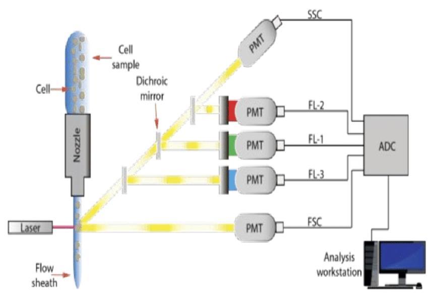
With analysis of the data of large numbers of cells in very short time(>1,000/sec), we can quickly obtain statistically valid information about cell populations.
Fig. 1. Mechanism of flowcytometry.
ISSN 2348-313X (Print)
International Journal of Life Sciences Research ISSN 2348-3148 (online)
Vol. 10, Issue 4, pp: (8-14), Month: October - December 2022, Available at: www.researchpublish.com
A cell suspension of blood, bone marrow aspirate, or lymph node is added to Fluorochrome-labeled monoclonal antibody solutions. For a short time, the tubes are incubated at room temperature. The suspension is passes through the cytometer. Many are automated but some are needed to operate manually and individually. Each cell passes a laser beam solitarily in a process named as single cell analysis. More than 10,000 cells are analyzed to produce statistically valid information. The fluorochrome in labeled monoclonal antibodies is excited by the laser to emit a light of a certain wavelength. The light also is scattered by cells at multiple angles. The emitted or scattered light is collected by the Photodetectors placed at a forward angle and at right angles to the axis of the laser beam. Multiparametric analysis detect forward and right-angle scatter signals, and five fluorochrome signals from every cell. These signals are digitized and passed to a computer for storage, display, and analysis. The data stored for future use (“list mode data storage”). Automation generation or operator-discrete of a variety of histograms for visual display. Data can be transferred to another computer. Different commercial cytometers use a standardized file format for list mode storage, many programs for data analysis and display.
In the present study 50 patients of acute leukemia were analyzed. Of these, 23 cases were of AML and 27 of ALL. ALL cases were further subdivided into B-ALL and T-ALL.
31(62%) cases had conventional immunophenotypes as they showed expression of lineage specific markers while 19 (38%) cases showed expression of CD antigens which were not of that specific lineage.
In 16 patients, lymphoid-lineage-associated antigens were present on AML cases while 14 cases showed myeloidassociated-antigens expression on ALL cases. These cases were considered as aberrant immunophenotypes.
In 23 patients with AML, we found CD7 expression in 6 cases (26%) and CD5 positivity in 3 patients (13 %). Five (22%) of these myeloid leukemia cases showed expression of TdT. One of 23 (4 %) patients expressed CD3 and CD 19. Among the 7 that were acute myeloblastic leukemia, 5 expressed CD7, 3 expressed TdT, 2 showed CD 5, and 1 was positive for CD19.
Thus, these markers were expressed early in hemopoietic ontogeny in the lesser differentiated acute myeloid leukemia subtypes, including FAB M0, M1, and M2. Whereas CD5, CD3 and TdT were each positive in 1 case of acute myelomonocytic leukemia (FAB M4 subtype), while CD 7, CD5 and TdT were aberrantly expressed in 1 case of monocytic leukemia (FAB M5 subtype).
The aberrant expression of CD7, CD5, CD 19 and CD3, representing the capacityof these leukemias for trilinear expression of leukocyte differentiation antigens pretending a poor prognosis.
In ALL myeloid markers CD13 and CD33 were frequent. In the present investigation, 20 cases of B - ALL were evaluated for the presence of CD13 and CD33. Of these 7 cases expressed myeloid markers. Among these 4 (20%) expressed CD13, 3 (15 %) expressed CD33, and 1 (5%) showed positivity for CD14 antigen. Seven cases of T- ALL were analyzed for myeloid markers CD13, CD33, and MPO. Of these 6 expressed myeloid markers, 2 (28%) were positive for CD13, 2 (28%) for CD33, 1(14 %) for CD19, and 1 (14%) for MPO expression.
It is suggested that leukemic cells may have aberrant markers because of their abnormal genetic program resulting in the expression of aberrant immunophenotypes.
Table 1: Frequency of each type of acute leukemia, according to age group
Types of acute children (n=24) Adult (n=21) Total (n=45) leukemia n (%) n (%) n (%) Acute Myeloid 3 (18.6%) 13 (81.2%) 16(35.5) leukemia Acute Lymphoblastic 22 (75.8%) 7(24.1%) 29(64.4%) leukemia
ISSN 2348-313X (Print)
International Journal of Life Sciences Research ISSN 2348-3148 (online) Vol. 10, Issue 4, pp: (8-14), Month: October - December 2022, Available at: www.researchpublish.com
Table 2: Total No. of male/ female ratio in AML/ALL: Males (n=33) Females Total(n=45) n(%) (n=12) n(%) n(%) AML 11(73.3%) 4(26.7) 15(33.3%)
ALL
T-cell ALL 6 (75%) 2 (25%) 8(26.7%) B-cell ALL 17(77.2%) 5 (22.7%) 22 (73.3%)
Table 3: Distribution of AML cases according to immunophenotype
AML FAB Subtype
No. Cases CD34 HLADR CD13 CD33 CyMPO CD14 CD 15 CD117
AML-M0 1 1 0 1 1 0 0 0 0 AML-M1 3 3 1 2 2 0 3 3 1 AML-M2 9 9 4 7 8 0 4 8 2 AML-M3 1 0 0 1 1 1 0 0 0 AML-M4 0 0 0 0 0 0 0 0 0
Table 4: Distribution of B and T cell ALL cases according to immunophenotype
B-Lineage No. Cases CD34 HLADR CD10 CyCD79a CyCD22 CD19 CD13 CD33 CD20 1. Common ALL (CD10+)* 16 16 14 16 11 6 8 9 1 3 2. Null-ALL (CD10-) 6 4 6 0 5 6 3 0 0 0 T-lineage No. cases CD34 CD3 CD7 CD10 CD13 CD33 HLADR CD4 CD7 8 3 6 7 1 0 0 0 7 0
Table 5: Frequency of type of acute leukemia according to the prognosis
CD20 +ve (Bad prognosis) CD20 –ve (good prognosis) B lineage 3(18.8%) 13(81.2%) CD45 (Bright expression) (Bad prognosis) CD45 ( Faint expression) (Good prognosis)
ALL (B and T cell) 8(26.7%) 22(73.3%)
We analyzed the data of 50 patients with acute leukemia in King Fahad Medical City in Riyadh city, in the period between November to December 2019. 62% of the patients had conventional immunophenotyping while the rest showed aberrant immunophenotyping. lymphoid-lineage-associated antigens were present on some AML cases and myeloid-associatedantigens was expressed on some ALL cases as well. An aberrant expression of CD7, CD5, CD 19 and CD3 was noticed in some patients with AML which indicates the capacity of AML for trilinear expression of leukocyte differentiation antigens that highlights poor prognosis. The same aberrant expression was noticed with ALL that indicates the genetic abnormality in these cells.
Aberrant phenotype canbe explained as an abnormal/loss ofexpressionofcell specific lineage marker due to genetic defects which may indicate unfavorable outcome. In leukemia, it is used to determine the minimal residual disease. Our study showed a 38% of aberrant phenotype that is close to another study that the national institute of blood and bone marrow transplantation where aberrant expressions occurred in32% of AMLpatients and 25% of ALLpatients. [12] These abberant expressions included CD5, CD7, CD64dim, CD10, CD117, CD25 and TdT that were in AML expressed while in ALL patients CD33, CD13, HLA-DR and CD3 were detected. [12]. We our study we noticed the expression of CD7, CD5, CD 19 and CD3 in AML and CD13, CD33, MPO, and CD 19 in ALL.
ISSN 2348-313X (Print)
International Journal of Life Sciences Research ISSN 2348-3148 (online)
Vol. 10, Issue 4, pp: (8-14), Month: October - December 2022, Available at: www.researchpublish.com
These markers were more frequently expressed early in the lesser differentiated AML subtypes which may suggest the capacity of trilineage differentiation that could highlight poor prognosis.
In a study of 150 acute leukemia patients, aberrant lymphoid phenotype in AML was an indicator for unfavorable hematological features such as total leukocyte count and greater blast percentage. . Nevertheless, aberrant myeloid phenotype in B-ALL is not associated with poor features [13]
These finding suggest that genetic changes in the cell's programs may play a role in the changes of the expression of these markers and consequently the prognosis of the leukemia.
Our limitation is that this is a retrospective study on a small number of patients with further smaller subgroups and heterogenic characteristics of patients and short period of study. We recommend larger scale studies that involve higher number of patients with longer duration.
The study has a wide variation of clinical implication that indicate the use of flowcytometry in helping in discrimination of prognosis, monitoring the response of treatment and lead the the intensity of therapies according to the sensitivity and response to the drugs.
[1] Leukemia". NCI. 1 January 1980. Archived from the original on 27 May 2014. Retrieved 13 June 2014. Cancer that starts in blood-forming tissue, such as the bone marrow, and causes large numbers of abnormal blood cells
[2] What You Need To Know About™ Leukemia". National Cancer Institute. 23 December 2013. Archived from the original on 6 July 2014. Retrieved 18 June 2014.
[3] Hoffbrand’s Essential Haematology
[4] Adult Acute Myeloid Leukemia Treatment". National Cancer Institute. 6 March 2017. Retrieved 19 December 2017.
[5] Childhood Acute Lymphoblastic Leukemia Treatment". National Cancer Institute. 8 December 2017. Retrieved 20 December 2017.
[6] www.iitk.ac.in
[7] Flow cytometry. By: Vrooman, Linda August, RN, BSN, OCN, Salem Press Encyclopedia of Health, 2019
[8] Lewis RE, Cruse JM, Sanders CM, Webb RN, Suggs JL. Aberrant expression of T-cell markers in acute myeloid leukemia..Exp Mol Pathol. 2007 Dec;83(3):462-3. Epub 2007 Sep 7.
[9] Shin S, Kahng J, Kim M, Lim J, Kim Y, Han K.Distribution of antigenic aberration in the bone marrow of acute leukemia in complete remission. Korean J Lab Med. 2008 Feb;28(1):1-7.
[10] McCoy P. 2002.
[11] Chapman GV. Instrumentation for flow cytometry. J Immunol Methods. 2000;243:3-12.
[12] Shahni, A., Saud, M., Siddiqui, S., & Mukry, S. N. (2018). Expression of aberrant antigens in hematological malignancies: A single center experience. Pakistan Journal of Medical Sciences, 34(2), 457-462. https://doi.org/ 10.12669/pjms.342.13996
[13] Mannelli, Francesco. Immunophenotyping of Acute Leukemias – from Biology to Clinical Application Www.intechopen.com, IntechOpen, 24 Aug. 2016, www.intechopen.com/chapters/49878.