ISSN 2348-313X (Print)
International Journal of Life Sciences Research ISSN 2348-3148 (online)
Vol. 8, Issue 1, pp: (41-51), Month: January - March 2020, Available at: www.researchpublish.com

ISSN 2348-313X (Print)
International Journal of Life Sciences Research ISSN 2348-3148 (online)
Vol. 8, Issue 1, pp: (41-51), Month: January - March 2020, Available at: www.researchpublish.com
Adegbuyi O.
Aderibigbe1 , Abimbola A.
Sowunmi1, Yusuf
Usman1, Elizabeth
1Deparment of Pharmacology & Therapeutics,University of Ibadan, Ibadan, Nigeria
2Deparment of Pharmacology, Afe Babalola University, Ado-Ekiti, Nigeria
Corresponding Author: Abimbola A. Sowunmi, sowunmiabimbola2@gmail.com
Olonode2
Abstract: Members of the plant genus Morus are widely used traditionally in Asia to treat depression, Morus mesozygia is the only member of the genus native to Africa. The study was carried out to evaluate the antidepressant-like activity of Ethanolic Extract of Morus Mesozygia Leaves (EEMM) in mice subjected to chronic unpredictable mild stress (CUMS). Materials and Methods: Male Swiss mice were randomly assigned to 6 different experimental groups and treated groups were given EEMM (2.5, 5 or 10 mg/kg, i.p) 30 minutes before exposure to different mild stressors in the CUMS paradigm for 14 days. They were then subjected to Sucrose Preference Test (SPT) and Forced Swim Test (FST). At the end of the behavioral studies, the levels of Nitrite, Malondialdehyde and Gluthathione in the homogenate of the brain tissues were determined spectrophotometrically. One-way ANOVA was used to statistically determine significant differences. Result: The results showed that EEMM at all doses significantly decreased immobility periods of stressed mice in the forced swim test without increasing the locomotor activity in open field test, and as well reversed the stress-induced anhedonia in the sucrose preference test. Biochemical analysis showed that EEMM significantly (p < 0.05) reduced the concentrations of Nitrite and MDA while the GSH level was elevated in the brains of mice exposed to CUMS. Conclusion: The findings provide significant evidences that EEMM has valid antidepressant-like activity, and might be useful in the management of stress-related disorders on account of its adaptogenic and neuroprotective effects as it prevents oxidative damage in brain tissues.
Keywords: Anhedonia, Stress, Neuroprotective, Depression, Medicinal plant.
Depression is a debilitating and life-threatening illness that negatively affects thoughts, behavior, feelings and sense of well-being. Depression usually presents as a triad of symptoms characterized by low or decreased mood, anhedonia and low energy or fatigue [27]. Depression can occur at any age from childhood to late life, oftentimes, if left untreated may result in fatal outcome with suicide as the hallmark ending [39]. The global prevalence of the disease is very high, reaching about 21% of the population [35]. In most cases, depression present as a complex heterogeneous disorder of diverse variants having more than one etiology, but more commonly, stressful life events have been identified as the major factor that precipitates most depressive episodes [36]. Studies combining behavioral, molecular and electrophysiological techniques reveal that major aspects of depression results from maladaptive stress-induced neuroplastic changes in the neural circuits and these neuroplastic alterations are reversible upon administration of antidepressants [11]. An identified mechanism is the stress-induced structural alteration resulting from excessive reactive oxygen species production causing oxidative damage to macromolecules including lipids, proteins and DNA, leading to neuronal dysfunction such as decrease release of the monoamine neurotransmitter, hence, the monoamine theory of
ISSN 2348-313X (Print)
International Journal of Life Sciences Research ISSN 2348-3148 (online)
Vol. 8, Issue 1, pp: (41-51), Month: January - March 2020, Available at: www.researchpublish.com
depression [23]. Additionally, stress-induced release of dynorphin which activates kappa opioid receptors and down regulates dopamine levels results in disruption of the brain reward circuitry, presenting as anhedonia, the characteristic feature of depression [20]. The exhibited despair and anhedonia are possible behavioral markers which are reversed after treatment with antidepressants [13]. Biochemical markers can as well predict antidepressant activity because they return to normal levels after successful treatment with antidepressants [5].
Numerous antidepressant compounds are available but they are not satisfactory because of a variety of side effects and the poor therapeutic effectiveness. Many of these antidepressant drugs such as Tricyclic Antidepressants (TCAs), Monoamine Oxidase Inhibitors (MAOIs), Selective Serotonin Reuptake Inhibitors (SSRIs) targets the neurotransmitters systems thereby persistently increasing the level of the neurotransmitters in the brain, thus having other associated effects of the elevated monoamine transmitters. These side effects of antidepressants are diverse, present at the minimal therapeutic dose and often defectively interfere with quality of life. Example is persistent dry mouth, constipation and sexual dysfunction associated with adrenergic pathway activation in TCAs. Therefore, majority of patients exhibits poor compliance and refuse to take medications [9]. Importantly, the cost of these antidepressants may also pose economical challenge for depressed patients especially in developing countries, thereby contributing to the residual symptomatology and relapse seen in almost 80% of patients treated with conventional antidepressants [6].
Being natural products, it is assumed that plants with potent antidepressant properties may offer lower side-effect profiles and may even serve as a cheaper alternative when compared to synthetic drugs [40]. Many plants have been identified to have antidepressant properties; examples include Bacopa monniera [34], Cimicifuga racemosa [42], Hypericum perforatum [26], Kaempferia parviflora [38], Olax Subscorpioidea [1], Ptychopetalum olacoides [30], Tinospora cordifolia [10], Zingiber officinale [44]. In Asia, plants belonging to the genus Morus have been identified to have antidepressant property and they are commonly used in Chinese medicine to treat brain and nervous disorders [33]. Morus mesozygia or African mulberry is the only member of the genus Morus native to Africa, growing as deciduous tree in the wild and the concoction/preparation is widely used traditionally across the subtropical Africa for treating mental illness, pain and many other illnesses [7]. Documented medicinal properties of crude extract and isolated compounds of Morus mesozygia include antimicrobial activity [21], antioxidant activity [18], antiplasmodial and cytotoxicity activity [45]. Emerging evidence have suggested that dietary phytochemicals, in particular flavonoids, may exert beneficial effects on the central nervous system by protecting neurons against stress-induced injury, suppressing neuroinflammation and improving cognitive function [17]. Morus mesozygia is rich in flavonoids; Moranoline, Albafuran, Albanol, Morusin, Kuwanol, Calystegine and Hydroxymoricin [18]. These isolated compounds from mulberry plants have many identified pharmacological profile including potential to reverse cell damage. Therefore, this study is to evaluate the antidepressantlike effect of ethanol extract of Morus mesozygia leaves (EEMM) in mice subjected to chronic unpredictable mild stress (CUMS).
Plant Authentication: The leaves of Morus mesozygia were collected at the Botanical Garden, University of Ibadan, Nigeria. The taxonomical identification and authentication of the plant was done at the herbarium section of the Forestry Research Institute of Nigeria (FRIN), Ibadan, Nigeria. A voucher specimen with identification number FHI:110387 was deposited and compared with the reference specimen.
Preparation of Extract: One hundred grammes (100g) of the air-dried leaves were pulverized and soaked in 50% ethanol (2L) for 48 hours. The filtrate was concentrated with a rotary evaporator to give a semisolid residue and evaporated to dryness to form solid residue. It was kept in the desiccator for further use. The dried extract was dissolved in distilled water and administered intraperitoneally.
Experimental Animals: Male Swiss mice (20-25g) obtained from the Central Animal House, University of Ibadan were used in the study. They were housed individually in plastic cages at room temperature and with access to rodent pellet food and water ad libitum. The animals were acclimatized for a period of 14 days before the study. The animals were handled in the study in accordance with the US National Institutes of Health (NIH) Guideline for the Care and Use of Laboratory Animals.
Chronic Unpredictable Mild Stress (CUMS): The CUMS protocol was carried out according to the previously described method, but with slight modifications [43]. After 2 weeks of acclimatization, mice were randomly assigned to 6 different experimental groups (n = 5), so that mean body weights were comparable in all groups. Thereafter, independent mice group received either EEMM (2.5, 5 or 10 mg/kg, i.p), Imipramine (15 mg/kg) or vehicle (distilled water, 10 ml/kg)
ISSN 2348-313X (Print)
International Journal of Life Sciences Research ISSN 2348-3148 (online) Vol. 8, Issue 1, pp: (41-51), Month: January - March 2020, Available at: www.researchpublish.com
30 min before daily exposure to different stressors in the CUMS paradigm as shown in Table 1. The CUMS procedure was repeated daily for 14 consecutive days and the stressors were applied in random order at varying times to maximize unpredictability. Mice subjected to CUMS procedure were called stressed mice while the unstressed mice were those not subjected to CUMS procedure but only treated with vehicle. Behavioral test employed for evaluation of antidepressantlike activity was Forced Swim Test (FST) and Sucrose Preference Test (SPT) for comparing CUMS-induced depressivelike phenotypes.
Table 1: The order of stressors used in CUMS paradigm
Days Monday Tuesday Wednesday Thursday Friday Saturday Sunday Week 1 I E W O F X T1 Week 2 E O F I X C W
I Immobilization for 10 mins; E Exposure to empty water bottles for 1 hour; W Wet bedding exposure for 24 hours; O overnight illumination; F Foot shock (0.2mA for 15s); X Tilted cage at 45 degrees for 7 hours; T1 Forced swim in cold water 4oC for 30 s.
Sucrose Preference Test (SPT): The SPT was used in the study as the bioassay for determining anhedonia, a core symptom of major depression in human. The test was carried out before and after the 14 days CUMS exposure between 9 a.m. - 12 noon which were the training and testing courses respectively. After 2 weeks of acclimatization before start of CUMS, mice were trained to consume 1% (w/v) sucrose solution. In the training course, mice were deprived of food and water for 24 hours and only exposed to 1% (w/v) sucrose solution. After 72 hours post training course, the animals were subjected to 1hr baseline test. In the test, mice were housed in individual cages with free access to two pre-weighted bottles, one containing 100 ml water and another containing 100 ml 1% (w/v) sucrose solution. After 1 hour both the amount of water and sucrose solution was measured and recorded. The ratio of amount of sucrose solution to that of total solution consumed within 1 hour represented the parameter of hedonic behavior. The sucrose preference was calculated according to the following formula:
sucrose solution intake (g)
Sucrose Preference = x 100 sucrose solution intake + water intake
The test was performed again 24 hr after the 14 days of CUMS exposure to evaluate the effect of stress as well as drug treatment on the depressive symptoms.
Forced Swimming Test (FST): On the second day post CUMS, 30 minutes after treatment as in the CUMS protocol, FST was carried out on mice according to the method of Porsolt [31]. The mice were placed individually in plexiglas cylinders (40 cm in height, 18 cm in diameter) filled with water (25 °C) up to 15 cm. A 2 min pre-swimming period was followed later by a 4 min test period during which the total immobility time was recorded. Mice were considered immobile when they made no further attempts to escape, except for necessary movements to keep their heads above the water. The period of immobility was recorded by an experimenter (not blind for the experimental conditions) using a stopwatch during the exposures. The water in the cylinders was changed before every trial and mice were towel dried and returned to their housing conditions after the swimming session.
Measurement of Locomotor Activity: To rule out the effects of various drug treatments on motor function, crossing activities of control and test animals were measured for a period of 5 min in an open field apparatus consisting of white plexiglas box (45 cm × 25 cm × 25 cm) with the floor equally divided into 16 equal squares marked with painted black grid. Ambulatory behavior was assessed as previously described [2]. The animals were placed singly in the center of the box; the number of squares crossed with all four paws was counted for 5 min. The cage was cleaned with 70% ethanol at 5 min interval when the animal is removed. Before each test, animals were kept in the test room at least 1 hour before open field test (OFT) for habituation. All animals were used only once in this test. These animals were different from those used in the CUMS procedure.
Biochemical Estimations: After subjecting unstressed and stressed mice to FST on the second day post CUMS, the animals were sacrificed by cervical dislocation. The brains were excised and quickly rinsed in ice-cold 1.15% KCl after which they were blotted. The brains were then minced with scissors in 10% w/v ice-cold 0.1M phosphate buffer, pH 7.4, and homogenized using a Tefflon homogenizer. The resulting homogenates were centrifuged at 10,000 rpm, 4°C for 10 minutes. The supernatant – post mitochondrial fraction (PMF) were collected and processed for biochemical estimations.
ISSN 2348-313X (Print)
International Journal of Life Sciences Research ISSN 2348-3148 (online) Vol. 8, Issue 1, pp: (41-51), Month: January - March 2020, Available at: www.researchpublish.com
Determination of Glutathione (GSH) Concentration: The concentrations of reduced GSH were measured in the aliquots of homogenates of individual mice brain of the respective treatment groups according to the method described by Moron [25]. Equal volume (0.4 ml) of tissue homogenate and 20% trichloroacetic acid (TCA) (0.4 ml) were mixed and then centrifuged using a cold centrifuge at 10,000 rpm at 4oc for 20 min. The supernatant (0.25 ml) was added to 2ml of 0.6mM DTNB reagent and the final volume was made up to 3ml with phosphate buffer (0.2M, pH 8.0). The absorbance was then read at 412 nm against blank reagent using a spectrophotometer. The concentrations of reduced GSH in the brain tissues were expressed as micromoles per milligram of protein.
Determination of Lipid Peroxidation: The malondialdehyde (MDA) content, a measure of lipid peroxidation determined by measuring the thiobarbituric acid reactive substances (TBARS) during lipid peroxidation, was estimated according to the method described by Okhawa [28]. Briefly, 0.5 ml of distilled water and 1.0 ml 10% TCA were added to 0.5 ml of each brain tissue homogenate and centrifuged at 3000 rpm for 10 min. Then 0.1 ml thiobarbituric acid (TBA) (0.375%) was added to 0.9 ml of supernatant. The mixture was placed in a water bath at 80ºC for 40 min and then cooled to room temperature. Upon cooling, the absorbance of the supernatant was measured at 532nm using a spectrophotometer. The thiobarbituric acid-reactive substances were quantified using an extinction coefficient of 1.56×10⁵ M-1 cm -1 and values were expressed as µmoles of MDA per gramme tissue. Tissue protein was estimated using the biuret method as previously described, and the brain malondialdehyde content was expressed as micromole of malondialdehyde per milligram of protein.
Estimation of Nitrite Levels: Nitrite levels in the aliquots of homogenates of individual mice brain of the respective treatment groups was measured by using the method described by Green [14]. A mixture of 1% (w/v) sulfanilamide in 5% aqueous solution of m-phosphoric acid (1 part) and 0.1% (w/v) N-(1-naphthyl) ethylenediamine dihydrochloride (1 part) was prepared and kept at 0 0C for 60 min. 0.5 ml brain homogenate was mixed with 0.5 ml of the above mixture and kept in dark for 10 min at room temperature. The absorbance was read at 546 nm using UV–visible spectrophotometer.
Data Analysis: All data were presented as Mean ± SEM. The results were analyzed by One-way Analysis of Variance (ANOVA) and post hoc test (Student’s-Newman-Keuls) was carried out to determine the source of significant main effect using GraphPad Prism Biostatistics software version 5.00. The level of significance for all tests was set at p < 0.05 (i.e. at 95% confidence interval).
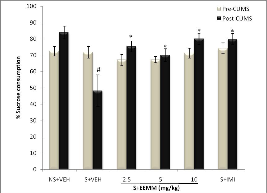
Effect of EEMM on CUMS-induced Anhedonia in Mice using the SPT: There was no significant difference in sucrose preference (%) among all the groups in the baseline test (pre-test). Exposure of the mice to stress for 14 successive days significantly (p < 0.05) decreased sucrose preference (%) in vehicle-treated stressed mice as compared to the unstressed control. EEMM (2.5, 5 and 10 mg/kg) and Imipramine (15 mg/kg) administered for 14 successive days significantly reverse the reduced sucrose preference (%) in stressed mice as compared to the vehicle-treated control (Fig. 1).
Fig. 1: Effect of EEMM on Sucrose preference in mice.
ISSN 2348-313X (Print)
International Journal of Life Sciences Research ISSN 2348-3148 (online) Vol. 8, Issue 1, pp: (41-51), Month: January - March 2020, Available at: www.researchpublish.com
The results are expressed as Mean +/- SEM (n=5). In the post-test, One way ANOVA revealed that there is significant [F (5, 24) = 6.414, p < 0.001] difference between various treatment groups.
# indicates significant difference from the control p < 0.05 (Student-Newman-Keuls test).
*indicates significant difference from the stressed vehicle treated p < 0.05 (Student-Newman-Keuls test).
NS+VEH: Non-stressed+Vehicle; S+VEH: Stressed+Vehicle; S+EEMM: Stressed+Ethanol Extract of Morus mesozygia; S+IMI: Stressed+Imipramine (15 mg/kg, i.p.).
Effect of EEMM on Immobility period in FST in mice subjected to CUMS: Chronic unpredictable mild stress (CUMS) significantly increased the immobility period in vehicle treated stressed mice (S+VEH) as compared to nonstressed vehicle treated (NS + VEH) control mice subjected to FST. EEMM (2.5, 5 and 10 mg/kg, i.p.) administered for 14 successive days significantly (p < 0.001) decreased the immobility period in stressed mice as compared to the stressed controls (Fig. 2).
Fig 2: Effect of EEMM on immobility period in FST in CUMS.
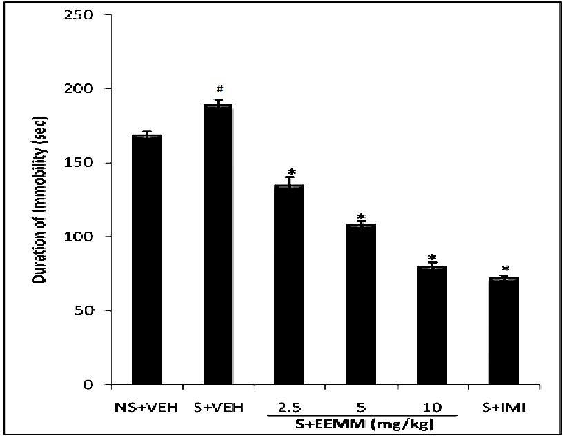
The results are expressed as Mean +/- SEM (n=5). One way ANOVA revealed that there is significant [F (5, 24) = 200.4, p < 0.001] difference between various treatment groups.
# indicates significant difference from the non-stressed control p< 0.05 (Student-Newman-Keuls test).
* indicates significant difference from the control p < 0.05 (Student-Newman-Keuls test).
NS+VEH: Non-stressed + Vehicle; S+VEH: Stressed + Vehicle; S+EEMM: Stressed + Ethanol Extract of Morus mesozygia; S+IMI: Stressed + Imipramine (15 mg/kg, i.p.).
Effect of EEMM on Locomotor Activity in Open Field: The administration of EEMM (2.5, 5 or 10 mg/kg, i.p.) showed a significant reduction [F (5, 24) = 137.7, P < 0.001] in the locomotor activity of mice when compared to vehicle (Table 2).
Table 2: Effect of EEMM on locomotor activity in open field
Locomotor Activity Test
Treatment (mg/kg)
Number of Square Crossing VEH 73.60±1.631
EEMM 2.5 57.60±1.568* 5 51.60±2.657* 10 46.60±1.661*
Values represent mean ± S.E.M for 6 animals per group. *P < 0.05 compared to Control (ANOVA followed by Newman Keuls test).
VEH: Vehicle; EEMM: Ethanol Extract of Morus mesozygia
ISSN 2348-313X (Print)
International Journal of Life Sciences Research ISSN 2348-3148 (online) Vol. 8, Issue 1, pp: (41-51), Month: January - March 2020, Available at: www.researchpublish.com
Effect of EEMM on brain Glutathione Levels: GSH levels were significantly (p < 0.001) decreased in brains of vehicle-treated stressed mice (S+VEH) as compared to non-stressed control (NS+VEH). EEMM (2.5, 5 and 10 mg/kg) and Imipramine (15 mg/kg) produced a significant (p < 0.001) increase in GSH levels in respective treated mice as compared to vehicle-treated stressed control (S+VEH) mice (Fig. 3).
Fig. 3: Effect of EEMM on GSH Levels in mice.
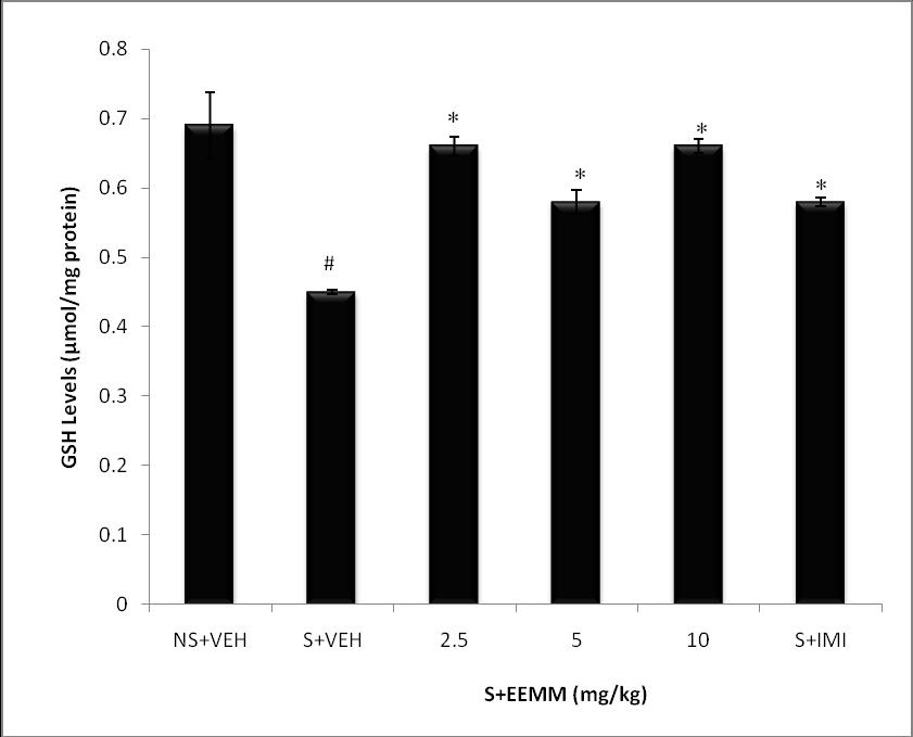
The results are expressed as Mean +/- SEM (n=5). One way ANOVA revealed that there is significant [F (5, 24) = 37.7, p< 0.001] difference between various treatment groups.
# indicates significant difference from the control p< 0.05 (Student-Newman-Keuls test).
*indicates significant difference from the stressed vehicle treated p< 0.05 (Student-Newman-Keuls test).
NS+VEH: Non-stressed+Vehicle; S+VEH: Stressed+Vehicle; S+EEMM: Stressed+Ethanol Extract of Morus mesozygia; S+IMI: Stressed+Imipramine (15 mg/kg, i.p.).
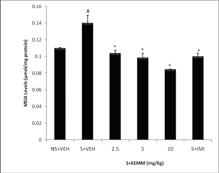
Effect of EEMM on brain Malondialdehyde Levels: Malondialdehyde levels were significantly (p < 0.001) increased in brains of vehicle-treated stressed mice (S+VEH) as compared to non-stressed control (NS+VEH). EEMM (2.5, 5 and 10 mg/kg) and Imipramine (15 mg/kg) produced a significant (p < 0.001) decrease in MDA levels in respective treated mice as compared to vehicle-treated stressed control (S+VEH) mice (Fig. 4).
Fig. 4: Effect of EEMM on MDA Levels.
ISSN 2348-313X (Print)
International Journal of Life Sciences Research ISSN 2348-3148 (online) Vol. 8, Issue 1, pp: (41-51), Month: January - March 2020, Available at: www.researchpublish.com
The results are expressed as Mean +/- SEM (n=5). One way ANOVA revealed that there is significant [F (5, 24) = 26.11, p < 0.001] difference between various treatment groups.
# indicates significant difference from the control p < 0.05 (Student-Newman-Keuls test).
*indicates significant difference from the stressed vehicle treated p < 0.05 (Student-Newman-Keuls test).
NS+VEH: Non-stressed+Vehicle; S+VEH: Stressed+Vehicle; S+EEMM: Stressed+Ethanol Extract of Morus mesozygia; S+IMI: Stressed+Imipramine (15 mg/kg, i.p.).
Effect of EEMM on Nitrite Levels: Brain nitrite levels were significantly (p < 0.001) elevated in vehicle treated mice subjected to CUMS. EEMM (2.5, 5 0r 10 mg/kg) administered for 14 successive days in CUMS significantly (p < 0.001) decreased nitrite levels in stressed mice as compared to vehicle-treated control mice (Fig. 5).
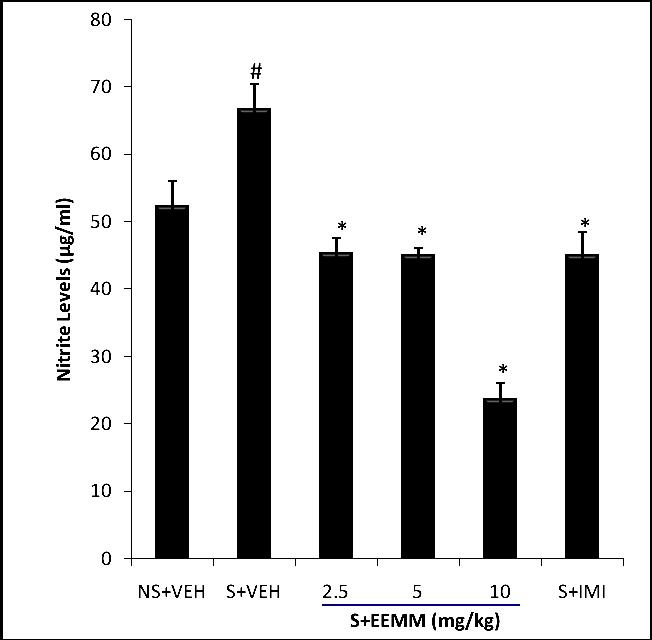
Fig. 5: Effect of EEMM on Nitrite Levels in mice.
The results are expressed as Mean ± SEM (n=5). One way ANOVA revealed that there is significant [F (5, 24) = 51.52, p< 0.001] difference between various treatment groups.
# indicates significant difference from the control p < 0.05 (Student-Newman-Keuls test).
*indicates significant difference from the stressed vehicle treated p < 0.05 (Student-Newman-Keuls test).
NS+VEH: Non-stressed+Vehicle; S+VEH: Stressed+Vehicle; S+EEMM: Stressed+Ethanol Extract of Morus mesozygia; S+IMI: Stressed+Imipramine (15 mg/kg, i.p.).
CUMS-induced depression model was used in this study as the chronic model of depression. Animals subjected to CUMS model exhibits characteristic depressive behavior similar to that observed in humans after a long-term exposure to multiple stressors, hence, the model satisfies most of the criteria of validity for an animal model of depression [41]. Additional studies have shown that CUMS can be used for evaluating the potential antidepressants by employing behavioral tests like FST and sucrose preference test [24]. Mice forced to swim in a restricted space from which they cannot escape are induced to a characteristic behavior of immobility. This prolonged immobility behavior reflects a state of despair similar to depression in humans and the immobility can be reduced in mice by several agents that are
ISSN 2348-313X (Print)
International Journal of Life Sciences Research ISSN 2348-3148 (online)
Vol. 8, Issue 1, pp: (41-51), Month: January - March 2020, Available at: www.researchpublish.com
therapeutically effective in human depression [32]. The sucrose preference test represents the anhedonia-like behavioral change, a hallmark core symptom of depression in humans, indicating loss of interest or pleasure. Anhedonia was modeled in mice by inducing a decrease in responsiveness to pleasure reflected by a reduced consumption of sucrose solutions. The behavioral and molecular changes induced by CUMS are reversed by treatment with antidepressant drugs after administration for weeks [22]. In this work, the CUMS mice model satisfactorily mimicked the depressive status “behavioral despair” as seen in human depression. The results of vehicle treated mice subjected to chronic stress in CUMS exhibited a significant prolongation of immobility time in FST and reduction of the sucrose solution consumption as compared to unstressed mice. The results reflect successful induction of depressive state in test mice by the applied CUMS protocol in this study. However, chronic administration of EEMM significantly decreased the duration of immobility in mice FST and prevented the reduced preference of sucrose solution which both indicates the antidepressantlike effect. The antidepressant-like activity of EEMM is specific and not false positive since the result of the locomotor activity indicated that EEMM at the respective treatment doses (2.5, 5, or 10 mg/kg) did not show CNS stimulant effect in mice subjected to open field test. Although CNS-depressant agents may be sedative, generally, the activity follows a doseresponse curve in the open field test [37]. The observed dose dependent reduction in locomotor activity in the EEMM treated mice is suggestive of a possible CNS depressant activity, however, the effect does not interfere-with or mask the antidepressant activity seen in the FST paradigm, thus, EEMM at the treatment doses is not sedative.
Reversal of CUMS-induced depressive-like phenotypes measured using the mouse FST and SPT has been reported as a valid measure of antidepressant property [3]. The findings here from the FST and SPT of mice subjected to CUMS indicate the face-validity of the antidepressant-like profile of action of EEMM. Additionally, SPT has been identified as a sensitive screening test for potential rapid-acting antidepressant-like drugs [13]. Therefore, the significant positive outcome of activities of EEMM on SPT reflects its potential rapid onset of antidepressant effect.
Physical and psychological stressors have also been shown to increase free radical production, cause oxidative damage and accelerate lipid peroxidation [15]. Repeated stress on a daily basis described as un-predictive increasing stress has been reported to enhance stress-induced tissue damage and malfunction [8]. Excessive reactive oxygen species production from repetitive stress can cause oxidative damage to macromolecules including lipids, proteins and DNA, leading to neuronal dysfunction and depression [12]. Reactive oxygen species play a role in the pathogenesis of neuropsychiatric disorders. Basically, derangement of the antioxidant defense system leads to neurotransmitter deficiencies, one of the most important mechanisms in major depression [19]. This free radical activity and the extent of tissue damage are often characterized by high level of malondialdehyde (MDA), high level of nitrite and a reduction of glutathione (GSH) concentrations. The high malondialdehyde level is a marker of lipid peroxidation which is characteristic of oxidative damage. Lipid peroxidation and antioxidant enzymes can be assessed as markers of major depression because they returned to normal levels after treatment with antidepressants [5]. Chronic unpredictable mild stress was found to impair the antioxidant status of brain tissue, presumably through production of excessive reactive oxygen species. In the present study, 14 days successive exposure of mice to different stressors resulted in increased lipid peroxidation and nitrite levels, and decreased endogenous antioxidant activity in mice. Chronic administration of EEMM showed a significant decrease in lipid peroxidation and increase in GSH in stress-exposed mice. Thus, EEMM showed a significant antioxidant activity in mice. The in vitro antioxidant activity of Morus mesozygia has been reported [18]. Stressful situations have also been shown to significantly increase nitrite levels [16]. EEMM significantly reduced nitrosative stress as indicated by reduction of the brain nitrite levels of EEMM treated stressed mice as compared to the vehicle treated stressed mice. Thus, EEMM showed a strong neuroprotective effect against oxidative stress and nitrosative stress that plays a key role in chronic unpredictable mild stress-induced depression. Hence, EEMM antidepressant-like activity in chronically stressed mice is probably through the decrease in plasma nitrite levels and due to its antioxidant activity. Importantly, when the production of reactive oxygen species exceeds the level which the normal body antioxidant defense mechanisms can cope with; there is a gradual breakdown of body’s stress adaptive system [4]. The results of this study may also suggest this inhibition of oxidative stress as an adaptogenic property of EEMM, which prevents oxidative breakdown of macromolecules such as DNA, proteins and lipids involved in body’s stress adaptive process. Generally, plant extracts having antioxidant properties have been experimentally shown to have neuroprotective effects [29]. They have also been shown to be useful in the management of various stress related metabolic and chronic diseases [39], which was also demonstrated in this study.
ISSN 2348-313X (Print)
International Journal of Life Sciences Research ISSN 2348-3148 (online) Vol. 8, Issue 1, pp: (41-51), Month: January - March 2020, Available at: www.researchpublish.com
The primary findings concluded that EEMM could improve the depressive-like symptoms induced by CUMS that may be related to regulation of oxidative and nitrosative stress. Hence, EEMM has a valid antidepressant property that could be rapid-acting, however, further studies needs be done to identify the active constituent in the plant and determine the modulation of the momoaminergic neurotransmitter systems by EEMM
Authors are thankful to the Head, Department of Pharmacology and Therapeutics, University of Ibadan, Prof. Ezekiel O. Iwalewa, for providing basic laboratory facilities and professional guidance during the course of this research study.
[1] Adeoluwa, O., Aderibigbe, A., Bakre, A., 2015. Evaluation of Antidepressant-like Effect of Olax Subscorpioidea Oliv. (Olacaceae) Extract in Mice. Drug Res (Stuttg) 65(06): 306-311
[2] Akanmu, M. A., Olowookere, T. A., Atunwa, S. A., Ibrahim, B. O., Lamidi, O. F., Adams, P. A., Ajimuda, B. O., Adeyemo, L. E., 2011. Neuropharmacological effects of Nigerian honey in mice. African Journal of Traditional Complementary Alternative Medicine, 8: 230‐249.
[3] Autry, A. E., Adachi, M., Nosyreva, E., Na, E. S., Los, M. F., Cheng, P. F., et al., 2011. NMDA receptor blockade at rest triggers rapid behavioral antidepressant responses. Nature 475: 91–95. Available from: http://www.intechopen com/ books/psychiatric-disorders-trends-and-developments/mouse-models-ofdepression
[4] Bartsch, H., and Nair, J., 2000. Ultrasensitive and specific detection of methods for exocyclic DNA adducts: markers for lipid peroxidation and oxidative stress. Toxicology, 153: 105-114.
[5] Bilici, M., Efe, H., Koroglu, M., et al., 2001: Antioxidative enzyme activities and lipid peroxidation in major depression: alterations by antidepressant treatments. J Affect Disord 64:43–51.
[6] Burcusa, S. L., Iacono W. G., 2007. Risk for recurrence in depression. Clin Psychol Rev, 27: 959-85.
[7] Burkill, H., 1985. “The Useful Plants of West Tropical Africa,” Economic Botany & Ethnobotany, Vol. 1, p. 319.
[8] Davydov, V., and Shvets, N., 2001. Lipid peroxidation in the heart of adult and old rats during immobilization stress. Exp. Gerontol., 36(7): 1155-1160.
[9] Demyttenaere, K., 1997. Compliance during treatment with antidepressants. J Affect Disord, 43:27–39
[10] Dhingra, D., Goyal, P. K., 2008. Evidences for the involvement of monoaminergic and GABAergic systems in antidepressant-like activity of Tinospora cordifolia in mice,Indian J Pharm Sci, 70: 761-765
[11] Duman, R., Malberg, J., Nakagawa, S., D'Sa, C., 2000. Neuronal plasticity and survival in mood disorders. Biol Psychiatry 48:732-739.
[12] Esch, T., Stefano, G. B., Fricchione, G. L., Benson, H., 2002. The role of stress in neurodegenerative diseases and mental disorders. Neuro Endocrinol Lett, 23:199– 208.
[13] Garcia, L.S., Comim, C. M., Valvassori, S. S., Reus, G. Z., Stertz, L., Kapczinski, F., et al., 2009. Ketamine treatment reverses behavioral and physiological alterations induced by chronic mild stress in rats. Prog. Neuropsychopharmacol.Biol. Psychiatry 33, 450–455.doi:10.1016/j.pnpbp.2009.01.004
[14] Green, L., Wagner, D., Glogowski, J., et al., 1982: Analysis of nitrate, nitrite, and [15N]nitrate in biological fluids. Anal Biochem 126:131–8.
[15] Han, S. G., Kim, Y., Kashon, M. L., Pack, D. L., Castranova, V., Vallyathan, V., 2005. Correlates of oxidative stress and free-radical activity in serum from asymptomatic shipyard welders. Am J RespirCrit Care Med, 172:1541-1548.
[16] Harvey, B., 1996. Affective disorders and nitric oxide: a role in pathways to relapse and refractoriness? Hum Psychopharmacol 113:309–19.
ISSN 2348-313X (Print)
International Journal of Life Sciences Research ISSN 2348-3148 (online) Vol. 8, Issue 1, pp: (41-51), Month: January - March 2020, Available at: www.researchpublish.com
[17] Jeremy, P. E., 2008. Flavonoids: modulators of brain function? British Journal of Nutrition 99, E-Suppl. 1, ES60–ES77).
[18] Kapche, W., Fozing, C., Donfack, J., Fotso, W., 2009. Prenylated arylbenzofuran derivatives from Morus mesozygia with antioxidant activity. Phytochemistry. 70: 216-221.
[19] Khanzode, S. D., Dakhale, G. N., Khanzode, S. S., Saoji, A., Palasodkar, R., 2003. Oxidative damage and major depression: the potential antioxidant action of selective serotonin re-uptake inhibitors. Redox Rep. 8(6): 365-370.
[20] Knoll, A. T., Carlezon Jr. W. A., 2010. Dynorphin, stress, and depression, Brain Res. 1314: 56–73.
[21] Kuete, V., Fozing, D., Kapche, W., Mbaveng, A., 2009. Antimicrobial activity of the methanolic extract and compounds from Morus mesozygia stem bark. Journal of Ethnopharmacology, 124: 551-555.
[22] Kumar, B., Kuhad, A., and Chopra, K., 2011. Neuropsychopharmacological effect of sesamol in unpredictable chronic mild stress model of depression: behavioral and biochemical evidences. Psychopharmacology 214:819–28.
[23] Manji, H. K., Duman, R. S., 2001. Impairments of neuroplasticity and cellular resilience in severe mood disorders: implications for the development of novel therapeutics. Psychopharmacol Bull; 35:45–9.
[24] Mao, Q., Ip, S., Ko, K., Tsai, S., Che, C., 2009. Peony glycosides produce antidepressant-like action in mice exposed to chronic unpredictable mild stress: effects on hypothalamic–pituitary–adrenal function and brain-derived neurotrophic factor. Prog Neuropsychopharmacol Biol Psychiatry 33: 1211–6.
[25] Moron, M. S., Depierre, J. W. and Mannervik, B. 1979. Levels of glutathione, glutathione reductase and glutathione S-transferase activities in rat lung and liver. BiochimicaetBiophysica ACTA 582: 67-78.
[26] Nathan, P. J., 2001. Hypericum perforatum (St John’s Wort): a non-selective reuptake inhibitor- A review of the recent advances in its pharmacology, J Psychopharmacol, 15: 47–54.
[27] Nina, D., Sandra, M. and Jan, M. 2011. Mouse Models of Depression, Psychiatric Disorders - Trends and Developments, Dr. Toru Uehara (Ed.), In Tech, pp: 185–224 ISBN: 978-953-307-745-1.
[28] Okhawa, H., Ohishi, N., and Yagi, K. 1979. Assay for Lipid Peroxides in Animal Tissues by Thiobarbituric Acid Reaction. Analytical Biochemistry 95(2): 351- 358.
[29] Parihar, M. S., and Hemnani, T., 2003. Phenolic antioxidants attenuate hippocampal neuronal cell damage against kainic acid induced excitotoxicity. J. Biosci, 28(1): 121-128.
[30] Piato, A. L., Rizon, L. P., Martins, B. S., Nunes, D. S., Elisabetsky, E., 2008. Antidepressant profile of Ptychopetalum olacoides Bentham (Marapuama) in mice, Phytother. Res, 23: 519-524.
[31] Porsolt, R., Anton, G., Blavet, N., Jalfre, M., 1978. Behavioural despair in rats: a new model sensitive to antidepressant treatments. Eur J Pharmacol 47: 379–391.
[32] Porsolt, R., Bertin, A., and Jalfre, M., 1977. Behavioural despair in mice: A primary screening test for antidepressants. Arch Int Pharmacodyn 229: 327–336
[33] Rupesh, K., Praveen, K., Suchita, M., 2013. Herbal Sources of Antidepressant Potential: A Review. Int. J. Pharm. Sci. Rev. Res., 18(1) 13, 86-91
[34] Sairam, K., Dorababu, M., Goel, R., Bhattacharya S., 2002. Antidepressant activity of standardized extract of Bacopa monniera in experimental models of depression in rats, Phytomedicine, 9(3): 207-211
[35] Schechter, L., Ring, R., Beyer, C. 2005. Innovative approaches for the development of antidepressant drugs: current and future strategies. NeuroRx, 2:590–611.
[36] Sickmann, H., Li, Y., Mork, A., Sanchez, C., Gulinello, M., 2014. Does stress elicit depression? Evidence from clinical and preclinical studies, Curr. Top. Behav. Neurosci. 18.
[37] Vogel, G., Vogel, W., 1997. Drug Discovery and Evaluation Pharmacological Assays: Psychotropic and Neurotropic activity. Springer USA. H, and H, (2nd Edition): 559-568.
ISSN 2348-313X (Print) International Journal of Life Sciences Research ISSN 2348-3148 (online) Vol. 8, Issue 1, pp: (41-51), Month: January - March 2020, Available at: www.researchpublish.com
[38] Wattanathorn, J., Pangpookiew, P., Muchimapura, K., Sripanidkuchai, B., 2007. Evaluation of the anxiolytic and antidepressant effects of alcoholic extract of Kaempferia parviflora in aged rats, African Journal of Animal and Biomedical Sciences, 2: 94-98
[39] WHO, 2012. World suicide prevention day. Available from: http://www.who.int/mediacentre/events/annual/world_ suicide_prevention_day/en/
[40] Willcox, J. K., Ash, S. L. and Catignani, G. L., 2004. Antioxidants and prevention of chronic disease. Crit Rev Food Sci Nutr, 44(4): 275-295.
[41] Willner, P., 2005: Chronic mild stress revisited: consistency and behavioral neurobiological concordance in the effects of CMS. Neuropsychobiology 52: 90– 110.
[42] Winterhoff, H., Spengler, B., Christoffel, V., Butterweck, V., Löhning, A., 2003. Modern Phytotherapy in Menopause: Cimicifuga racemosa (Klimadynon, Menofem) Pharmacological and Clinical Data, 2002, Berlin. Cimicifuga extract BNO 1055: reduction of hot flushes and hints on antidepressant activity. Maturitas 44: S51-S58
[43] Yalcin, I., Aksu, F., and Belzung, C., 2005. Effects of desipramine and tramadol in a chronic mild stress model in mice are altered by yohimbine but not by pindolol. European Journal of Pharmacology 514: 165–174.
[44] Yi, L. T., Xu, Q., Li, Y. C., Yang, L., Kong, L. D., 2009. Antidepressant-like synergism of extracts from magnolia bark and ginger rhizome alone and in combination in mice, Prog Neuropsychopharmacol Biol Psych, 33: 616–624
[45] Zelefack, F., David, G., Alexis, V., René, C., 2012. Antiplasmodial and cytotoxic activities of flavonoids and arylbenzofuran derivatives from Morus mesozygia. Greener Journal of Biological Sciences 2 (2): 020-024.