International Journal of Healthcare Sciences ISSN 2348-5728 (Online)
Vol. 10, Issue 2, pp: (154-158), Month: October 2022 - March 2023, Available at: www.researchpublish.com

International Journal of Healthcare Sciences ISSN 2348-5728 (Online)
Vol. 10, Issue 2, pp: (154-158), Month: October 2022 - March 2023, Available at: www.researchpublish.com
1 Internship doctor in RSU KabupatenTangerang Hospital, Tangerang, Banten
2 Pediatric Orthopedic Surgeon, DepartementofSurgery, RSUKabupaten Tangerang Hospital,Tangerang, Banten
DOI: https://doi.org/10.5281/zenodo.7488448
Published Date: 28-December-2022
Abstract: Fractures of humeral shaft in children are relatively rare injuries, accounting for less than 3% of all childhood fractures. Although these injuries are rare, it is important to know the clinical approach and definitive treatment to get a better result in pediatric humeral fracture. This paper provides an overview of diaphyseal fractures of the humerus in children, including the clinical approach and definitive treatment. Fractures of the humerus midshaft can be caused by direct trauma to the arm, a fall, a car accident, or a sports injury. Older children usually have pain, swelling, and bruising. For most pediatric humeral diaphyseal fractures, x-ray images with standard anterior-posterior (AP) and full-length lateral views are sufficient. The definitive treatment of the humeral fracture can be conservative or need a surgical treatment. Most of humeral midshaft fractures in children can be treated conservatively. But, surgical treatment is mainly used for older children, adolescents, and some indications, like showed in this case report. In this case, patient favorORIF over IMN as the optimal treatment decision for a traumatic humeral shaft fracture. Plating has been shown to have better overall results compared to the interlocking nails in treatment of closed humeral shaft fractures. In this report, we present a case of midshaft humerus fracture in a 15-Year-Old Adolescent.
Keywords: Midshaft humerus fracture,Clinical approach, Definitive treatment
Fractures of humeral shaft in children are relatively rare injuries, accounting for less than 3% of all childhood fractures. Although these injuries are rare, it is important to know the clinical approach and definitive treatment to get a better result in pediatric humeral fracture. This paper provides an overview of diaphyseal fractures of the humerus in children, including the clinical approach and definitive treatment. Describes etiology, clinical findings, imaging, fracture patterns, classification, associated injuries, management (non-surgical and surgical), andcomplications. As there is an increasing tendency to treat these injuries with surgical fixation, overall, there is ample evidence that the majority of humeral diaphyseal fractures inchildren and adolescents can be successfully treated conservatively. However, surgical treatment has proven beneficial with good clinical results and low complication rates, especially when clinically indicated in older children and adolescents as presented in this case. Fractures of the humerus diaphysis can be caused by direct trauma to the arm, a fall, a car accident, or a sports injury [1,2]. Up to 56% of humeral diaphyseal fractures in older children are due to falls [3] .
Symptoms depend on the age of the child and the mechanism of injury. Children older than usually have pain, swelling, and bruising. The child may press his arm against his bodyto limit his mobility. Examinationmayreveal obvious palpation deformities and pain or crepitus [4] However, most humeral diaphyseal fractures show no visible deformation, and x-ray images show minimal displacement [5] .
International Journal of Healthcare Sciences ISSN 2348-5728 (Online)
Vol. 10, Issue 2, pp: (154-158), Month: October 2022 - March 2023, Available at: www.researchpublish.com
For most pediatric humeral diaphyseal fractures, x-ray images with standard anterior- posterior (AP) and full-length lateral views aresufficient. Most humeral trunk fractures in children can be treated conservatively, There are several fixing techniques for children and adolescents, such as slings and / or capes, bell paw bandages, collars and cuffs, adjustable sprints (eg Sugarton sprints), and hanging arm casts [6,7,8]. Surgical treatment is mainly used for older children and adolescents. Surgical treatment of humeral trunk fractures in children includes open fractures, fractures with associated vascular injury, bilateral fractures, floating elbows, crushed fractures, compartment syndrome, and closed fractures (acceptable dislocation or keratosis is non- surgical) (If it cannot be treated with) is applied to the method [9]
Complications associated with non- surgical and surgical management of pediatric humeral diaphyseal fractures can be deformities or nonunions and differences inlimb length
The purpose ofthis workis to outline a clear description of the clinical approachand the definitive treatment for the humeral midshaft fracture. Describes etiology, clinical findings, imaging, fracture patterns, classification, management, and complications. Our goal is to raise awareness about the clinical approach and definitive treatment of such injuries in standard acute trauma units and refer them to aspecialized pediatric tertiary ward. As there is an increasing tendency to treat these injuries with surgical fixation, we have reviewed more recent literature available to evaluate these surgical options.
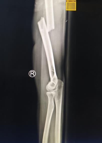
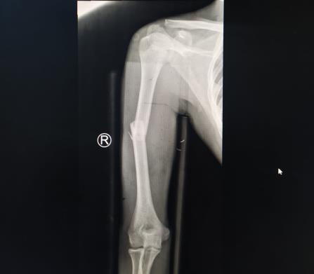
A 15-year-old patient came to the emergency room with pain in his right arm. Hefell in a motorcycle accident, collided with another motorcycle and landed on the right side. When he was hospitalized, he was conscious and had normal vital functions. His right arm was injured, swollen, and deformed. He had a hard time moving his arm. On examination there was pain on palpation of theright humerus with swelling and pain at any ofmotion. His neurovascular examination was intact. In the Emergency room, the patient got the wound toilet management, fluids of ringer lactate, and the ketorolac for the pain management. As soon as the secondary clinical survey was completed the patient, the patient got the medicine, he scheduled to take an x ray of his right humerus because of the suspect humerus fracture to the due of the deformity and the range of movement of his arm. Radiographs showed a transverse fractureof the middle third of the humeral shaft on the right side. The arm was not immobilized yet inthe emergency room.
International Journal of Healthcare Sciences ISSN 2348-5728 (Online)
Vol. 10, Issue 2, pp: (154-158), Month: October 2022 - March 2023, Available at: www.researchpublish.com
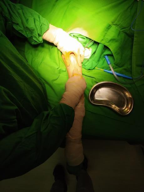
As soon as the secondary clinical survey was completed the patient was carried to the room to get the pain medicine and preoperation medicine to wait the operatingtheatre. In the Operating theatre the surgeon preferred to attempt stabilization of the shaft fracture with the (ORIF) open reduction internal fixation to the reduction. Access by the anterior, direct lateral, or posterolateral approach depends on the fracture pattern and the surgeon's preference.
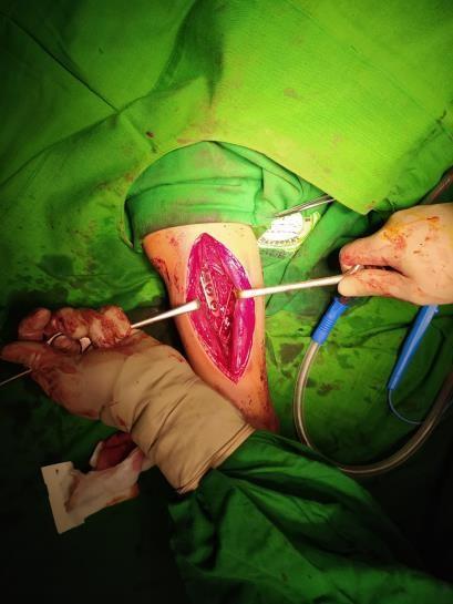
For this case, the surgeon preferred to attempt the anterior approach. After the patientand the family got the sufficient information and sign the informed consent, The patient was placed in a supine position under general anesthesia After the timeout and aseptic management, the surgeon does the deep dissection through internervous plane of brachialis muscle and put the plate and seven screw. Care must be taken not to damage the radial nerve or pinch the nerve under the plate.The operation went well, the patient got the antibiotic, pain management, and fluid afterthe operation.
Fractures of the humeral shaft in children are relatively rare injuries, accountingfor less than 3% of all childhood fractures and 10% of all humerus fractures. Fractures of the humerus diaphysis can be caused by direct trauma to the arm, a fall, a car accident, or a sports injury [1,2] Up to 56% of humeral diaphyseal fractures in older children are due to falls The examination can determine tenderness, humeral stem instability, and crepitus at the fracture site. Children older than usually have pain, swelling, and bruising. Examination may reveal obvious palpation deformities and pain or crepitus [2] However, most humeral diaphyseal fractures show no visible deformation, and x-ray images show minimal displacement [4].
In this case the patient came to the emergency room with pain in his right arm. From the history of the mechanism injury he fell in a motorcycle accident, collided with another motorcycle and landed on the right side. When he was hospitalized, he was conscious and had normal vital functions. His right arm was injured, swollen, and deformed. He had a hard time moving his arm. On examination there was pain on palpation of theright middle of the humerus with minimal swelling, crepitation sound, and pain at any of motion of the arm. these findings are in line with common clinical findings of humeral fracture.
For most pediatric humeral diaphyseal fractures, x-ray images with standard anterior- posterior (AP) and full-length lateral views aresufficient. The upper and lower joints need to be properly mapped, otherwise a special shoulder and elbow series should be performed [8]. It is noteworthy that prominent vascular grooves are often found in the distal humerus. This is a normal finding and should not be confused with a fracture [9] When reviewing such images, care must be taken to rule out the underlying cause, such as bone cysts in pathological fractures. In these situations, more detailed imaging may be needed [3] .
International Journal of Healthcare Sciences ISSN 2348-5728 (Online)
Vol. 10, Issue 2, pp: (154-158), Month: October 2022 - March 2023, Available at: www.researchpublish.com
Traditionally, there has been no generally accepted pediatric classification system for humeral diaphyseal fractures. The standard classification was based on the principles of diaphyseal fractures, including:
(1) Anatomical position (proximal, central, or distal one-third). (2) Fracture pattern (spiral, diagonal, lateral, or crushed); (3) Degree of displacement; (4) Opening and closing [3.23]. The AO Pediatric Comprehensive Classification System (AO PCCF) for long bone fractures was introduced in 2007 based on the location and morphology of the fracture.. Diaphyseal humerus fractures are coded as "12D" (1 for humerus, 2D for diaphysis).
In this case the radiographs, x-ray images with standard anterior-posterior (AP) and lateral view showed a transverse fracture of the middle third of the humeral shaft on the right side, the underlying cause, such as bone cysts in pathological fractures can be excludedwith there is no evidence in the history of the patient’s mechanism of injury, medical history and x ray. These findings are in line with the theory of x-ray results of right humeral fracture.
Most of the humeral trunk fractures in children can be treated conservatively, There are several fixing techniques for children and adolescents, such as slings and / or capes, bell paw bandages, collars and cuffs, adjustable sprints (eg Sugarton sprints), and hanging arm casts [5,6,7,8]
Surgical treatment is mainly used for older children and adolescents. Surgical treatment of humeral trunk fractures in children includes open fractures, fractures with associated vascular injury, bilateral fractures, floating elbows, crushed fractures, compartment syndrome, and closed fractures (acceptable dislocation or keratosis is non- surgical) (If it cannot be treated with) is applied to the method [9] .
In this case, the patient had a close humeral fracture that usually goes to be treatedconservatively. But in this case there are evidences that the alignment of the bone fragment is not acceptable, and the evidence said that the surgical intervention had a better result in the older children or adolescent. The patient in this case is 15 years old male which is an older children. Surgical options include open reduction internal fixation (ORIF), intramedullary (IM) nailing, or external fixation. The latter is primarily indicated for patients with extensive soft tissue or open fractures with bone loss, or severe polytrauma [6] The ORIF can be run on 3.5 or 4.5 mm plates, depending on the size of the patient [3] Access by the anterior, direct anterolateral, or posterolateral approach depends on the fracture pattern and the surgeon's preference. Care must be taken not to damage the radial nerve or pinch the nerve under the plate. For stability reasons, it is advisable to fix six cortex above and below the fracture [3]. ORIFis also the recommended surgical choice in thepresence of associated vascular injury to avoid fracture displacements that can interfere with vascular anastomosis and good for the transverse fracture pattern [3] IM nailing is a surgical option for lateral or short diagonal fractures (length-stable fractures). This option is advantageous because the indirect reduction is performed first and the surgical incision is smaller than the surgical incision . Prograde or prograde access should be considered (depending on the pattern of destruction), taking care to avoid physique to prevent growth arrest [7] .
International Journal of Healthcare Sciences ISSN 2348-5728 (Online)
Vol. 10, Issue 2, pp: (154-158), Month: October 2022 - March 2023, Available at: www.researchpublish.com
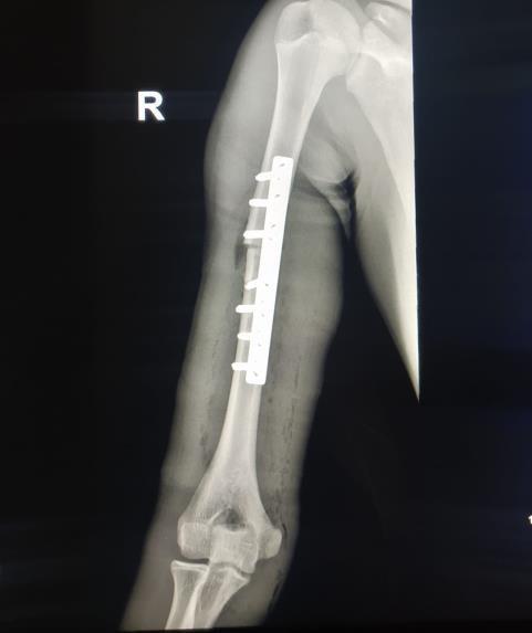
In this case the surgeon preferred to choose the open reduction internal fixation for the reduction. This is in line with the theory that it could be useful for the midshaft humerus fracture, patients favor ORIF over IMN as the optimal treatment decision for an acute traumatic humeral shaft fracture. Plating has been shown to have better overall results compared to the interlocking nails in treatmentof closed humeral shaft fractures.
Clinical approach and definitive treatment is very important to get a better result in pediatric humeral fracture. The pitfall diagnosis can occur because of the other differential diagnosis. To confirm this doubtful situation, beside the anamnesis of the patient’s medical history, physical examination, the x-ray of the humerus could help to get the diagnosis. The definitive treatment of the humeral fracture can be conservative or need a surgical treatment. It depends on the indication of the fracture itself.
[1] O’Shaughnessy MA, Parry JA, Liu H, Stans AA, Larson AN, Milbrandt TA. Management of paediatric humeral shaft fractures and associated nerve palsy. J Child Orthop. 2019;13(5): 508-15.[PMID:31695818];[PMCID:PMC680807 3]; [DOI: 10.1302/1863-2548.13.190012]18054670]; [DOI: 10.1016/j.hcl.2007.09.002]
[2] Rush J. Pediatric Orthopaedic Society of North America (POSNA): Humeral Shaft Fractures. 2021.
[3] Herring JA, Ho C. Upper Extremity Injuries. In: Herring JA, ed. Tachdjian’s Pediatric Orthopaedics. 5th ed. Philadelphia: Elsevier Saunders; 2014: 1262-4
[4] Ryan LM, Boutis K, Wiley J. UpToDate: Midshaft Humeral Fractures in Children. 2021.
[5] Bae DS. Humeral Shaft and Proximal Humerus, Shoulder Dislocation. In: Flynn JM, Skaggs DL, Waters PM, eds.Fractures in Children. 8th ed. Philadelphia: Wolters Kluwer; 2015: 784-99.
[6] Asche G. Use of external fixation in pediatric fractures. Zentralbl Chir 1986; 111: 391-7. [PMID: 3716669]
[7] Lieber J, Schmittenbecher P. Developments in the treatment of pediatric long bone shaft fractures. Eur J Pediatr Surg. 2013; 23(6): 427-33. [PMID: 24327219]; [DOI: 10.1055/s-0033-1360460]
[8] Shrader MW. Proximal Humerus and Humeral Shaft Fractures in Children. Hand Clin. 2007; 23(4): 431-5. [PMID:
[9] Singisetti K, Ambedkar M. Nailing versus plating in humerus shaft fractures: a prospective comparative study. Int Orthop. 2010;34(4):571-576. doi:10.1007/s00264-009- 0813-2