International Journal of Healthcare Sciences ISSN 2348-5728 (Online)
Vol. 10, Issue 2, pp: (1-6), Month: October 2022 - March 2023, Available at: www.researchpublish.com

International Journal of Healthcare Sciences ISSN 2348-5728 (Online)
Vol. 10, Issue 2, pp: (1-6), Month: October 2022 - March 2023, Available at: www.researchpublish.com
2
Dewi Arimas Ni MadeCinical Pathology Laboratory
Buleleng District General Hospital, Bali, Indonesia
DOI: https://doi.org/10.5281/zenodo.7138946
Published Date: 03-October-2022
Abstract: Tuberculosis is an infectious disease caused by the Mycobacterium tuberculosis. Indonesia ranks third in the world as the country with the most tuberculosis cases. Bacteriology is the standard of examination for the diagnosis of tuberculosis. Culture examination takes a long time, TCM must use a sputum sample and special examination tools. Interferon-γ release assays immunological examination (IGRA) is quite expensive and requires special equipment. Hematological examination can be used as an option for supporting examination in establishing the diagnosis of tuberculosis by immunology. The purpose of this study is a journal review to identify the potential (MLR) Monocyte Lymphocyte Ratio as a support in diagnosing tuberculosis. Article searches were conducted online from the NCBI, PubMed and Science Direct databases. Sorting articles using the PRISMA flow. Finally, eligible articles were selected based on the criteria for patient, intervention, comparison, outcome, and study (PICOS), namely tuberculosis patients, MLR, tuberculosis diagnosis, and the original study. The results of this study obtained seven articles from the initial number of 9,234 articles found from key words. Two of the seven articles stated that MLR could be used as a support for the diagnosis of tuberculosis. Two articles stated that monocytes and lymphocytes could be markers of bacterial infections including tuberculosis. One article mentions the association of tuberculosis with decreased production of monocyte and lymphocyte cytokines. Two articles stated that MLR was not associated with tuberculosis cases. One article mentions the MLR value limit of 0.378 to support the diagnosis of tuberculosis. Observational research on MLR to support the diagnosis of tuberculosis in Indonesia still needs to be done, especially the assessment of the MLR value limit.
Keywords: Diagnosis, hematology, lymphocytes, monocytes, MLR, tuberculosis.
Tuberculosis is an infectious disease that is mostly found in the lungs, but can also be foundin other organs. Mycobacterium tuberculosis (M. tuberculosis) is the bacterium that causes tuberculosis.1 Most tuberculosis cases were found in Asia, namely 44% in 2018. India ranks first with the most tuberculosis cases in the world and Indonesia ranks third with the most tuberculosis cases in the world.2
Currently, bacteriology and immunology are the most commonly used methods for diagnosing tuberculosis. Bacteriological diagnosis using BTA, TCM and bacterial culture, and immunologically using interferon-γ release assays (IGRA) and TST. TCM examination is fast and accurate, but requires sputum as a sample and requires special reagents. BTA sputum is less sensitive and bacterial culture takes a long time, which is more than one day, and the IGRA examination is expensive. Therefore, it is necessary to have a fast, accurate and low-cost supporting examination in diagnosing tuberculosis.2
Analysis of the ratio of monocytes and lymphocytes has the potential to be used as a supporting examination in diagnosing tuberculosis. Research by Sibley et al.3 on monkeys in 2019showed that the Monocyte Lymphocyte Ratio (MLR) increased when infected with tuberculosis. The research was conducted on the Indian genotype (RM), Chinese genotype (CCM), and Mauritian genotype (MCM). RM and MCM were vulnerable groups in this study, and CCM was used as a control. MLR in MCM and RM before M. tuberculosis infection was higher than CCM. This study showed a significant increase in MLR in the group of monkeys infected with M. tuberculosis. These changes in MLR in the MCM and RM groups indicate that MLR can be a tuberculosis biomarker 1
International Journal of Healthcare Sciences ISSN 2348-5728 (Online)
Vol. 10, Issue 2, pp: (1-6), Month: October 2022 - March 2023, Available at: www.researchpublish.com
Research on MLR in tuberculosis patients is indeed interesting and has been widely carried out as a solution for establishing the right diagnosis. Based on this explanation, researchers are interested in examining MLR (Monocyte Lymphocyte Ratio) as a supporting examination in establishing a diagnosis in tuberculosis patients. This research was conducted by reviewing several literatures using the scoping review method.
Article searches were conducted on three databases, namely Highwire, PubMed and Science Direct. There are differences in inclusion criteria and keywords in each database (Table 1).
Table 1: Search Articles in Three Databases
Database Keywords Inclusion Highwire MLR on diagnostic TB 2010-2020, free articles full text PubMed Tuberculosis”[Mesh]) AND “Blood Cell Count”[Mesh] 2010-2020, RCT, CT, free full text Science Direct Tuberculosis MLR 2015-2022 free articles
In Highwire the keywords are "MLR on TB diagnostics" with the inclusion criteria of free articles full text 2015-2022. In PubMeduse thekeywords Tuberculosis”[Mesh]) AND “Blood Cell Count”[Mesh],with2015-2022inclusioncriteria,RCT, CT, free full text. Tuberculosis MLR are keywords in Science Direct, with inclusion criteria for 2015-2022 free articles. Exclusion criteria in this study were duplicate articles, articles that were not fully accessible, and articles with a discrepancy between the title and abstract.
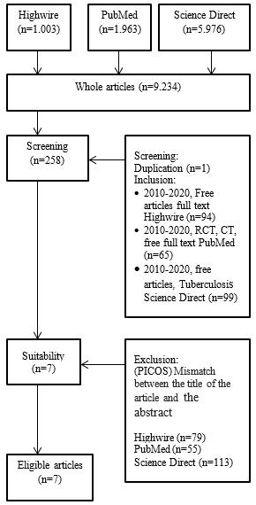
The search results of several articles from the three databases were then filtered using the PRISMA method described in Image The total number of articles obtained in the database is 9,323 articles. After inclusion, there were 268 articles. Eligible articles are selected based on (PICOS) criteria for patient, intervention, comparison, outcome, and study. The PICOS criteria selected in this study were TB patients, monocyte lymphocyte ratio, TB diagnosis, and the original study. A total of seven eligible articles were obtained after the screening stage using the PICOS criteria.
Image 1: Search and Selection Stages Articles
2348-5728 (Online)
Vol. 10, Issue 2, pp: (1-6), Month: October 2022 - March 2023, Available at: www.researchpublish.com
This study involved seven articles from studies in various countries including South Korea, Kenya, Pakistan, Sweden, Thailand, Tanzania and Norway (Table 2)4-10. In this articles, three types of research designs are used, including clinical trials, observationalandcohorts. Clinical trial research was conducted byJanols etal.6 and Lee et al.9 Observationalresearch was conducted by Laghari et al.10 The cohort study was conducted by Rees et al.4,
Janols et al.6 Clinical trial
Patients were measured for leukocytes, Creactive protein (CRP) using an automated immunoturbidimetric assay system, soluble TNF alpha receptors (sTNFR) assessed by quantitative ELISA, and blood analysis using a flow cytometer.
New findings suggest that lymphocytes and monocytes can be markers for bacterial infections such as neuroborreliosis or TB.
Lee et al.9 Clinical trial
Laghari et al.10 Prospective observational
Concentrations of all cytokines except plasma IL-1β were calculated by Bio-plex Multiplex immunoassay systems. Plasma IL1β was measured by ELISA kit. PGE2 was measured with the EIA kit.
The child was diagnosed with TB with clinical symptoms, contact history, positive TST, PPA scoring chart, chest x-ray, culture, TCM. Laboratory data obtained in the questionnaire.
Cases of active TB are associated with decreased production of gamma IFN by monocytes and cytokines produced by TH1 in response to mycobacterial antigens.
TB positive patients experienced more decreased lymphocytes (121 patients) than increased lymphocytes (113 patients). TB positive patients experienced more monocyte increase (9 patients) than monocyte decrease (8 patients).Thereisarelationshipbetween positive TB cases with NLR and erythrocyte sediment rate (ESR).
Rees et al.4 Prospective cohort
This study used IGRA test and measurement of CBC count with white blood cell differential count. IGRA tests were performed on days 0, 60, 420, and 720. CBC was assessed on days 0, 60, 90, and 720.
The value of monocyte to lymphocyte ratio (MLR), neutrophil to lymphocyte ratio (NLR), and platelet to lymphocyte ratio (PLR) were not associated with TB cases. The number of red blood cells, hemoglobin, and hematocrit decreased significantly in TB patients.
Choudhary et al.5 Longitudinal cohort
Diagnosis of TB is done by observing symptoms, physical examination, tuberculin skin test (TST), Ziehl-Neelsen staining and bacterial culture. Blood specimens were examined for full blood count and differential count. Patients were given combination antiretroviral therapy (abacavir and lamivudine with nevirapine, efavirenz, or lopinavir/ritonavir).
The median MLR valuein theconfirmed TB group was higher than in the unconfirmed or unlikely TB group. The cut-off value for MLR is 0.378.
Miyahara et al.7
Prospective cohort study
The NLR was calculated as the absolute number of neutrophils divided by the absolute number of lymphocytes.
Naess et al.8 Cohort prospective Patients were grouped based on duration of fever before hospital admission and final diagnosis (bacterial infection,viral infection, clinically diagnosed infection, no infection, no diagnosis). Age, sex, temperature, and CRP results were recorded at enrollment. WBC examination and differential cell counts were checked using an instrument.
MLR was higher in positive TB cases than in negative TB cases. NLR is associated with active TB and the NLR threshold value for TB is 2.
NLR and MLR were significantly higher in patients with bacterial infection than without infection. NLR and MLR in bacterial infections are lower than viral infections.
International Journal of Healthcare Sciences ISSN 2348-5728 (Online)
Vol. 10, Issue 2, pp: (1-6), Month: October 2022 - March 2023, Available at: www.researchpublish.com
Choudhary et al.5, Miyahara et al.7, and Naess et al.8 In the research conducted in various countries, the respondents were varied between 39 people up to 1,118 people.7
Article Janols et al.6 conducted research in the form of identification of lymphocyte and monocyte immunophenotyping as specific markers in diagnosing disease. This study involved 39 respondents who were conducted in Sweden in 2010. This study found that monocytes and lymphocytes can be used as markers of neuroborreliosis orTuberculosis bacterial infection.
Article Lee et al.9 conducted a study to detect the production of monocyte and lymphocyte cytokines in the face of mycobacterial antigens. This study involved 49 respondents who were conducted in South Korea in 2015. This study found adecreaseintheproductionofmonocyteandlymphocytecytokinesin casesofactivetuberculosis.Thisoccursasaresponse of monocytes and lymphocytes in the face of mycobacterial antigens.
Article Laghari et al.10 conducted a study to detect the ratio of monocytes and lymphocytes in tuberculosis patients. This study involved 508 respondents who were conducted in Pakistan in 2019. This study found that patients with positive tuberculosis had a decrease in lymphocytes and an increase in monocytes. However, the increase in monocytes was not significant, because eight of them experienced a decrease in monocytes, and nine people experienced an increase in monocytes. Another finding from this study was that there were significant changes in ESR and NLR in some cases of tuberculosis.
Article Rees et al.4 conducted a study to detect the ratio of monocytes, neutrophils and platelets to lymphocytes in tuberculosis patients. This study involved 145 adolescents conducted in Tanzania in 2020. This study found that the MLR, NLR and PLR results did not show significant differences in positive or negative IGRA results. So the conclusion is MLR, NLR and PLR have no significant relationship with changes in IGRA results. There are other findings on the examination of red blood cells, namely the hematocrit and hemoglobin decreased significantly on IGRA results that turned positive.
The article Choudhary et al.5 conducted a study to find the relationship between MLR and cases of active tuberculosis in children. This study compared MLR in confirmed, unconfirmed and non-tuberculosis-like groups. This study involved 160 children withHIVconductedinKenyain2019.ThemedianstudyresultsfromMLRinchildren withconfirmedtuberculosis were higher than those without. The MLR value is 0.378, if it is below this value, it is negative for tuberculosis, and it is said to be positive for tuberculosis if it is above this value.
Article Miyahara et al.7 conducted a study to find the relationship between NLR in cases of active tuberculosis within one year after screening. This study involved a large number of respondents, namely 1,118 people who were conducted in Thailand in 2018-2019. This study obtained the results of NLR associated with cases of active tuberculosis. In addition to comparing NLR, this study also compared MLR, absolute monocytes and lymphocytes between patients with negative and positive tuberculosis. The absolute number of lymphocytes in the positive tuberculosis patients was lower than the negative ones, while the MLR and the absolute number of monocytes were higher in the positive than the negative tuberculosis patients. This automatically shows the relationship between MLR and tuberculosis cases.
Article Naess et al.8 conducted a study of MLR and NLR functions to distinguish whether the cause of fever was due to infection or not. This study involved 299 respondents conducted in Norway in 2017. In this study, the results in the form of MLR and NLR values in patients with bacterial infections showed a significant increase compared to patients without bacterial infections, with lower MLR and NLR values in bacterial infections. compared to infections caused by viruses. The role of MLR in tuberculosis cases is described in four articles. Articles that mention the role of MLR can be used as a supporting examination in diagnosing tuberculosis, namely articles by Choudhary et al.5 and Miyahara et al.7 The article which states that there is no relationship between MLR, NLR, and PLR on tuberculosis is Rees et al.4, while in the article byLagharietal.10 itis stated thatMLRchangesarenotsignificantintuberculosiscases.Therearethreearticlesthatexamine MLR, but there is no analysis of the relationship with the diagnosis of tuberculosis, namely in the articles of Naess et al.8, Lee et al.9, and Janols et al.6 In a study conducted by Naess et al.8 and Janols et al.6 explains the results that monocytes and lymphocytes can be used as markers of bacterial infection including tuberculosis cases, but are not specific markers in diagnosing tuberculosis. Lee et al.9 described the association of active tuberculosis cases with decreased monocyte and lymphocyte cytokine production. In a study conducted by Choudhary et al.5, the MLR limit value was shown to be 0.378.
The results of a review of seven articles from various countries found that the studies of Choudhary et al.5 and Miyahari et al.7 showed that MLR could be used as a support in diagnosing tuberculosis. In the articles of Naess et al.8 and Janols et al.6, monocytes and lymphocytes can be used as markers of bacterial infection, including tuberculosis. Lee et al.9 mentioned
International Journal of Healthcare Sciences ISSN 2348-5728 (Online)
Vol. 10, Issue 2, pp: (1-6), Month: October 2022 - March 2023, Available at: www.researchpublish.com
a decrease in cytokine production in tuberculosis cases. In the study of Rees et al.4 and Laghari et al.10, MLR was not associated with cases of tuberculosis. In the study by Choudhary et al.5, the limit of the MLR value to support the diagnosis of tuberculosis was 0.378.
This study is in accordance with a review article on protective biomarkers against tuberculosis conducted by Basu Roy et al.11 The study was published in 2019 which examined the vulnerability and protection of children against tuberculosis. The studyused PubMedasadataandarticlesearch.Theresultsofhisreviewstated thatMLRcouldbeariskforthedevelopment of tuberculosis. The development of tuberculosis is characterized by an increase in MLR. So it is said that there is a relationship between MLR and tuberculosis. The review article did not mention the MLR value limit for tuberculosis cases.
The difference in results is found in a review conducted by Russell et al.12 The 2019 study examined the functions of NLR, MLR, and PLR as markers of infection. In this study, PubMed, Embase, and Cochrane were used to collect articles. The study examined the relationship of NLR, MLR, and PLR to bacterial, viral, and malarial infections. In most cases assessed by NLR alone, while cases assessed by MLR were Clostridioides difficile and respiratory virus, while tuberculosis was assessed by NLR. The results of the review mentioned the relationship between influenza virus and MLR, while NLR had a relationship with tuberculosis. The conclusion is that the leukocyte ratio can be used as a marker of infection in diagnosing bacteremia and influenza infection.
This study has limitations, namely the lack of articles that mention the relationship between MLR and tuberculosis cases and the selection of keywords that are less than optimal, the absence of articles obtained from paid journals and several studies that are not specific for tuberculosis
The conclusion there are two articles mention that MLR has a relationship with tuberculosis cases, two articles have no relationship. Three articles did not specifically state the association between MLR and tuberculosis. The MLR limit value is found in one article, which is 0.378. Neutrophil lymphocyte ratio (NLR), monocyte and lymphocyte cytokine production, red blood cell count, hemoglobin, and hematocrit have a relationship with tuberculosis cases. This study can be used as a considerationinestablishingthediagnosisoftuberculosis.TheresultoftheMLRexaminationcanbecombinedwithclinical and radiological symptoms so that a false positive or false negative diagnosis does not occur. After this, its is hope that it will open up insight about MLR as a supporting examination in diagnosing tuberculosis. Hoped that this research can be used as the basis for conducting next research on MLR in diagnosing tuberculosis.
[1] Husain AN. Lung. In: Kumar V, Abbas AK, Aster JC, editors. Robbins basic pathology. 10th edition. Philadelphia: Elsevier; 2017.p. 459– 516.
[2] World Health Organization. Global tuberculosis report 2019. Geneva: World Health Organization; 2019.
[3] Sibley L, Gooch K, Wareham A, Gray S, Chancellor A, Dowall S, dkk. Differences in monocyte: lymphocyte ratio and tuberculosis disease progression in genetically distinct populations of macaques. Sci Rep. 2019;9(1):3340.
[4] Rees CA, Pineros DB, Amour M, Munseri P, Said J, Magohe A, dkk. The potential of CBC-derived ratios (monocyteto-lymphocyte, neutrophil-to-lymphocyte, and platelet-to-lymphocyte) to predict or diagnose incident TB infection in Tanzanian adolescents. BMC Infect Dis. 2020;20(1):609.
[5] Choudhary R, Wall K, Njuguna I, Pavlinac P, Lacourse S, Otieno V, dkk. Monocyte-to-lymphocyte ratio is associated with tuberculosis disease and declines with anti-TB treatmentin HIV-infected children. J Acquir ImmuneDefic Syndr. 2019;80(2):174–81.
[6] Janols H, Bredberg A, Thuvesson I, Janciauskiene S, Grip O, Wullt M. Lymphocyte and monocyte flow cytometry immunophenotyping as a diagnostic tool in uncharacteristic inflammatory disorders. BMC Infect Dis. 2010;10:205.
[7] Miyahara R, Piyaworawong S, Naranbhai V, Prachamat P, Kriengwatanapong P, Tsuchiya N, dkk. Predicting the risk of pulmonary tuberculosis based on the neutrophil-to-lymphocyte ratio at TB screening in HIV-infected individuals. BMC Infect Dis. 2019;19(1):667.
International Journal of Healthcare Sciences ISSN 2348-5728 (Online) Vol. 10, Issue 2, pp: (1-6), Month: October 2022 - March 2023, Available at: www.researchpublish.com
[8] Naess A, Nilssen SS, Mo R, Eide GE, Sjursen H. Role of neutrophil to lymphocyte and monocyte to lymphocyte ratios in the diagnosis of bacterial infection in patients with fever. Infection. 2017;45(3):299–307.
[9] Lee JY, Jung YW, Jeong I, Joh JS, Sim SY, Choi B, dkk. Immune parameters differentiating active from latent tuberculosis infection in humans. Tuberculosis (Edinb). 2015;95(6):758–63.
[10] Laghari M, Sulaiman SAS, Khan AH, Memon N. A prospective study of socio-demographic, clinical characteristics and treatment outcomes of children with tuberculosis in Sindh, Pakistan. BMC Infect Dis. 2019;19(1):82.
[11] Basu Roy R, Whittaker E, Seddon JA, Kampmann B. Children and Mycobacterium tuberculosis: a review of susceptibility and protection. Lancet Infect Dis. 2019;19(3):e96–108.
[12] Russell CD, Parajuli A, Gale HJ, Bulteel NS, Schuetz P, de Jager CPC, dkk. The utility of peripheral blood leucocyte ratios as biomarkers in infectious diseases: a systematic review and meta-analysis. J Infect. 2019;78(5):339–48.