ISSN 2348-313X (Print)
International Journal of Life Sciences Research ISSN 2348-3148 (online)
Vol. 8, Issue 2, pp: (29-37), Month: April - June 2020, Available at: www.researchpublish.com

ISSN 2348-313X (Print)
International Journal of Life Sciences Research ISSN 2348-3148 (online)
Vol. 8, Issue 2, pp: (29-37), Month: April - June 2020, Available at: www.researchpublish.com
1 ,
Ashok K. Ghosh2
1(P.G.Department of Environmental Sciences, A.N.College/Patliputra University,Patna, India) 2(Chairman, Bihar State Pollution Control Board, Patna, Bihar, India)
Abstract : The soil acts as a reservoir for millions of microorganisms. Microorganisms play a vital role in maintaining soil fertility and plant health. They can act as biofertilizers. This study aimed at isolating and characterizing plant growth-promoting bacteria associated with the paddy (Oryza sativa, variety - Sugandha), as well as assessing their ability to promote plant growth. Samples were collected from near river Ganga Sites were dry field, banana field, wetland and stagnant pond areas. Five sampling sites namely S1 (Banana field soil, Hazipur), S2 (Near Gandhi Setu, wetland area), S3 (Sediment pond), S4 (Vaishali) and S5 ( Sonepur, stagnant pond area) were selected for isolation of microbial strains and total of 111 microbial strains (bacteria) were isolated. The strains were selectively isolated from the population of microbes growing on the nutrient agar medium. Strains which showed the best result were tested for its’ biofertilizer potential. These strains were S1 (C), S2 (F) and S3 (R). The isolate S2 (F) was selected on the basis of their luxuriant growth on NA with yellow colour characteristics of colony. This isolate showed a wide range of temperature for growth between 4ْC to 55ْ C which suggests its potential as biofertilizer. The isolate was Bacillus although molecular characterization (16s rRNA studies) was done to confirm the genus and species of the selected isolate. On the basis of BLAST alignment of the sequenced nucleotides the isolate S2 (F) was identified as Brevibacillus borstelensis. The reported literatures suggest that the B. borstelensis acts as PGPR.
Keywords: Microbes, Bacteria, Biofertilizer potential, Oryza sativa, Molecular characterization, Brevibacillus borstelensis.
Soil is a dynamic, living matrix that is essential part of the terrestrial ecosystem. It is a critical resource not only for agricultural production and food security but also towards maintenance of most life processes. The functions of soil biota are central to decomposition processes and nutrient cycling. Soil is considered a store house of microbial activity, though the space occupied by living microorganisms is estimated to be less than 5% of the total space. Therefore soil is rich in micronutrient and major microbial activity is confined to the ‘hot-spot’, i.e. aggregates with accumulated organic matter, rhizosphere (RS) [1-2]. Microorganisms play important roles in several biological activities that can be beneficial to mankind and all plants [3]. Microbial population in soil counts for a huge mass of organic matter on earth. Importance of microorganism in maintaining human habitat on earth is now beyond a question of discussion. These microorganisms are of very diverse that includes bacteria, archaea, yeast, fungi, algae, and protozoa. Microorganisms can live in environment along with human and in extreme conditions such hot springs, miles deep in the ocean, inside rocks and in extreme cold
ISSN 2348-313X (Print)
International Journal of Life Sciences Research ISSN 2348-3148 (online)
Vol. 8, Issue 2, pp: (29-37), Month: April - June 2020, Available at: www.researchpublish.com
temperature. In the light of exploring the utility and importance of bacteria as plant growth promoting rhizobacteria (PGPR)/biofertilizers, the present investigation was aimed towards isolation and purification of the strains from soil, their characterization and identification and further establishment of their biofertilizer potential on rice (Oryza sativa) plant. Thus, the Government of India has been constantly trying to promote practices involving use of biofertilizers along with fertilizers. Searching novel sources of the microbes having biofertilizer or plant growth promoting potential are therefore necessary to meet the escalating demand of sustainable farming. The ecological niches inhabiting a variety of such microorganisms are Western Ghats, the Himalayan ranges, marine ecosystem, fresh water ecosystem, mangroves, coral reefs, sand dunes, industrial effluents, contaminated soil, refineries, activated sludge, fertile agricultural land and several other natural sources. Further, the microorganisms found in the different habitats show a great diversity in their growth requirements, morphology, and bio-chemical characteristics. Biofertilizer has been identified as an alternative to chemical fertilizer to increase soil fertility and crop production in sustainable farming. These are products containing living cells of different types of microorganisms, which are bio control agents and have an ability to convert nutritionally important elements from unavailable to available form through biological processes [4]
The utilization of microbial products are considered safer than many of the chemicals now in use. Bacteria of several taxonomic classes are found in crop rhizosphere and soil. They have biofertilizer potential and can increase plant growth and productivity [5] Increase in plant productivity occurs through different mechanisms such as symbiotic N2 fixation [6], solubilization of mineral phosphate and other nutrients [7], production of plant hormones [8] and control of phytopathogenic microorganisms [9]. The term plant growth promoting rhizobacteria (PGPR) was coined for the bacterial bio-control agents of rhizosphere [10] Recently, the term plant growth promoting bacteria (PGPB) has been proposed to encompass rhizobacteria, which enhance plant growth by other means [11] The interest on PGPR and PGPB has increased due to the prospect of use of their efficient strains as bio-inoculant (biocontrol and biofertilizer) components in organic agriculture, which is emerging as an alternative to chemical inputs (pesticides and inorganic fertilizers) in intensive agriculture [12] Efficient rhizobacteria have been successfully used in many cereal crops [13] Plant growth promoting rhizobacteria (PGPR) represent a wide variety of soil bacteria which, when grown in association with a host plant, result in stimulation of growth of their host. For PGPR to have a beneficial effect on plant growth via an enhancement of the nutrient status of their host, there obviously needs to be an intimate relationship between the PGPR and the host plant. The state of Bihar specially requires much attention for its microbial biodiversity assessment. A number of important rivers arise from Himalayas in Nepal and join the Ganges at different sites making the areas flood prone and wetland but soil highly fertile and best suited for paddy, wheat, maize, pulses and sugarcane. Thus the Gangetic planes of Bihar need immediate attention to isolate and screen microbes that help in enhanced soil fertility and decomposition of agricultural wastes. This will not only help generating the database of indigenous microbes but also play a major role in poverty alleviation through involvement of rural masses. A detailed assessment of the potentials of selected microorganisms is likely to throw light on their commercialization as biofertilizer. In the light of current scientific knowledge, it is important to identify and document well characterized strains of diverse microorganisms that can be beneficially utilized in agriculture and industry. The rationale of the present study is to characterize, identify and prepare a database of diverse microorganisms as a soil fertility inhabiting unexplored wetland, rain fed ecosystems of Bihar. Such a co-coordinated effort is likely to create awareness about the characteristics of microbes that can be utilized for enhanced agricultural production through development of biofertilizers, biomineralizers or biocontrol (biopesticides) agents. Some of the earlier works from our group established the successful isolation and characterization of microbes having phosphate solubilizing potentials from Gangetic planes of Bihar [14] However, most microorganisms are not visible to the naked eye and they need to be studied either microscopically or on the basis of biochemical parameters. The culturable microorganism can be identified provided they sporulate but the biotrophs which are not amenable to culturing in growth media, go unnoticed. It is, therefore, imperative that there is a huge paucity in our knowledge of the diversity of all microbes.
The isolates were obtained from the soil of Gangetic region of North Bihar. The next step was the screening of isolates to select bacterial strains with biofertilizer potential. They are mostly differentiated on the basis of structure and development of cells, morphology, responses to culturing, physiology and biochemistry. Their colony on agar media produce spores, the later representing the preparative phase. The majority of microorganisms, when grown on solid
ISSN 2348-313X (Print)
International Journal of Life Sciences Research ISSN 2348-3148 (online)
Vol. 8, Issue 2, pp: (29-37), Month: April - June 2020, Available at: www.researchpublish.com
media, grow into colony representing a massive cluster of cells of organisms which are capable of growing together in a single complex colony. Microscopic examination of the organisms from a colony should reveal only a single type of cells. Differential staining procedures, such as Gram stains are useful for establishing that the colony does not contain a mixture of different microbial types [15]
All the media, chemicals and reagents for culture growth and biochemical tests were obtained from Hi Media (India) unless otherwise specified. Peptone and urea agar were obtained from Merck (India).
Morphological characterization of the selected bacterial isolates
Colony morphology
Purified colonies were allowed to grow on Petri plate in NA. Morphological features like configuration, margin, elevation, surface, pigment, opacity were observed. Many of these features were observable with naked eyes but were confirmed by looking under a microscope. The characteristics for the freshly grown cultures of the isolates (S1(C), S2 (F) and S3(R)) were then tabulated.
A thin smear of the bacterial fresh culture was made on a clear glass slide. The smear was covered with crystal violet and allowed to stand for 20 sec. The stain was briefly washed off with distilled water. The smear was then covered with Gram’s iodine and allowed to stand for one minute. Gram’s iodine was poured off and the smear was flooded with 95% ethyl alcohol for 10 to 20 sec. The action of alcohol was stopped by washing the slide under running distilled water for a few sec. Subsequently, the smear was covered with safranin for about 20 sec. Gentle washing followed by blotting with bibulous (blotting) paper was done and the slide was allowed to dry at room temperature. The slides were immediately examined under microscope.
A thin smear of the bacterial fresh culture was made on clear glass slide and was heat fixed. The smear was covered with 5% malachite green solution and heated over a spirit lamp for 1 to 2 min. More stain was added, if the stain boils off. Slide was cooled then rinsed in water for 30 sec. The smear was covered with counterstain safranin for about 15 to 30 sec. Gentle washing followed by blotting with bibulous (blotting) paper was done and allowed to dry at room temperature. These slides were immediately examined under microscope.
A dilute suspension of the culture in normal saline was made and a drop of the suspension was added on a cover slip. Cover slip was inverted over a cavity slide making sure that the drop was hanging over the cavity. These slides were immediately examined under microscope in oil immersion.
Physiological characterization of the selected bacterial isolates
The fresh culture of the test isolates namely S1(C), S2 (F)and S3(R) were streaked on NA plate. They were incubated aerobically at different temperatures such as 4˚C, 12˚C, 15˚C, 25˚C, 30˚C, 37˚C, 42˚C and 55˚C. The data were recorded on the 4th day of growth.
NA plates containing varying NaCl concentration (2.0%, 4.0%, 6.0%, 10.0% and 12.0%) were prepared. With the help of loop, the plates were inoculated with light suspension of the cultures of the selected test isolates namely, S1(C), S2 (F) and S3(R). They were incubated aerobically at 37˚C ± 2˚C.
NA plates of varying pH (5 to11) were prepared. The plates were inoculated with light suspension of the cultures with the help of a loop. The plates were incubated aerobically at appropriate temperature. The data were recorded on the 4th day of growth.
ISSN 2348-313X (Print)
International Journal of Life Sciences Research ISSN 2348-3148 (online) Vol. 8, Issue 2, pp: (29-37), Month: April - June 2020, Available at: www.researchpublish.com
In McCartney bottle, 5 ml medium (peptone water because of its high tryptophan content) was distributed and sterilized by autoclaving. Selected isolates namely S1(C), S2 (F) and S3(R) were inoculated and incubated at 37± 2˚C for 2 to 3 days. To this 600 µl of Kovac’s reagent was added and left for 1 minute. Appearance of deep golden red ring indicated +ve reaction.
Methyl Red (MR) and Voges Proskauer (VP) test
MR-VP medium was sterilized by autoclaving and 5 ml was poured in a sterile tube. The MR/VP medium contains buffered peptone as carbon and nitrogen source for general growth requirement whereas; dextrose is used as a fermentable carbohydrate. The culture was inoculated into two tubes and incubated aerobically for 18 to 24 hours at 37 ± 2˚C. For MR test, 2 to 3 drops of methyl red indicator solution was added and +ve reaction was indicated by red coloration. For VP test, 600 µl each of reagent A and B was added and +ve reaction was indicated by pink or red colouration.
The test was performed by inoculating the microorganism into Simmon’s Citrate slant by means of stab and streak, where, sodium citrate was the only source of carbon and energy. One tube was kept uninoculated which served as the control. All slants were incubated at 37± 2˚C for 48 hours. Bromothymol blue was used as the indicator. When the citric acid was metabolized, CO₂ was generated and combined with sodium and H₂O to form Na₂CO₃, an alkaline product, which changed the colour of the indicator from green to blue.
Triple sugar iron agar slants were prepared by distributing 5 ml of media into each test tube and sterilized by autoclaving at 121 ˚C for 20 min. The culture was inoculated by stabbing the slant with a needle and also streaking on the slope of the slant. The tubes were incubated aerobically at 37±2 ˚C for 4 days. Appearance of black precipitate along the line of inoculation indicated the production of hydrogen sulphate.
Tubes of sterile purple broth base with inverted Durhams tubes were taken. 1 ml of filter sterilized solution of glucose (10% wt/vol.) was added to purple broth tubes. These media already contained a protein source to support microbial growth and a pH indicator. Purple broth base contain bromocresol purple, which turns from purple to yellow at acidic pH. Glucose supplemented tube was inoculated with selected isolates and incubated at 37˚C. Colour of the tubes was observed after 72 hours of incubation. Yellow colour indicated acid production by test organisms. Partial or complete filling of the Durhams tube indicated gas production.
All the selected isolates S1(C), S2 (F) and S3(R) were streaked on milk agar medium plate and incubated for 7 days 37±2 ˚C. Appearance of a clear zone around the colony indicated the presence and extent of proteolytic property of the selected isolates.
The test was performed by inoculation of different isolates into different nutrient gelatin stabs. One uninoculated tube was kept as the control. All stabs were incubated at 37± 2˚C for 7 days. After incubation, all tubes were chilled in refrigerator and examined carefully. No solidification of gelatin stabs showed the hydrolysis of gelatin by isolates.
Amylase production was tested by growing organisms on starch agar (SA). After incubation, the SA was flooded with Lugol’s iodine. The iodine reacts with starch to produce a dark purple or brown colour. All three selected test isolates namely S1(C), S2 (F) and S3(R) were streaked on SA plates and kept in incubator for 5 to 7 days at 37 ± 1˚C. Iodine solution was flooded over 7 days old culture in order to test the starch utilization or amylase production. Formation of a transparent zone shows the presence of amylase.
ISSN 2348-313X (Print)
International Journal of Life Sciences Research ISSN 2348-3148 (online)
Vol. 8, Issue 2, pp: (29-37), Month: April - June 2020, Available at: www.researchpublish.com
Urea broth (Stuart’s Urea Base) medium was sterilized by autoclaving and 5ml was dispensed in each tube. To this medium filter sterilized urea solution 40% (w/v) was added so as to make a final concentration of 2% (w/v) Culture of all five selected isolates were incubated aerobically for 3 to 4 days. Microorganisms which metabolized urea and released ammonia, made the medium alkaline as indicated by a change of colour of phenol red from yellow to red or pink- red.
The phosphate solublizing ability of the selected test isolates namely S1(C), S2(F) and S3(R) were checked by streaking them on Petri plates containing Pikovskaya’s medium. The plates were kept in incubator for 6 to 7 days. A visible clear zone formation around the colony indicates the phosphate solublising ability of the microorganism.
In order to test NO₃ - reduction isolates were incubated in separate nitrate medium and incubated for 7 days at 37± 2˚C. After incubation a nitrate disc was placed over the colony of each isolate. Appearance of red/ pink colour indicated the reduction of nitrate into nitrite. The colour was intensified by addition of rehydrating reagents.
All selected test isolates namely S1(C), S2 (F) and S3(R) were streaked on NA plates for 5 to 7 days and incubated at temperature 37±2 ˚C. Hydrogen peroxide (3% w/v) was added by dropper on 7 days old culture. Release of gas bubbles showed the presence and the extent of catalase production.
The test solution N, N, N, N- tetra methyl- p- phenylenediamine dihydrochloride was prepared by adding loop full of oxidase test reagent in 5ml sterile distilled water. The solution was mixed well. One drop of oxidase test solution was added on all test isolates namely S1(C), S2 (F) and S3(R).
5 ml of Thornley’s medium was distributed in tubes and sterilized by autoclaving. One isolate at a time was inoculated in each tube and incubated for 4 days. Results were recorded after incubation for 3 to 4 days and +ve reaction was shown by a colour change (red).
Culture suspension of selected isolates was inoculated into 5 ml sterilized medium. Using a flame sterilized forceps, one paper disc impregnated with a particular carbohydrate was placed in each tube containing 5 ml medium. They were incubated aerobically for 3 days. Acid production was indicated by change in colour from purple to yellow.
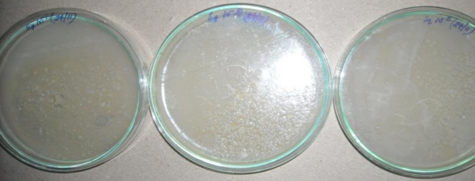
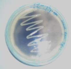
Fig. 1.1 (A, B and C) NA plates showing colonies of selected isolates S1(C), S2(F) and S3(R), respectively.
ISSN 2348-313X (Print)
International Journal of Life Sciences Research ISSN 2348-3148 (online) Vol. 8, Issue 2, pp: (29-37), Month: April - June 2020, Available at: www.researchpublish.com
Fig. 1.2 Light microscopic pictures of Gram’s stained isolate S2 (F)
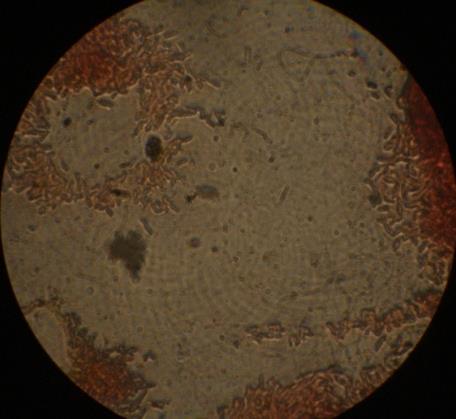
Fig. 1.3 Acid formation and gas production by one of the test isolate S2 (F) as observed by change in the color of medium to yellow and gas bubble in inverted Durham’s tube, respectively. A B C
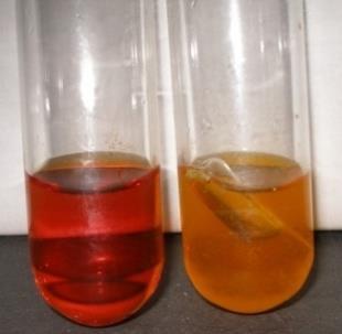
Fig. 1.4 All the tested isolates except isolate S1(C) (A in this figure) showed amylase production as confirmed by formation of clear zone around the single colony.
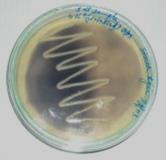
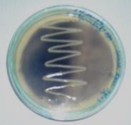
Fig. 1.5 Positive catalase test by one of the test isolate S2 (F) as observed by the production of gas bubbles with the addition of 2-3 drops H2O2 in the test tube.
ISSN 2348-313X (Print)
International Journal of Life Sciences Research ISSN 2348-3148 (online) Vol. 8, Issue 2, pp: (29-37), Month: April - June 2020, Available at: www.researchpublish.com
Table 1.1.1 Morphological characterization of bacterial isolates.
Methods Properties Bacterial isolates S2(F)
Gram’s staining Gram’s reaction +ve Cell shape Rods Size (µm) 3 µm in length Arrangement occurring singly as well as in chains
Endospore staining Spores +ve Shape Oval Position Central Sporangia -ve
Light microscopy Motility +ve
-ve: negative, +ve: positive
Table 1.1.2 Physiological characterization of bacterial isolates. Parameters Bacterial isolates S2(F) Temperature (°C) 4121525 + 30 + 37 + 4250NaCl concentration (%) 1 + 2 + 4681012pH 5 + 6 + 7 + 8 + 9 + 10 + 11 +
-: Absent, +: Present
ISSN 2348-313X (Print)
International Journal of Life Sciences Research ISSN 2348-3148 (online) Vol. 8, Issue 2, pp: (29-37), Month: April - June 2020, Available at: www.researchpublish.com
Table 1.1.3 Biochemical characterization of bacterial isolates.
Parameters Bacterial Isolates S2(F)
Indole test -ve
Methyl red test -ve
Voges Proskauer test -ve
Citrate Utilisation -ve
H₂S production -ve Gas production from Glucose +ve
Casein hydrolysis -ve
Gelatin hydrolysis -ve Starch hydrolysis +ve
Urea hydrolysis -ve
Phosphatase -ve
Nitrate reduction -ve Catalase test +ve
Oxidase test +ve Arginine dihydrolase -ve Ornithine decarboxylase +ve Acid from Trehalose -ve Acid from Arabinose -ve Acid from Galactose -ve Acid from Glucose +ve Acid from Mannitol -ve Acid from Raffinose -ve Acid from Salicin -ve Acid from Xylose +ve Acid from Sucrose -ve Acid from Fructose +ve
-ve: negative, +ve: positive, ND: Not determined
Out of 111 isolates, 3 isolates were selected for their morphological, physiological and biochemical characterizations on the basis of their best growth in NA.
1. The bacterial isolates from soil varied widely in their colony morphology, spore and pigment production.
2. The isolate was Gram +ve.
3. Most of them could grow in wide range of temperature but 37°C was the optimum temperature for growth. However, the isolate S2 (F) shows a wide range of temperature for growth which is quite interesting property and suggests its potential as biofertilizer in field conditions where the organism has to survive under wide range of natural temperature fluctuations.
4. The isolate grew in presence of low concentration of NaCl in the medium.
5. Neutral and alkaline conditions were favorable for the growth of isolate
6. Only isolate S1(C) could be used as nitrate reducer whereas none of the isolates acted as phosphate solubilizers.
7. The strain S2 (F) has shown luxuriant growth on starch solution.
ISSN 2348-313X (Print)
International Journal of Life Sciences Research ISSN 2348-3148 (online) Vol. 8, Issue 2, pp: (29-37), Month: April - June 2020, Available at: www.researchpublish.com
On the basis of above findings the test isolate was the species of genus Bacillus showing promising results for their applications as biofertilizers/PGPR.
BLAST alignment of the sequenced nucleotides of the isolate S2 (F) identified as strain of Brevibacillus borstelensis.
As per the literature B. borstelensisis is a plant growth promoting bacteria. Thus, the bacterial strain isolated from Gangetic soil can act as PGPRs. It has the potential to be used as biofertlizer which has already been established by our previous results.
Description of S2 (F) Brevibacillus borstelensis MTCC 10642:
This strain is gram +ve and shows +ve spore staining and motility property. It grows well at temperature 25 °C to 37 °C but not at 12 °C and 42 °C, pH for growth is 5.2 to10, growth occurs at 1% to 2% NaCl but not from 5% to 10%. Starch and casein are hydrolyzed. Citrate is not utilized and Gelatin is not liquefied. H2S production, MR-VP, nitrate reduction, indole, catalase, oxidase and urea production tests are –ve. Acid is produced from glucose, xylose and fructose and not produced from others like mannitol, sucrose and rhamnose.
We thank Department of Science and Technology (DST), Govt. of India for financial support to carry out this research.
[1] Lynch, J.M. (1990). Introduction: some consequences of microbial competence for plant and soil. In the Rhizosphere ( Ed. Lynch, J.M.), John Wiley & Sons, Chichester, UK, p. 1-10
[2] Pinton, R., Varanini, Z. &Nannipieri, P. (2001). The Rhizosphere: Biodiversity and Organic Substances at the SoilPlant Interface, Marcel Dekker, New York.
[3] BHA Rehm, Microbial production of biopolymers and polymer precursors,Applications andperspectives,Caister Academic Press2008.
[4] Vessey J K (2003) Plant growth promoting rhizobacteria as biofertilizers. Plant and Soil 255: 571–586
[5] Hassan U, Mirza MS, Mehnaz S, Rasul G, Malik KA (1998) Isolation and identification of diazotrophic bacteria from rice, wheat and kallargrass.In Nitrogen Fixation with Non-legumes
[6] Boddy RM, Dobereiner J (1995) Nitrogen fixation associated with grasses and cereals: Recent progress and perspectives for the future. Fert Res 42:241–250
[7] Richardson AE (2003) Making microorganisms mobilize soil phosphorus. In First Virtual International Meeting on Microbial Phosphate Solubilization, http://webcd.usal.es/web/psm/abstracts/Richardson2.htm., pp 1–5
[8] Arshad M, Frankenberger Jr WT (1991) Microbial production of plant hormones. Plant Soil 133:1–8
[9] Rangarajan S, Saleena LM, Vasudevan P, Nair S (2003) Biological suppression of rice diseases by Pseudomonas spp. under saline soil condition.Plant Soil 251:73–82
[10] Kloepper JW, Leong J, Teintze M, Scrhorth MN (1980) Enhanced plant growth by siderophores produced by plant growth promoting rhizobacteria. Nature 286:885–886
[11] Bashan Y (1998) Inoculants of plant growth-promoting bacteria for use in agriculture. Biotechnol Advances 16: 729-770
[12] Ryder MH, Stephens PM, Bowen GD (eds) (1994) Improving Plant Productivity with Rhizosphere Bacteria, Commonwealth Scientific and Industrial Research Organization, Adelaide, Australia
[13] Okon Y, Labandera-Gonzalez CA (1994) Agronomic applications of Azospirillum. In: Improving Plant Productivity with Rhizosphere Bacteria (eds Ryder MH, Stephens PM, Bowen GD, Commonwealth Scientific and Industrial Research Organization, Adelaide, Australia, pp 274–278
[14] Singh A, Poonam, Ghosh AK (2011) Characterization, identification and cataloguing of agriculturally important microorganisms isolated from selected wetland and rain-fed ecosystem of Bihar. Asian J Exp BiolSci 2:575-582
[15] Atlas RM, Brown AE, Dohara KW, Miller L (1984) Experimental microbiology: fundamentals and application, Mac Millan Publishing Company NY