(Online)
Vol. 8, Issue 1, pp: (153-157), Month: April 2020 - September 2020, Available at: www.researchpublish.com

(Online)
Vol. 8, Issue 1, pp: (153-157), Month: April 2020 - September 2020, Available at: www.researchpublish.com
Abstract: Keratitis is mostly caused by adenovirus infection to the eye. Superficial epithelial infiltrates (SEI) and superficial punctate keratitis (SPK) are pathognomonic for the diagnosis of adenoviral conjunctivitis. There are no specific diagnostic methods and treatments for adenoviral conjunctivitis which has resulted in its incorrect diagnosis and unnecessary antibiotic use. In our case series, study of 10 misdiagnosed patients, 70% suffering with diabetes mellitus healed after an accurate diagnosis of adenoviral keratitis and treatment within 4-6 weeks of regular follow-up. Eye ointment, eye drop, and combination medicines, which are used to relieve pain, can act as the appropriate treatment options for SEI and SPK.
Conjunctivitis is commonly known as a pink eye. It may be an infectious or non-infectious inflammation of the conjunctiva. The conjunctiva is a thin clear membrane that covers the anterior part of the sclera (bulbar conjunctiva) and covers the rear part of the eyelid (palpebral conjunctiva) [1].
Usually, the patient's symptoms, history, and inspection of the eyes, assist doctors in determining whether a bacterium, virus, or allergen is causing conjunctivitis. Eye redness or swelling, and discharge are the common clinical symptoms in conjunctivitis but symptoms may be variable depending on the etiology of infection. Viral conjunctivitis is the most prevalent type with 80% of the acute cases of conjunctivitis [2]. Keratitis or acute peribulbar infections are viral conjunctivitis cases mostly caused by adenovirus infection to the eye [3].
Keratitis mainly presents with symptoms such as unilateral itching, photalgia or photophobia, tearing, burning, and foreign body sensation [3]. On the surface of the cornea, tiny round vesicles appear which are filled with fluid and cellular debris. They are known as epithelial microcysts and are usually observed at the initial stages of keratitis. Anterior stromal infiltrates may be observed and persist from a few months to a year. Tabery HM reported cases with recurrent erosions and sterile anterior stromal infiltrates [4].
Superficial epithelial infiltrates (SEI) are considered as the important symptoms of keratoconjunctivitis. The presence of SEIs is considered pathognomonic for the diagnosis of adenoviral conjunctivitis. These infiltrates are typically observed within seven to ten days after the onset of the initial signs of infection [5]. According to a study by Oudova et al [6], superficial punctate keratitis (SPK) is mostly bilateral and might affect either eye. This disease was frequently misdiagnosed and treated incorrectly. The treatment by corticosteroids might curtail subjective symptoms but did not cure the disease [6]. Though there are a number of diagnostic methods for identification of viral keratitis such as viral culture, real-time polymerase chain reaction (RT-PCR), and detection of viral antigen, the widely accepted practice is initiation of treatment on clinical judgment [7-9].
We came across 10 misdiagnosed patients in the last 1 year which were included in the study. The patients were examined with torchlight and slit lamp. A slit-lamp provides a bright source of light and magnification to detect the character and severity of keratitis. Follicles can be observed in a variety of conditions, including inflammation caused by pathogens
International Journal of Healthcare Sciences ISSN 2348-5728 (Online)
Vol. 8, Issue 1, pp: (153-157), Month: April 2020 - September 2020, Available at: www.researchpublish.com
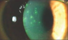
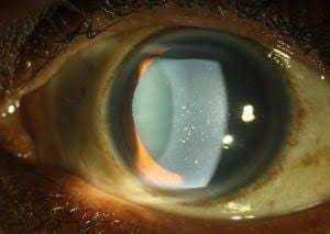
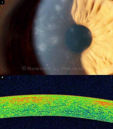
such as bacteria, viruses, topical medications, and toxins [1]. So, we followed a protocol to avoid misdiagnosis (Figure 1). SEI and SPK were observed in some cases (Figures 2 and 3, respectively) examined under the slit-lamp.
Torchlight examination revealed signs such as conjunctival congestion, lid swelling, matting of lashes, conjunctival chemosis and congestion, conjunctival and sub-conjunctival hemorrhage, and corneal haze in most of the cases. In some patients, we also observed preauricular lymph nodes, fever, corneal scarring, upper respiratory tract infection (URTI), and hypertension. Fluorescein staining, a grading method for SPK magnitude was performed (Figure 4). Refraction or a vision test was also performed and visual acuity was noted before the start of treatment as mentioned in table 1.
Laboratory investigations were performed mainly for blood sugar levels and conjunctival swabs were sent for evaluation, if conjunctivitis lasted more than 4 weeks which showed normal conjunctival flora. A total of 7 out of 10 patients were diabetic. The symptoms, clinical history, and diagnosis of patients are mentioned in table 1. All the patients were in the age group of 40 to 55 yrs, and both genders were equally affected. The average time for the appearance of symptoms was 4-5 days. A total of 8 cases showed signs like conjunctival chemosis and congestion.
Treatment was almost similar for all cases having vision below 6/12. All the misdiagnosed patients were given treatment with various drugs such as Nafzol eye drops, Moxifloxacin eye drops, paracetamol tablet, Ciplofloxacin eye drops, Neosporin eye ointment, Moxifloxacin-dexamethasone eye drops, flurometholone (FML) eye drops, Bepofree eye drops, Pyrimon eye drops, Lotepred, Winolap, Nepastar eye drops, and Tears plus eye drops. Most of the medications were administered for bacterial keratitis until we accurately diagnosed the patients. We administered medication to patients according to the clinical diagnosis of the disease with only Acivir eye ointment, tear substitutes, nepafenac eye drops, oral acyclovir, anti-inflammatory drugs in most cases with vision less than or equal to 6/12 and oral steroids (Deflazacort) were administered in a case which had coexisting iritis. In cases with good vision, symptomatic treatment was given consisting of decongestant eye drops, tear substitutes, and lubricating ointments as these cases underwent self-resolution in 2 to 3 weeks. The patient was recalled for a follow-up examination. In most of the cases, symptomatic improvement and reduction in SEI and SPK were observed within 1-2 weeks of treatment. Vision improvement was seen in all the cases and the cases recovered completely after 5-6 weeks of treatment.
Adenoviruses are the most common reason for viral conjunctivitis causing infection in 65-95% of cases but the viral conjunctivitis is usually misdiagnosed as bacterial or allergic conjunctivitis [2]. According to the study by Statham et al [10], the primary health care provider was able to diagnose only 35.9% of the acute eye diseases correctly. Even ophthalmologists are unable to diagnose the diseases correctly and only 48.2% of patients were correctly diagnosed by them. As there is no specific licensed treatment for adenoviral conjunctivitis, so it is necessary to find reliable diagnostic methods other than clinical evaluation to diagnose them correctly A proper diagnosis and treatment may assist in the faster recovery of patients.
In these cases, the patients were earlier misdiagnosed and eventually received improper treatment. When they visited our hospital, they were successfully diagnosed with the help of torchlight, slit lamp and complete clinical examination. The protocol for follicles assisted to ensure the accurate diagnosis of disease.
The patients with DM are more prone to bacterial keratitis whereas non-diabetic patients are more likely to suffer from herpes simplex virus keratitis (HSK) [11]. SPK, dry eye, and persistent epithelial defects are considered as potentially sight-threatening conditions and show delayed wound healing [12-14]. Although the patients cannot be cured completely of the adenovirus keratitis [6]; however, in our case study, 7 out of 10 patients having DM, healed within the few weeks of treatment and the vision acuity was recovered
To conclude, a protocol should be followed to differentiate the symptoms of adenovirus infection for correct diagnosis. Eye ointment, eye drop, and combination medicines used to relieve pain are appropriate treatment options for SPK and SEI. The patients should be followed-up for up to six weeks for viral keratitis. Thus, regular follow-ups have been planned for these cases. Nevertheless, it should be noted that this treatment helped in the recovery of almost all patients with DM within 4 to 6 weeks which should be regarded as a success.
International Journal of Healthcare Sciences ISSN 2348-5728 (Online)

Vol. 8, Issue 1, pp: (153-157), Month: April 2020 - September 2020, Available at: www.researchpublish.com
Case Symptoms and comorbidities
Slit lamp diagnosis Vision
1 Watering and pain, DM SPK in RE
2 Photophobia, swelling, pain, mucopurulent discharge both eyes (BE), DM, URTI
6/9RE, 6/6 LE
SPK diffuse all over cornea 6/24 BE
3 Pain, photophobia, watering,blurring of vision SEI, mild iritis 6/18 LE
4 Grittiness,watering and pain in BE, URTI
Few SPK mainly pericentriic 6/9 BE
5 Photophobia, swelling, pain and mucoid discharge, blurring BE, DM SEI in BE diffuse all over cornea 6/12 BE
6 Pain, photophobia, watering,blurring of vision in right eye (RE), DM Scanty SEI pericentric 6/12RE
7 Grittiness,watering, photophobia,and pain BE, DM SPK in BE 6/12 RE6/18 LE
8 Photophobia, swelling, pain, grittiness, watering, blurring of vision re, DM Severe SEI RE 6/24 RE 6/18 LE
9 Swelling, pain and mucopurulent discharge both eyes, DM, HT SEI BE 6/12 BE
10 Blurring and pain RE, URTI Scanty SPK RE 6/9 RE
International Journal of Healthcare Sciences ISSN 2348-5728 (Online) Vol. 8, Issue 1, pp: (153-157), Month: April 2020 - September 2020, Available at: www.researchpublish.com
[1] Solano D, Czyz CN. Viral Conjunctivitis. In: StatPearls. Treasure Island (FL): StatPearls Publishing; 2020.
[2] Azari AA, Barney NP. Conjunctivitis: a systematic review of diagnosis and treatment. JAMA 2013; 310(16):1721‐9.
[3] Bialasiewicz A. Adenoviral keratoconjunctivitis. Sultan Qaboos Univ Med J 2007; 7(1):15‐23.
[4] Tabery HM. Corneal stromal infiltrates in patients with recurrent erosions. Acta OphthalmolScand 1998; 76(5):589‐592.
[5] Pihos AM. Epidemic keratoconjunctivitis: A review of current concepts in management. J Optom 2013; 6(2):69‐74.
[6] Oudová P, Filipec M. Thygeson's keratitis-clinical characteristics and therapy. CeskSlovOftalmol 2004;60(1):17‐23.
[7] Athmanathan S, Bandlapally SR, Rao GN. Collection of corneal impression cytology directly on a sterile glass slide for the detection of viral antigen: an inexpensive and simple technique for the diagnosis of HSV epithelial keratitis - a pilot study. BMC Ophthalmol 2001;1:3.
[8] Gupta N, Tandon R. Investigative modalities in infectious keratitis. Indian J Ophthalmol 2008;56(3):209‐213.
[9] Hoffman J. Adenovirus: ocular manifestations. Community Eye Health. 2020;33(108):73‐75.
[10] StathamMO, Sharma A, Pane AR. Misdiagnosis ofacute eye diseases byprimary health care providers: incidence and implications. Med J Aust 2008; 189(7):402‐404.
[11] Wang B, Yang S, Zhai HL, et al. A comparative study of risk factors for corneal infection in diabetic and non-diabetic patients. Int J Ophthalmol 2018; 11(1):43‐47.
[12] Achtsidis V, Eleftheriadou I, Kozanidou E, Voumvourakis KI, Stamboulis E, Theodosiadis PG, Tentolouris N. Dry eye syndrome in subjects with diabetes and association with neuropathy. Diabetes Care 2014; 37(10):e210‐e211.
[13] Ye H, Lu Y. Corneal Bullous Epithelial Detachment in Diabetic Cataract Surgery. Optom Vis Sci 2015; 92(7):e161‐e164.
[14] Kaji Y. Prevention of diabetic keratopathy. Br J Ophthalmol 2005; 89:254-255.