ISSN 2348-313X (Print)
International Journal of Life Sciences Research ISSN 2348-3148 (online)
Vol. 8, Issue 3, pp: (1-7), Month: July - September 2020, Available at: www.researchpublish.com

ISSN 2348-313X (Print)
International Journal of Life Sciences Research ISSN 2348-3148 (online)
Vol. 8, Issue 3, pp: (1-7), Month: July - September 2020, Available at: www.researchpublish.com
Abstract: A probiotic is a live microbial food supplement which beneficially affects the host animal by improving its intestinal microbial balance. In the present study, we isolated probiotic Lactobacillus salivarius from cow curd. Pour plate technique was used to isolate the organisms. Samples were used directly and also diluted to 10-1, 10-2 and 10-3 using sterile peptone water. 1 ml aliquot of the samples and dilutions were plated into MRS (Man, Rogosa and Sharpe) agar (Oxoid LTD, Basingstoke, England) selective medium according to Dave and Shah,. The plates were incubated at 37°C for 3 days under aerobic conditions. All isolates were sub-cultured by streaking method to get pure cultures. Pure culture of specific probiotic isolate from the sample was identified on the basis of biochemical tests such as catalase, oxidase and IMVIC (indole, methyl-red, voges proskaur, citrate utilization) tests. Gram-staining was carried out. Identification of isolates was carried out based on the method described by Sakazaki and Shimad Collins et al., (1989). The identified Lactobacillus species was confirmed through PCR reaction using genus specific primer. The curd has suggested as a source of potential probiotic strain which has probiotic properties.
Keywords: Lactobacillus, probiotics, biochemical tests, rRNA sequence.
Probiotics are commonly defined as mono or mixed cultures of live microbes that when applied to an animal or human posses a beneficial effect on the health of host by improving the balance of the indigenous microflora, and also improved nutrition, growth and prevention of various gastrointestinal disorders. (R Fuller 1999). Probiotics are widely used to prepare fermented dairy products like curd, yogurt, and these are important source of probiotic Lactobacillus (Kamrunnahar Islam et al., 2015). These are a group of bacteria that are frequently used as probiotics. They have a long history in their use as probiotics in the food industry and the Lactobacillus strains are “generally recognized as safe” (GRAS). They are commonly found in the environment such as soil, water, decaying plant materials, as well as in the normal microflora of the gastrointestinal tract (GIT) of animals ( Kizerwetter – Swida and Binek, 2005).
L Salivarius found in the mouth and small intestine has antimicrobial activities and breaks down proteinase and produces vitamin B, enzymes and Lactic acid. L salivarius helps to inhibit Salmonella and ulcer Lactobacillus causing by bacteria H. Pylori. Lactobacillus salivarius belongs to the terrabacteria group in the class Firmicutes. It is in the class Bacilli, of the order Lactobacillales, and belongs to the family Lactobacillus (“European Bioinformatics” 2018). Lactobacillus salivarius is a well characterized, rod-shaped Gram-positive species of probiotic bacteria (Chapot-Chartier et al., 2014). L salivarius is characterized as a lactic acid bacterium (LAB), which denotes its ability to produce lactic acid by fermentation (Kuratsu et al., 2010).
Cow curd sample was collected from the local area. Pour plate technique was used to isolate the organisms. Samples were used directly and also diluted to 10-1, 10-2 , 10-3, using sterile peptone water. 1 ml aliquot of the samples and dilutions were plated into MRS (Man, Rogosa and Sharpe) agar (Oxoid LTD, Basingstoke, England) according to Dave and selective medium (Shah, 1996). The plates were incubated at 37°C for 3 days under aerobic conditions. All isolates were sub-cultured by streaking method to get pure cultures.
ISSN 2348-313X (Print)
International Journal of Life Sciences Research ISSN 2348-3148 (online) Vol. 8, Issue 3, pp: (1-7), Month: July - September 2020, Available at: www.researchpublish.com
Bacterial Identification: Gram-staining was carried out by using the isolated organisms. Identification of isolates was carried out by using the following biochemical tests based on the method described by Sakazaki and Shimad (1986), Collins et al., (1989) and Cheesbrough (2002). Following Biochemical tests were used for the identification of bacteria
Catalase test: The Tryptone soy agar medium was sterilized and slants were prepared which were inoculated with the culture and incubated at 37oC for 48 h. After the incubation period, 3 or 4 drops of H2O2 were allowed to flow over the growth of the culture on the slant and observed for evolution of O2 as bubbles which indicate as positive reaction.
Oxidase test: A smear of the fresh culture, 24 h-old culture was prepared on Whatmann’s filter paper impregnated with 2-3 drops of 1% oxidase reagent which was reduced with sodium thionate and left for 10 sec. If purple color appears within 10 sec, it was a positive test for oxidase. But if the color was produced between 10 and 60 sec, it was delayed positive reaction while absence of color was a negative reaction.
Indole and hydrogen sulfide test: The medium was distributed in 5 ml portions in test tubes, sterilized 121oC for 15 min, and cooled (40o C). The test tubes were inoculated with culture suspension and incubated at 35oC for 48 h. After 48 h of incubation, 1ml of Kovac’s reagent was added to each tube including control. Uninoculated tubes served as control. The tubes were gently shaken after 10-15 min intervals and the tubes were allowed to stand to permit the reagent to come to top.
Methyl red and Voges-Proskauer tests: MRVP broth (5 ml/test tube) was sterilized, cooled, inoculated with the culture suspension and incubated at 37oC for 48 h. Five drops of methyl red indicator was added to the test tubes. Development of red color indicates positive reaction for MR test and no change indicates negative result. Another set of MRVP tubes was also inoculated with the same culture but incubated for 96 h.
Urease test: Urease agar slant surface was streaked with a portion of a well-isolated colony or inoculate slant with 1 to 2 drops from an overnight brain-heart infusion broth culture. Leave the cap on loosely and incubated the tube at 35°-37°C in ambient air for 48 hours to 7 days.
Gelatin hydrolysis test: Gelatin agar slants were prepared, inoculated with the bacterial culture and incubated over night. If the medium liquefies, it indicates positive test.
Citrate utilization test: Simmon’s citrate agar (pH 6.9) slants were prepared and inoculated with the culture by means of a stab-and-streak inoculation and incubated at 37oC for 48 h. Un inoculated slants served as control. The slants were observed for change in color. Change in color from green to blue is indicative of positive reaction while no change in color was negative reaction.
Disc diffusion method: It was used to test the penicillinase activity. The pure samples were spread on the Nutrient Agar plate. The 3 different concentrations of Penicillin discs were prepared. The individual Samples were spread on the plate and different concentrations (10mg/ml, 20mg/ml, 50mg/ml, 100mg/ml) of Penicillin discs were placed on the plates .the microbes were allowed to grow on the plate for 48 hours. The Cultures which grown with higher resistance were isolated and were used for further studies. The isolated colonies were then tested for the zone of inhibition.
Bacterial identification based on 16s ribosomal RNA gene sequence analysis Four steps were required to reach bacterial identification through 16S rRNA sequencing: DNA extraction, PCR amplification, nucleotide sequencing, and data base homology search and reporting.
Isolation of DNA from the samples preparation of genomic DNA from bacteria: Bacteria from a saturated liquid culture were lyses and the proteins were removed by digestion with the proteinase K. Cell wall debris, polysaccharides, and remaining proteins were removed by selective precipitation with CTAB (Cetyl Trimethyl Ammonium Bromide), and high-molecular-weight DNA was recovered from the resulting supernatant by isopropanol precipitation.
Quantification of DNA:The isolated DNA was quantified by measuring absorbance at 260 and at 280 nm. Ratio of absorbance 260/280 was used to determine the quality of isolated DNA. The concentration was calculated using the following formula:
Concentration of the DNA = OD260 X 50 X dilution factor = g of DNA /ml
ISSN 2348-313X (Print)
International Journal of Life Sciences Research ISSN 2348-3148 (online) Vol. 8, Issue 3, pp: (1-7), Month: July - September 2020, Available at: www.researchpublish.com
Selection of 16 S rRNA primers:
Primers were selected from the published literature which recognizes most of the bacterial species.
Polymerase chain reaction (PCR) : Polymerase chain reaction was performed using primers that were designed from the specific gene of the Shigella sp primers were commercially obtained from Bio-serve India pvt, Ltd. All the primers designed were 20 base pairs in length with GC content in the range of 40-80%. Tm was calculated according to the formula. The annealing temperature for each pair of PCR primers was optimized experimentally.
Tm(C) = 2(A+T) + 4(G+C) 5
PCR amplification was carried out with following reaction parameters:
Table-1: PCR reaction mixture composition
S.No Components Volume
1
DNTPs 200µM
2 PCR reaction buffer 1X
3 Magnesium chloride 1.5
4 Primer (forward) 2.5 mM
5 Primer (reverse) 10 pmoles
6 Template DNA 100 ng
7 Taq DNA polymerase 1U
The DNA was visualized by placing on a UV light source and photographed directly by gel documentation unit.
Purification of PCR product by gel elution:
Spin column method: Assay Procedure:
1. The separated DNA was cut with a new sterilized blade and transformed in to a sterile 1.5 ml tube.
2. Binding buffer 750 µl i.e., three times the weight of the gel piece) was added.
3. The eppendorf tube was then kept in a water bath that was preset at 60°C for about 10 min.
4. The entire mix was then slowly transferred to the column placed in 2.0ml collection tube and was spun at 10,000 rpm for 2 min at room temperature.
6. The eluted solution was discarded and 750 µl of wash buffer was added to the column and centrifuged at 10,000 rpm at room temperature for 2 min.
7. Centrifugation step was repeated to remove excess isopropanol from the sample.
8. The column was then transferred to a fresh collection tube and 50 µl of autoclaved, distilled water was added and spun at 10000 rpm at room temperature for 2 min.
9. Again 10 ul of autoclaved distilled water was added and centrifuged at 10,000 rpm.
10. The eluted product was transferred in to clean 1.5 ml eppendorf tube and preserved at -20°C.
DNA sequencing was performed with forward primer of the 16s rRNA gene to know the first 600bp sequencing by Sanger’s DNA sequencing method performed at Eurofins sequencing company.
Blast analysis:
The result obtained on sequencing was subjected for blast analysis for the identification of microorganism.
ISSN 2348-313X (Print)
International Journal of Life Sciences Research ISSN 2348-3148 (online) Vol. 8, Issue 3, pp: (1-7), Month: July - September 2020, Available at: www.researchpublish.com
Based on the results of morphological studies, Gram staining and biochemical tests conducted (Table: 2) and following the Bergey’s Manuals (Aneja, 2003), the isolate was tentatively identified as Lactobacillus sp.
Fig-1 Gram staining (Gram Positive Bacterium)
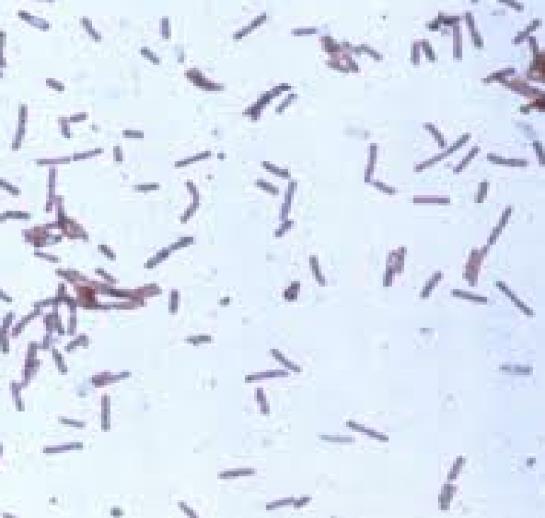
Table-2: Morphological and biochemical tests of the bacterial isolate Test Result
Gram staining Positive Catalase Positive Oxidase test Positive Indole test Negative Hydrogen sulphide production Negative Methyl red Positive VogesProskauer Positive Urease Positive Gelatin hydrolysis Positive Citrate utilization Positive
PCR result
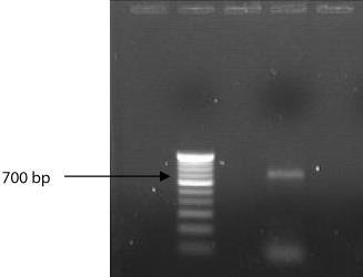
Amplified product was shown in the following figure-1
Figure-2 Lane2: 100 bp and ladder Lane 4: 700 bp PCR amplified product.
ISSN 2348-313X (Print)
International Journal of Life Sciences Research ISSN 2348-3148 (online) Vol. 8, Issue 3, pp: (1-7), Month: July - September 2020, Available at: www.researchpublish.com
16S ribosomal RNA (or 16S rRNA) is the component of the 30S small subunit of a prokaryotic ribosome that binds to the Shine-Dalgarno sequence. The genes coding for it are referred to as 16S rRNA gene and are used in reconstructing phylogenies, due to the slow rates of evolution of this region of the gene. In our investigation the isolate confirmed as Lactobacillus sps. (Fig: 1)
FASTA format of the Sequence obtained:
TAGAGTTTGATCCTGGCTTAGGACGAACGCTGGCGGCGTGCCTAATACATGCAAGTC
GAACGAAACTTTCTTACACCGAATGCTTGCATTCACCGTAAGAAGTTGAGTGGCGGA
CGGGTGAGTAACACGTGGGTAACCTGCCTAAAAGAAGGGGATAACACTTGGAAACA
GGTGCTAATACCGTATATCTCTAAGGATCGCATGATCCTTAGATGAAAGATGGTTCT
GCTATCGCTTTTAGATGGACCCGCGGCGTATTAACTAGTTGGTGGGGTAACGGCCTA
CCAAGGTGATGATACGTAGCCGAACTGAGAGGTTGATCGGCCACATTGGGACTGAG
ACACGGCCCAAACTCCTACGGGAGGCAGCAGTAGGGAATCTTCCACAATGGACGCA
AGTCTGATGGAGCAACGCCGCGTGAGTGAAGAAGGTCTTCGGATCGTAAAACTCTG
TTGTTAGAGAAGAACACGAGTGAGAGTAACTGTTCATTCGATGACGGTATCTAACCA
GCAAGTCACGGCTAACTACGTGCCAGCAGCCGCGGTAATACGTAGGTGGCAAGCGT TGTCCGGATTTATTGGGCGTAAAGGGAACGCAGGCGGTCTTTTAAGTCTGATGTGAA
AGCCTTCGGCTTAACCGGAGTAGTGCATTGGAAACTGGAAGACTTGAGTGCAGAAG
AGGAGAGTGGAACTCCATGTGTAGCGGTGAAATGCGTAGATATATGGAAGAACACC
AGTGGCGAAAGCGGCTCTCTGGTCTGTAACTGACGCTGAGGTTCGAAAGCGTGGGT
AGCAAACAGGATTAGATACCCTGGTAGTCCACGCCGTAAACGATGAATGCTAGGTG
TTGGAGGGTTTCCGCCCTTTAGTGCCGCAGCTAACGCAATAAGCATTCCGCCTGGGG
AGTACGACCGCAAGGTTGAAACTCAAAGGAATTGACGGGGGCCCGCACAGGCGGTG
GAGCATGTGGTTTAATTCGAAGCAACGCGAAGAACCTTACCAGGTCTTGACATCCTT
TGACCACCTAAGAGATTAGGTTTTCCCTTCGGGGACAAAGTGACAGGTGGTGCATGG
CTGTCGTCAGCTCGTGTCGTGAGATGTTGGGTTAAGTCCCGCAACGAGCGCAACCCT
TGTTGTCAGTTGCCAGCATTAAGTTGGGCACTCTGGCGAGACTGCCGGTGACAAACC
GGAGGAAGGTGGGGACGACGTCAAGTCATCATGCCCCTTATGACCTGGGCTACACA
CGTGCTACAATGGACGGTACAACGAGTCGCAAGACCGCGAGGTTTAGCTAATCTCTT
AAAGCCGTTCTCAGTTCGGATTGTAGGCTGCAACTCGCCTACATGAAGTCGGAATCG
CTAGTAATCGCGAATCAGCATGTCGCGGTGAATACGTTCCCGGGCCTTGTACACACC
GCCCGTCACACCATGAGAGTTTGTAACACCCAAAGCCGGTGGGGTAACCGCAAGGA
GCCAGCCGTCTAAGGTGGGACAGATGATTGGGGTGAAGTCGTAACAAGGTAGCCGT
AGGAGAACTTGCGGCTGGATCACCTCCTTAAATC
ISSN 2348-313X (Print)
International Journal of Life Sciences Research ISSN 2348-3148 (online) Vol. 8, Issue 3, pp: (1-7), Month: July - September 2020, Available at: www.researchpublish.com
BLAST analysis data: (Alignment view using combination of NCBI) was represented in the following figure-2
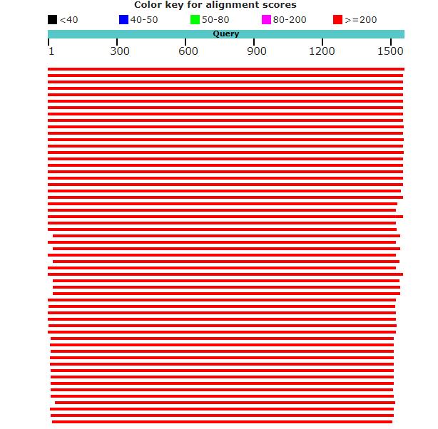
Fig-3: BLAST analysis data
Based on the above tests and results the bacteria isolated from curd sample was identified as Lactobacillus salivarius, which was a probiotic bacteria. This probiotic Lctobacillus salivarius strain inhibit 4NQO- induced oral cancer due to the protection of DNA against oxidative damage and down regulating cyclo oxygenase 2 expression. (Chaves et al., 2017). Probiotics like Lactobacillus salivarius play an important role in glycenic regulation.
[1] R L. Dave, R.L, N.P. Shah, “Evaluation of media for selective enumeration of Streptococcus thermophilus, Lactobacillus delbrueckii ssp. bulgaricus Lactobacillus acidophilus and Bifidobacterium spp,” Journal of Dairy Science, Vol 79 PP 1529-1536, 1996.
[2] Collins, A., Brown, J. S., and Newman, S. E. (1989). “Cognitive Apprenticeship: Teaching the Craft of Reading, Writing and Mathematics. In L. B. Resnick (ed.) Knowing, Learning, and Instruction: Essays in Honor of Robert Glaser. Hillsdale”, Journal of Nutrition: 1989.
ISSN 2348-313X (Print)
International Journal of Life Sciences Research ISSN 2348-3148 (online) Vol. 8, Issue 3, pp: (1-7), Month: July - September 2020, Available at: www.researchpublish.com
[3] Rolfe, R.D, “The role of probiotic cultures in the control of gastrointestinal health ”, Journal of Nutrition Vol 130, PP 396S– 402S 2000
[4] R. Fuller, “Probiotics for farm animals,” in Probiotics. A Critical Review, G. W. Tannock, Ed., pp. 15–22, Horizon Scientific Press, Wymondham, UK, 1999.
[5] Kamrun Nahar Islam, Touaha Akbar, Fahmida Akther , Nazneen Naher Islam, “Characterization and Confirmation of Lactobacillus spp. from Selective Regional Yoghurts for Probiotic and Interference with Pathogenic Bacterial Growth” Asian Journal of Biological Sciences, Vol 9 (1-2) PP 1-9, 2016.
[6] Kizerwetter-Swida M, Binek M. Protective effect of potentially probiotic Lactobacillus strain on infection with pathogenic bacteria in chickens. Pol. J. Vet. Sci. 2009;12:15–20
[7] Chapot-Chartier MP, Vinogradov E, Sadovskaya I, Andre G, Mistou M-Y, Trieu-Cuot P, Furlan S, Bidnenko E, Courtin P, Péchoux C (2010) Cell surface of Lactococcus lactis is covered by a protective polysaccharide pellicle. J Biol Chem 285:10464–10471.