ISSN 2348-313X (Print)
International Journal of Life Sciences Research ISSN 2348-3148 (online)
Vol. 8, Issue 4, pp: (18-22), Month: October - December 2020, Available at: www.researchpublish.com

ISSN 2348-313X (Print)
International Journal of Life Sciences Research ISSN 2348-3148 (online)
Vol. 8, Issue 4, pp: (18-22), Month: October - December 2020, Available at: www.researchpublish.com
Shrawan Kumar1, Prakash Singh1 , G.B.Chand1*
1Aquatic Toxicology Laboratory, Department of Zoology, Patna University, Patna- 800 005
Email: gbchand@rediffmail.com
Abstract: Arsenic is the most noxious metalloid affecting the aquatic organism by inducing oxidative stress and histopathological abnormalities in vital organs. Bihar is emerging as the hot spot to the arsenic toxicity next to West Bengal in India. Most of the districts of Bihar have the prevalence of arsenic in the ground water. In the present study the freshwater air breathing fish Clarias batrachus (Linn.) has procured from Phulwarisharif Fish farm, Patna and acclimatized in the laboratory for 15 days. 96 hours and 48 hours LC50 of As2O3 for fish was calculated as per standard APHA method. The fishes were exposed to 2.5 ppm, 5.0 ppm and 7.5 ppm of As2O3 for one week. After the termination of exposure, the autopsy was done and the fish kidney were extracted and fixed in 10% NF and paraffin spread sections were prepared for light microscopy The major histopathological anomalies incurred in kidney were – infiltration of neutrophils, fibrin and pus cells in the lumen of collecting tubules, oedematous, deposition in the lumen of PCT and DCT, infiltration of plasma cells in intertubular space, metastatic calcification of glomeruli etc. The findings of the present study shows that the nephrotoxicity generated in fish due to Arsenic is dose & duration dependent which ultimately lead to the renal failure of the fish. A close monitoring of the health of the fish is the need of hours for their conservation and enhancement of aquaculture sector of the State.
Keywords: Clarias batrachus, Nephrotoxicity, As2O3, LC50, renal tissues.
Heavy metals and pesticides contamination of the aquatic system has attracted the attention of researchers all over the world. The natural aquatic systems may extensively be contaminated with heavy metals released from domestic, industrial and other human activities. They may have devastating impact on the ecological balance of the recipient environment and a diversity of aquatic organisms including fish.
Arsenic toxicity has numerous deleterious impacts on the experimental model as well as in the human beings. Several different organ systems are affected by arsenic including skin, respiratory, reproductive, gastrointestinal and nervous systems.
The investigation of histological changes in the organs of fish is an accurate way to assess the effects of xenobiotic compounds in experimental studies. Hence this study was undertaken to examine the effect of arsenite at sublethal concentration on histology of kidney of common air breathing fish Clarias batrachus
Since the kidney, being the major route of arsenic excretion, is seriously affected by arsenic poisoning. Various sites of arsenic damages in the kidney include capillaries, tubules and glomeruli[1], [2]. Other serious implications of arsenic poisoning at renal level are mitochondrial damage in prominent epithelial cells leading to proteinuria, shock, dehydration and ultimate renal failure[3]
Banerjee & Bhattacharya[4] have studied histopathological abnormalities in the kidney of Channa punctatus exposed to chronic nonlethal level of elsan, mercury and ammonia. They have shown marked acute tubular epithelial degeneration,
ISSN 2348-313X (Print)
International Journal of Life Sciences Research ISSN 2348-3148 (online)
Vol. 8, Issue 4, pp: (18-22), Month: October - December 2020, Available at: www.researchpublish.com
karyolysis, dilation and shrinkage of Bowman’s capsule & glomerulus. Long term exposure to arsenic has been shown to affect head kidney & impair humoral immune responses of Clarias batrachus[5].
Present study is designed to observe the histopathological alterations caused in kidney of Clarias batrachus due to different level of arsenic exposure within one week.
Same age groups of Clarias batrachus, ranging from 50-80 gm and size between 18-20 cms were collected during prespawning season (March-May). The fishes were brought to the laboratory, disinfected with 0.1% KMNO4 solution and were acclimated for 15 days in the laboratory condition. After acclimation, the fishes were transferred to plexi glass aquaria of 50 litre capacity @ 20 fish each having dechlorinated aerated tap.
Here in the experimental protocol, the 96 hrs. mean lethal concentration (LC50) of AS2O3 for Clarias batrachus was calculated as 15 0 mg/l (as 11 4 mg/l). Three doses considered in the experimental protocol were 2 5 mg/l, 5 0 mg/l and 7 5 mg/l and accordingly stock solutions were prepared by dissolving appropriate amount of AS2O3 in the deionized water.
After termination of each exposure, different groups of anaesthetized fish were sacrificed and 10 pieces of kidney samples of each group were collected and fixed in 10% neutral buffered formalin for 24 hrs. Tissue slides were prepared using double staining method for light microscopic study.
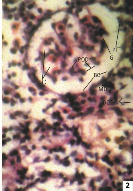
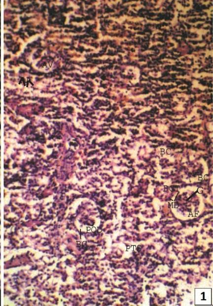
Normal Kidney of Clarias batrachus is dividing into two parts - head kidney and trunk kidney. Head kidney is generally made up of lymphoid, hematopoietic, inter-renal and chromaffin tissues and is devoid of renal corpuscles and tubules, while trunk kidney contains specific structures related with excretion. A typical nephron of freshwater teleost consists of a renal corpuscle, containing well vascularized glomerulus, a ciliated neck region of variable length, connecting the renal corpuscle with the tubule, an initial proximal segment with prominent brush border and numerous prominent lysosomes, a second proximal segment with numerous mitochondria but less well developed brush border, a narrow ciliated intermediate segment, a distal segment with relatively clear cells and elongated mitochondria and a collecting duct system (Fig. 1 & 2).
Fig. 1: showing prominent arcuate artery (AA) & arcuate vein (AV) at the junction of renal cortex (Cor) & medulla (MR). Several renal corpuscles are seen with normal features.
ISSN 2348-313X (Print)
International Journal of Life Sciences Research ISSN 2348-3148 (online) Vol. 8, Issue 4, pp: (18-22), Month: October - December 2020, Available at: www.researchpublish.com
Fig. 2: A magnified view (x400) of renal corpuscles (RC) with prominent parietal layer lined by simple squamous epithelial cell, visceral layer of Bowman’s capsule (BC) lined by podocytes (PoD), glomerular capilleries (C), urinary space (asterisk) and macula densa (MD) of distal convoluted tubule (DCT).
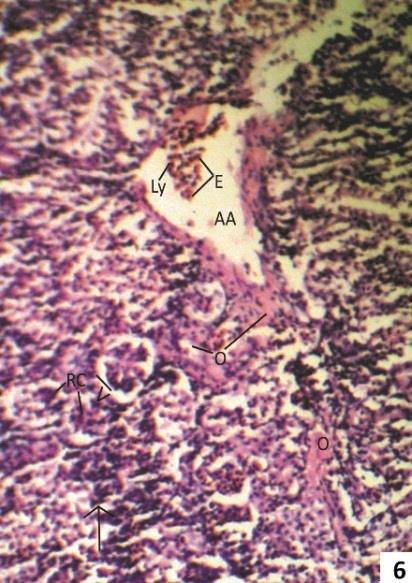
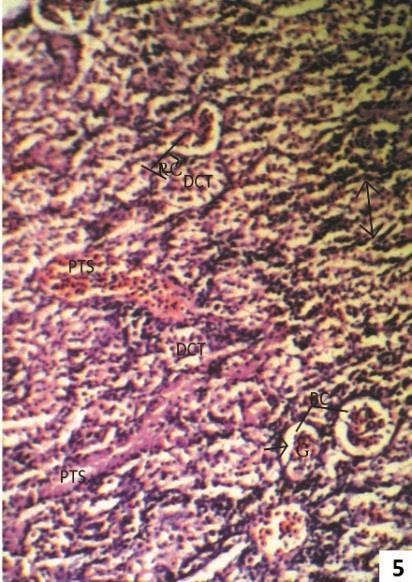
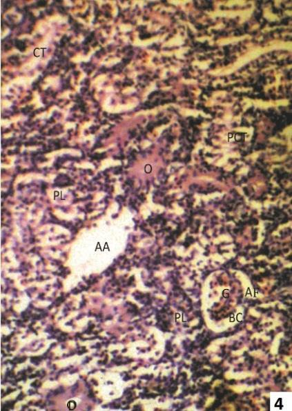
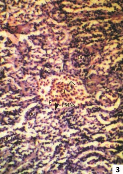
Fig. 3 & 4: Photomicrographs of transverse sections of kidney of one week 2.5 ppm arsenic treated Clarias batrachus stained with Haemotoxyline &Eosin.
Fig. 3: Section of 2.5 ppm Arsenic trioxide treated kidney for one week showing condition of acute pyelonephritis, as marked by collecting tubules filled with pus (arrow). The lumen of collecting duct shows infiltration of polymorpho nuclear cells (PMNS) and fibrin (F).
Fig. 4: Degeneration in tubules of nephrons as marked by edematous (O) deposition in distal convoluted tubule (DCT), proximal convoluted tubule (PCT) as well as collecting tubule (CT). Note mass infiltration of plasma cells (PL) in the intertubular space (ITS) are also prominent.
The 2.5 ppm arsenic treated kidney showed lumen of collecting tubule filled with pus cells, neutrophils & fibrin (Fig.3), edoematous deposition in proximal and distal convoluted tubule and infiltration of plasma cells in the intertubular space (Fig. 4).
Fig. 5 & 6: Photomicrographs of transverse sections of kidney of one week 5.0 ppm arsenic treated (Group II) Clarias batrachus stained with Haemotoxyline &Eosin.
ISSN 2348-313X (Print)
International Journal of Life Sciences Research ISSN 2348-3148 (online)
Vol. 8, Issue 4, pp: (18-22), Month: October - December 2020, Available at: www.researchpublish.com
Fig. 5: Section of 5.0 ppm Arsenic trioxide treated kidney for one week shows widening of urinary space (arrow), intermingling of visceral and parietal layer of Bowman’s capsule (asterisk), massive infiltration of lymphocyte (Ly) and plasma cell (PL). The peritubular space (PTS) is filled with edematous fluid (O).
Fig. 6: Section showing beginning of metastatic calcification with marked fusion of glomeruli (arrow head), calcification of elastic lamina (arrow) and presence of lymphocyte (Ly) and eosinphils (E) in lumen of arcuate artery and oedematous fluid in peritubular space (PTS).
After one week treatment of 5.0 ppm As2O3 fish kidney showed widening of urinary space intermingling of visceral and parietal layer of Bowman’s capsule, and infiltration of lymphocytes, plasma cells and deposition of oedomatous fluid in peritubular space (Fig. 5). Massive metastatic calcification of glomeruli were also seen (Fig. 6).
Fig. 7 & 8: Photomicrographs of transverse sections of kidney of one week 7.5 ppm arsenic treated (Group II) Clarias batrachus stained with Haemotoxyline &Eosin.
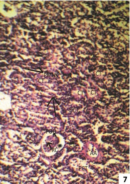
Fig. 7: Section of 7.5 ppm Arsenic trioxide treated kidney for one week showing widening of Bowman’s space (asterisk), predominating collecting tubules (CT), necrotic PCT and DCT (arrow) surrounding corpuscles and massive infiltration of polymorpho nuclear cell (PMNS) in intertubular space (ITS).
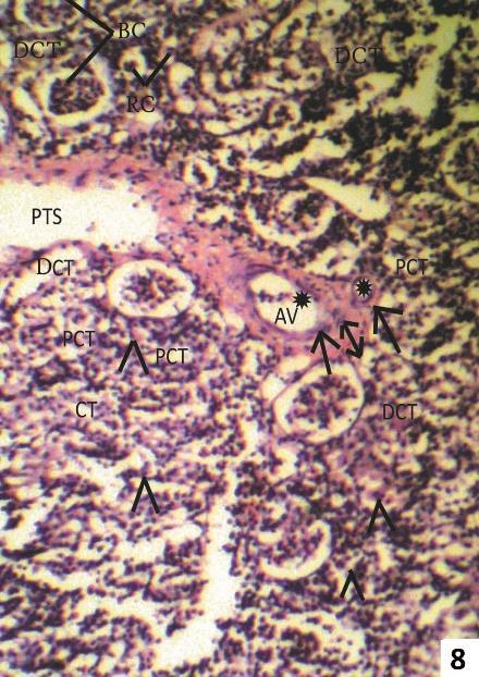
Fig. 8: Showing Thickened fibrous intima (double arrow) and constricted lumen (asterisk) of arcuate artery (AA) and arcuate vein (AV). Oedematous fluid (O) in glomerular capillaries.
At higher dose of 7.5 ppm As2O3, fish kidney showed widening of Bowman’s capsule and necrosis in renal tubules (Fig. 7 & 8).
Histopathological and cytological alterations may be considered as first sign of stress condition, which is later reflected in biochemical and hematological studies.
The central theme of the present investigation is to explore a systematic deleterious impact of arsenic trioxide (As2O3) on renal tissues of fresh water catfish Clarias batrachus
The major histopathological changes in fish kidney due to lower sub lethal exposure of arsenic were shrinkage and constriction of glomerular tuft, inflammation of podocytes, widening of urinary space, increased incidence of necrotic renal tubules as well as massive fibrosis of arcuate artery and arcuate vein. Besides, massive deposition of edematous fluid in the lumen of proximal, distal and collecting tubule was prominent. Infiltration of lymphocytes and plasma cells within peritubular space was also marked.
At higher dose of arsenic, the necrosis in renal tubules was very intense and great reduction in the number of PCT & DCT was recorded in contrast to collecting tubules.
ISSN 2348-313X (Print)
International Journal of Life Sciences Research ISSN 2348-3148 (online)
Vol. 8, Issue 4, pp: (18-22), Month: October - December 2020, Available at: www.researchpublish.com
Das & Mukherjee[6] reported dilation of tubules and various other necrotic changes characterized by karyorvhexis and karyolysis at the nuclei of affected cells of L.rohita exposed to hexachloro-cyclohexane. Oritz et al.[7] showed tubular necrosis, desquamation and vacuolization of tubular epithelial cells in kidney of fish exposed to lindane. Roy and Bhattacharya[8] also reported similar results in fish exposed to arsenic.
The higher bioaccumulation of heavy metals in the kidney alters specific metabolic process. Mesengial cells are key cells in kidney dealing with foreign materials and cellular debris[9]. This macrophage constitutes lipofuscin and hemosiderin pigments. The probable presence of hemosiderin pigment in majority of heavy metals toxicated kidney supported by exogenous environmental factors like contaminants, influenced pigment composition of macrophage[9], [10]
The circulatory disorders recruit numerous macrophages and inflammatory cells which develop necrosis around the border of tissue. It is probably the main cause for change in shape of kidney[11]
Light microscopic observations of kidney of different groups of fish marked various histopathological anomalies likeshrinkage and constriction of glomerular tuft, inflammation of podocyte, widening of urinary space, increased incidence of necrotic renal tubule, massive fibrosis of arcuate artery and arcuate vein, lumen of PCT, DCT and CT filled with edematous fluid, lymphocytes and plasma cells and prominent perivenular fibrosis etc.
In conclusion it can be convincingly stated that arsenic (III) generates histopathological alterations in the renal tissues leading to altered scenario of the cellular structure in the fish.
The authors are thankful to the Head, Department of Zoology, Patna University, Patna for providing infrastructural facilities.
[1] Schoolmeester, W.L. and White D.R. (1980): Arsenic poisoning. South Med. J. 73: 198-208.
[2] Winship, K.A. (1984): Toxicity of inorganic arsenic salts. Adv. Drug. React. Acute Poisoning Rev. 3: 129-160.
[3] Giberson, A., Vaziri, N.D., Mirahamadi, K and Rosen, S. (1976): Hemodialysis of acute arsenic intoxication with transient renal failure. Arch. Intern. Med. 136: 1303-1304.
[4] Banerjee, S. and Bhattacharya, S. (1994): Histopathology of kidney of Channa punctatus exposed to chronic nonlethal level of Elsan, mercury and ammonia. Ecotoxicol Environ. Saf. 29: 265-75.
[5] Ghosh, D., Datta, S., Bhattacharya, S. and Mazumder, S. (2007): Long-term exposure to arsenic affects head kidney and impairs humoral immune responses of Clarias batrachus. Aquatic Toxicology. Vol. 81: 79-89.
[6] Das, B.K. and. Mukherjee, S.C (2000): A histopathological study of carp (Labeo rohita) exposed to hexachlorocyclohexane. Veterinarski Arhiv., 70: 169-180.
[7] Oritz, J.B., De Canales, M.L.G. and Sarasquete, C. (2003): Histopathological changes induced by lindane (gamaHCH) in various organs of fish. Sci. Mar., 67: 53-61.
[8] Roy, S. and Bhattacharya, S. (2006): Arsenic-induced histopathology and synthesis of stress proteins in liver and kidney of Channa punctatus. Ecotoxicol Environ Saf., 65(2): 218-29.
[9] Blazer, V. S., Facey, D. E., Fournie, J.W., Coutrney, L.A. and Summers, J.K. (1994): Macrophage aggregates as indicators of environmental stress. In: Modulators of fish immune response, SOS Publications, Ed: Stolen, J.S., Fletcher, Fair Haven, New Jersey. 1: 169-185.
[10] Kruger, R., Pietrock, M., Meinneit, T., Yoshida, T., Steffens, W. and Steinberg, C. (1996): Distribution of macrophage centres in bream (Abramis brama L.) liver from the Oder river (Germany/Poland) within the nature reserve “Unteres Odertal” near the town of Schwedt, Int. Rev. Ges. Hydrobiol., 81(4): 635-644.
[11] Vinodhini, R. and Narayanan, M. (2009): Heavy metal induced histopathological alterations in selected organs of the Cyprinus carpio L. (common carp). Int. J. Environ. Res., 3(1) : 95-100.