ISSN 2348-313X (Print)
International Journal of Life Sciences Research ISSN 2348-3148 (online)
Vol. 8, Issue 4, pp: (27-31), Month: October - December 2020, Available at: www.researchpublish.com

ISSN 2348-313X (Print)
International Journal of Life Sciences Research ISSN 2348-3148 (online)
Vol. 8, Issue 4, pp: (27-31), Month: October - December 2020, Available at: www.researchpublish.com
Lecturer at Department of Biology Faculty of Education
Alberoni University, Kapisa, Afghanistan asajaad2018@gmail.com
Abstract: The development of in vitro models of toxicity is required to decrease the dependence on potentially misleading in vivo animal studies. Research suggests that for certain systems, animal models are insufficient for toxicology testing. It is tedious to study all the functions of the liver in vivo, because of other organs interventions. Hence, the hepatocytes were cultured in vitro in order to study the different activities of the liver. Cadmium is one of the heavy metals which have toxic effects on the various organs and systems of the body. This paper evaluates whether cadmium induces alterations in the activity of alkaline phosphatase (AKP), aspartate aminotransferase (AST) and alanine aminotransferase (ALT) changes in chicken hepatocytes. The obtained results indicate that cadmium induced hepatocyte proliferation at different concentrations.
Keywords: Hepatocytes, Cadmium, Aspartate transferase, Alanine transferase, Alkaline phosphatase, Trypan blue dye.
Liver is considered as a biochemical factory responsible for most of the synthesis, metabolism, excretion, and detoxification processes. In addition, it also produces a variety of proteins, including blood proteins, enzymes, hormones, clotting and immune factors. Unlike in other animals, in poultry, liver is the major site of de novo fatty acid biosynthesis (Leveille, 1966, 1969; Pearce, 1977). Liver functions are impaired due to liver injuries and damage which in turn lead to many health hazards. The known immediate consequences of either damage result in dysfunction of metabolic activities. However, it is difficult to study specific liver functions in vivo due to influences from the adjacent organs which also has the ability to metabolize drugs (Lee et al., 2014).
Cadmium (Cd) as a toxic heavy metal has been distributed widely and uniformly and with small amounts throughout the earth's crust. It is not considered as essential element for living organisms, therefore its presence in organism tissues is considered as contamination (Kramarova et al., 2005; Rehman et al., 2012).
Cd was absorbed by gastrointestinal tract and lungs, transported by blood and accumulated in various tissues and organs in the body especially kidney and liver. This ability of liver and kidneys to accumulate high concentration of cadmium is a common feature not only to chickens but also in other animals. Its accumulation and amount in different organs depends on the interval of exposure, the quantity ingestion, the production and reproduction phases of the animals, as well as their age and breed and the rate of elimination from these organs is relatively low (Baykov et al., 1996; Koréneková et al., 2002). Several researchers studied its distribution pattern in liver and kidney of animals (Baykov et al., 1996; Doganoc and Gacnik, 1995; Sharma et al., 1979; Swaileh et al., 2009; White and Finley, 1978).
ISSN 2348-313X (Print)
International Journal of Life Sciences Research ISSN 2348-3148 (online)
Vol. 8, Issue 4, pp: (27-31), Month: October - December 2020, Available at: www.researchpublish.com
High levels of cadmium accumulation has been shown to reduce certain biomarkers such as alanine aminotransferase, aspartate aminotransferase and lactate dehydrogenase in both liver and skeletal muscles (Maxwell and Iwegbue, 2008)
The aim has been to investigate the morphological and biochemical changes observed following exposure of hepatocytes to cadmium.
The main objective of this research project is to investigate the effect of heavy metal on biochemical functions in chicken hepatocytes.
1. To determine the cadmium induced morphological changes of hepatocytes.
2. To determine the cadmium induced biochemical changes on hepatocyte proliferation in chicken liver.
3. To establish the correlation between enzyme activity and hepatocyte proliferation.
The term "cell culture" refers to the culturing of cells derived from multicellular eukaryotes especially animal cells, in contrast to plant tissue culture, fungal culture, and microbial culture. Therefore, cell culture is the process by which cells derived from specialized types of tissues are grown under in vitro conditions. Cell cultures provide well-defined conditions for both cytotoxic and metabolic studies. They can be used to screen for toxicity both by estimation of the basal functions of the cell or by tests on specialized cell functions (Ekwall, 1983b).
Various methods have been employed to isolate and hepatocytes which forms the primary culture. These include the three basic methods i.e., enzymatic dissociation of tissues, mechanical dissociation method or explant culture method (Bols and Lee, 1991). Sometimes combination of all these methods can also be employed. However, it was very difficult with the mechanical and chemical methods to isolate hepatocytes with sufficient viability (Wang et al., 1985).
Cell growth/proliferation can be measured by several methodologies including cell count, DNA content, protein content, or enzyme activity (Costa, 1979). Another index of toxicity is cell viability. Cell viability is measured by using vital dyes such as trypan blue which enters dead cells only or neutral red which enters only living cells (Guess et at., 1965).
Heavy metals are metallic elements having a relatively high atomic weight compared to water. Many heavy metals are used as trace elements. All heavy metals, in spite of being essential trace elements, have toxic effects on living organisms. Because the heaviness and toxicity are inter-related heavy metals are able to induce toxicity at even at low level of exposure. (Duffus, 2002)
Cadmium (Cd) is an abundant, non-essential and harmful heavy metal of the natural and occupational environment. It is a rare metal in the Earth's crust having great motility in soil, high water solubility and superior toxicity, even at low level and does not have any positive biological function in living organisms (Tkalec et al., 2008; Berg et al., 2008).
Toxicity of cadmium on experimental animals is well known and widely reported ( ). Cd produces hepatocyte injury through the generation of ROS and lipid peroxidation, which alters hepatic functions (Amara et al., 2006; Lasfer et al., 2008).
Several scientific evidence have reported that heavy metals induce toxicity in birds through altering their reproductive success, behavior, immune response, and biochemical processes (Binkowski et al., 2013).
The function of liver is determined by the concentration of certain liver marker enzymes. Aspartate aminotransferase (AST), alkaline phosphatase (ALP) and alanine aminotransferase (ALT) are enzymes, involved in the gluconeogenesis and amino acid metabolism. Diseases, such as liver metabolic syndrome, atherogenesis and type I and II diabetes can elevate the activity of enzymes (Zareej et al., 2017; Dunne et al., 1990; Roohbakhsh et al., 2015; Nadeau et al., 2006; Shafiee-Nick et al., 2012)
ISSN 2348-313X (Print)
International Journal of Life Sciences Research ISSN 2348-3148 (online) Vol. 8, Issue 4, pp: (27-31), Month: October - December 2020, Available at: www.researchpublish.com
Hank’s Balanced Salt Solution:
Balanced salt solution (BSS) can provide an environment that maintains the structural and physiological integrity of cells in vitro. Solutions most commonly include sodium, potassium, calcium, magnesium, and chloride. Balanced salt solutions are used for washing tissues and cells and are usually combined with other agents to treat the tissues and cells. Hanks' salts is a collective group of salts rich in bicarbonate ions, formulated in 1940 by the microbiologist John H. Hanks.
Primary cell cultures usually need to digest and dissociate the tissue to cell suspension. Continuous passage cultures need to digest the adherent cell from culture dishes. The common digestion solutions are trypsin, EDTA solution and collagenase solution. In a tissue culture lab, trypsin is used to re-suspend cells adherent to the cell culture dish wall during the process of harvesting cells.
Antibiotics:
Antibiotics and antibiotics were added to prevent contamination. The common ones are penicillin and streptomycin (P/S).
The stock solution of cadmium was made in double distilled water and sterilized using a filter. A working solution in the corresponding media (1:10 v/v) was prepared to give the concentrations of 1, 10 and 100μM).
There are three main method that can isolate and culture the cells: enzymatic method, Mechanical method and Explant method. Mechanical method uses for soft cells like liver, kidney and nervous cells, most useful method is enzymatic method. Enzymatic method uses when the viability of cells will be less.
Although there are different classifications for cytotoxicity and cell viability assays, in this chapter, cell viability through Trypan blue dye exclusion assay to determine the number of viable and/or dead cells in a cell suspension. Trypan blue dye exclusion assay is based on the principle that live cells possess intact cell membranes that exclude this dye, whereas dead cells do not. Trypan blue is a large negatively charged molecule.
In this assay, the cells are incubated with test compounds for specific time. After the compound treatment, cells are washed and suspended. Cell suspension is mixed with dye and then visually examined to determine whether cells take up or exclude dye. Viable cells will have a clear cytoplasm, whereas dead cells will have a blue cytoplasm.
In recent years, much attention has been made towards the toxicity of agents, particularly cadmium, lead, arsenic, and chromium (Daud et al., 2013), has threatened living beings, causing adverse effects on health. During recent years, and cadmium is one of them. Cd is a very toxic metal that have many negative effect.
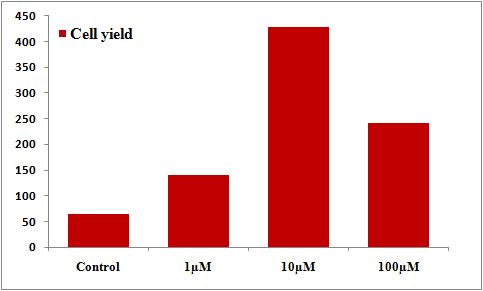
The effect of Cd has been characterized as dose dependent both in vitro and in vivo (Xiao et al., 2009; Geoffroy-Siraudin et al., 2012; Lafuente, 2003; Shao, 2014). The effects of cadmium on cultured Chicken hepatocytes were studied over a concentration range (1, 10 and 100μM) for 24 h (Figure )
On exposure to cadmium of different concentrations for 24h, it was found that at 1μM the cell count markedly decreased but at concentrations of 10μM and 100μM it was found that the cell count was similar but reduced when compared with the control.
ISSN 2348-313X (Print)
International Journal of Life Sciences Research ISSN 2348-3148 (online) Vol. 8, Issue 4, pp: (27-31), Month: October - December 2020, Available at: www.researchpublish.com
Figure: Effect of the different concentrations of Cd on the viability of chicken hepatocytes. At 24 h of culture, Chicken hepatocytes were treated with varied concentrations of Cd in the presence (1, 10 and 100μM). After 24 h, cell viability was evaluated using the Trypan Blue assay.
The liver is the largest internal organ in the body and the most important and vital biological functions related to metabolism take place in liver. Liver is the most frequently targeted organ in terms of cytotoxicity. The main achievements of this initial work are summarized in the following point:
Chicken hepatocyte proliferation has shown that cadmium had negative effects on hepatocyte proliferation (Figures).
[1] Abe, T., Hara, Y., Abe, Y., Aida, Y., Maeda, K., 1998.Serum or growth factor deprivation induces the expression of alkaline phosphatase in human gingival fibroblasts. Journal of Dental Research, 77(9), 1700–1707.
[2] Amara, S., Abdelmelek, H., Garrel, C., Guiraud, P., Douki, T., Ravanat, J.L., Favier, A., Sakly, M., Ben Rhouma, K., 2006. Influence of static magnetic field on cadmium toxicity: study of oxidative stress and DNA damage in rat tissues. J. Trace. Elem. Med. Biol. 20, 263–269.
[3] Anteau, M.J., Afton, A.D., Custer, C.M., Custer, T.W., 2007. Relationships of cadmium, mercury, and selenium with nutrient reserves of female lesser scaup (Aythyaaffinis) during winter and spring migration. Environ. Toxicol. Chem. 26, 515–520.
[4] Apel, K.; Hirt, H. Reactive oxygen species: Metabolism, oxidative stress, and signal transduction. Annu. Rev. Plant Biol. 2004, 55, 373–399.
[5] Baykov B.D., Stoyanov M.P., Gugova M.L. (1996). Cadmium and lead bioaccumulation in male chickens for high food concentrations. Environmental Toxicology and Chemistry. 54: 155-159.
[6] Berg, T., Aas, W., Pacyna, J., Uggerud, H.T., Vadset, M., 2008.Atmospheric trace metal concentrations at Norwegian background sites during 25 years and its relation to European emissions. Atmos. Environ. 42, 7494–7501.
[7] Berry, M.N., Friend, D.S., 1969. High-yield preparation of isolated rat liver parenchymal cells. A biochemical and fine structural study .J. Cell Biol. 43,506-520.
[8] Berry, M.N., 1976. The development of techniques for the preparation of hepatic parenchymal cell suspensions – a history and rationale. In use of isolated liver cells and kidney tubules in metabolic studies,J-31-138. Eds., Tager, J. l'1'.,Söling, H-D., Williamson, J. R.North Holland. Publishing Co., Amsterdam.
[9] inkowski, . ., awicka- apusta, ., zarek, ., trzy ewska, ., elsmann, M., 2013. istopathology of liver and kidneys of wild living Mallards Anasplatyrhynchos and Coots Fulicaatra with considerable concentrations of lead and cadmium. Sci. Total Environ. 450–451, 326–333.
ISSN 2348-313X (Print) International Journal of Life Sciences Research ISSN 2348-3148 (online) Vol. 8, Issue 4, pp: (27-31), Month: October - December 2020, Available at: www.researchpublish.com
[10] CAvuZZI,D. M., V. ROa~aMAN, and S. MARGOLIS. 1971. Simplified method for isolation of intact avian and rat liver parenchymal cells. Biochem. Biophys.Res. Commun. 45:421-429.
[11] Chen, Q.M., Bartholomew, J.C., Campisi, J., et al., 1998. Molecular analysis of H2O2-induced senescent-like growth arrest in normal human fibroblasts: p53 and Rb control G1 arrest but not cell replication. Biochem. J.332:43–50.
[12] Clark,D. G., Rornstad, R., and Kmz, J., 1974. Lipogenesis in rat hepatocytes. J. Biol. Chem. 249:2028-2036.
[13] Claus, T. H., S. J. PILraS, and C. R. Park. 1975. Stimulation by glucagon of the incorporation of U- 14Clabeledsubstrates into glucose by isolated hepa- tocytes from fed rats. Biochim. Biophys. Acta. 404:110-123.
[14] Cram,M. C., and Porter, J.W., 1973. Synthesis of fatty acid synthetase by isolated liver cells ob- tained from rats in different nutritional or hormonal states. Arch. Biochem. Biophys. 159:606-614.
[15] Daud, M.K., Ali, S.,Variath, M.T., et al., 2013.Differential physiological, ultra-morphological and metabolic responses of cotton cultivars under cadmium stress. Chemosphere, 93(10), 2593–2602.
[16] David, G. 8., Galbraith' W., Geyer, Koether, Pai,mer' N- F- and Ptxler, J. (1975). Improved isolation, separation and. Cytochemistry of living cells. Prog. Histochem. Cytochem.7, L-49-
[17] Dickens, B.F., Weglicki, W.B., Li, Y.S., Mak, I.T., 1992. Magnesium deficiency in vitro enhances free radicalinduced intracellular oxidation and cytotoxicity in endothelial cells. FEBS.Lett. 311, 187–191.
[18] Duffus, J.H., 2002. Heavy metals-a meaningless term? Pure Appl Chem. 74(5):793–807.
[19] Dunne MJ, Yule DI, Gallacher DV, Petersen OH., 1990. Effects of alanine on insulin-secreting cells: patch-clamp and single cell intracellular Ca2+ measurements. BiochimicaetBiophysicaActa (BBA)-Molecular Cell Research. 1055(2):157-64.
[20] Ekwall,, B., 1983b. Screening of toxic compounds in mammalian cell cultures. Ann. N.Y. Aead.Sci.,407, 64-77.
[21] El-Sokkary, G.H., Nafady, A.A., Shabash, E.H., 2010. Melatonin administration ameliorates cadmium-induced oxidative stress and morphological changes in the liver of rat. Ecotoxicol. Environ. Saf.73, 456–463.
[22] Finkel, T., Holbrook, N.J.,