
Vol. 8, Issue 2, pp: (37-51), Month: October 2020 - March 2021, Available at: www.researchpublish.com
ASTIGMATISM AMONG STUDENTS IN MEDICAL FACULTY OF UDAYANA UNIVERSITY
SHARLEEANNA MECDONAH
PROGRAM STUDI PENDIDIKAN DOKTER FAKULTAS KEDOKTERAN UNIVERSITAS UDAYANA, DENPASAR
Abstract: Astigmatism is frequently present during childbirth and may happen in blend with myopia or farsightedness. Astigmatism most of the time happens with other vision conditions like nearsightedness which is partial blindness (myopia) and hyperopia which is farsightedness. From the result of this study it can be concluded that the number of female and male who have astigmatism is almost similar. Study found that there are is no significant difference on gender when it comes to astigmatism. Most of the astigmatism students have some first degree relatives the diagnosis of astigmatism. Only a small percentage of students with astigmatism wear contact lenses. Most of the students with astigmatism spend more than 14 hours on phone, 7-14 hours on reading and less than 7 hours for watching TV.
Keywords: Astigmatism, students, (myopia), watching TV.
I. INTRODUCTION
1.1 Background
Astigmatism is a refractive error that happens when parallel rays of light entering the non accommoding eye are not centered around the retina. Astigmatism most of the time happens with other vision conditions like nearsightedness which is partial blindness (myopia) and hyperopia which is farsightedness. Together these vision conditions are alluded to as refractive mistakes since they influence how the eyes twist or "refract" light. The curvature of the cornea and focal point twists the light entering the eye keeping in mind the end goal to concentrate it absolutely on the retina at the back of the eye.1,2,3
In astigmatism, the surface of the cornea or lens has a somewhat different curvature. The surface of the cornea is shaped more like an egg rather than round like a ball, the vision can't concentrate light beams to a solitary point. Vision winds up noticeably out of center at any separation. Light beams refracted by this cornea are definitely not conveyed to a solitary point center, and retinal pictures from objects both far off and close are obscured and may show up widened or lengthened. This refractive blunder is called astigmatism.2
What's more, the shape of the focal point inside the eye can change, bringing about an expansion or reduction in astigmatism. This change habitually happens in adulthood and can go before the improvement of cataract. Now and again astigmatism may create following eye damage or eye surgery.1,2
Add up to astigmatism can be partitioned into three which one of it is corneal or keratometric astigmatism in which the cornea turns out to be continuously thinner and cone shaped. 2,3
This outcomes in a lot of astigmatism, which causes poor vision that can't be plainly amended with eyeglasses. Individuals with keratoconus more often than not require contact focal points for clear vision and in the end may require a
International Journal of Healthcare Sciences ISSN 2348-5728 (Online)
Page | 37 Research Publish Journals
International Journal of Healthcare Sciences ISSN 2348-5728 (Online)
Vol. 8, Issue 2, pp: (37-51), Month: October 2020 - March 2021, Available at: www.researchpublish.com
corneal transplant. Astigmatism additionally can be separated into lenticular astigmatism, and retinal astigmatism. Most astigmatism is corneal in source. 1,2 Lenticular astigmatism is a consequence of uneven curvature and contrasting refractive lists inside the crystalline focal point.2
1.2
PROBLEM STATEMENT
1.2.1 What is astigmatism?
1.2.2 What are the treatment of astigmatism?
1.3
AIM OF RESEARCH
To know and learn more about astigmatism among students in Faculty of Medicine in Udayana University.
1.4
BENEFIT OF RESEARCH
1.4.1
Academical benefits
To add research material into previous studies on this topic. This research will be a guideline for future studies.
1.4.2
Benefit for public
To build a generation of people whom are more concerned about their health especially in eye which is astigmatism. To educate the public the importance of astigmatism knowledge.
II. LITERATURE REVIEW
2.1
Definition
Astigmatism (from the Greek “a” meaning absence and “stigma” meaning point) is a refractive error (ametropia). Astigmatism happens when either the front surface of your eye (cornea) or the focal point, inside your eye, has crisscrossed curves. Rather than having one curve like a round ball, the surface is egg shaped. This causes blurred vision at all separations. 1,2,3,
Astigmatism is frequently present during childbirth and may happen in blend with myopia or farsightedness. Regularly it's not sufficiently articulated to require remedial activity. When it is, treatment alternatives are restorative focal points or surgery. 3
2.2 EPIDEMIOLOGY
Out of 3,475 selected peoples, 2,635 took place in the study in which the response rate was 75.8%. After the achieving the admittance and elimination criteria, 52% of females were analyzed from the data of 2124 peoples. It was 32.1 (±19.5) mean of the age of the peoples with the age between 1- 90 years old. The prevalence of astigmatism with cylinder power is more than 0.5 D was 32.2%. The prevalence of male was 31.1% and female was 33.3% for females. There was no any significant relationship between the gender and the prevalence of astigmatism according to the logistic regression. There is a increase of linearly with age of the prevalence of astigmatism. 2,4,7
The prevalence of astigmatism was 14.3% in the members more young than 15 years old and demonstrated a significant increment up to 67.2% in the members more than 65-years of age, hence, every 1-year increment in age improved the probability of astigmatism by 1.04 times (P < 0.001). As indicated by the consequences of this investigation, the commonness of astigmatism with barrel control more than 1, 2 and 3 D was 15.6% (95% CI: 14.0-17.1), 4.2% (95% CI: 3.3-5.0), and 1.5% (95% CI: 1.3-2.1), individually.4,7
2.3 ANATOMY
Vision is our dominant sense: some 70% of all the sensory receptors in the body are in the eyes and nearly half of the cerebral cortex is involved in some aspect of visual processing. 10
The adult eye is a sphere with a diameter of 2.5 cm (1 inch). Only the anterior one –sixth of the eye‟s surface is visible; the rest is enclosed and protected by a cushion of fat and the walls of the bony orbit. The fat pad occupies nearly all of the orbit not occupied by the eye itself. The eye is a complex structure and only a small portion of its tissues are actually involved in photoreception.10
Page | 38 Research Publish Journals
International Journal of Healthcare Sciences ISSN 2348-5728 (Online)
Vol. 8, Issue 2, pp: (37-51), Month: October 2020 - March 2021, Available at: www.researchpublish.com
2.3.1 Structure of the Eyeball
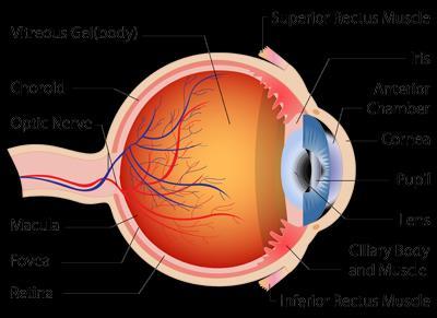
The eye itself, commonly called the eyeball, is a slightly irregular hollow sphere (Figure 2). Because the eyeball is shaped roughly like the globe of the earth, it is said to have poles. Its most anterior point is the anterior pole; its most posterior point, the posterior pole. Its wall is composed of 3 coats, or tunics, the fibrous, vascular and sensory tunics. Its internal cavity is filled with fluids called humors that help to maintain its shape. The lens, the adjustable focusing apparatus of the eye, is supported vertically within the internal cavity, dividing it into anterior and posterior segments or cavities.10
Figure 1: Internal structure of the eye (sagittal section)
In a normal eye, the cornea and lens focus light rays on the retina. 1
In astigmatism, images focus in front of and beyond the retina, causing both close and distant objects to appear blurry.1
Figure 2
2.4 ETIOLOGY
In spite of broad research, the correct reason for astigmatism is as yet not known one conceivable clarification of the etiology of astigmatism is that astigmatic refractive mistakes are hereditarily decided. Various examinations have been attempted to research the impact of hereditary qualities on astigmatic advancement. Be that as it may, the examinations
Page | 39 Research Publish
Journals
International Journal of Healthcare Sciences ISSN 2348-5728 (Online)
Vol. 8, Issue 2, pp: (37-51), Month: October 2020 - March 2021, Available at: www.researchpublish.com
into hereditary qualities and astigmatism display some clashing outcomes. Certain examines demonstrate some level of heritability of astigmatism and furthermore tend to support an autosomal overwhelming method of legacy. 2,3,7,4
Different investigations support a more grounded environmental impact. No doubt both hereditary and ecological components have parts in the advancement of astigmatism. The correct idea of these components is still not completely understood. Other conceivable causes incorporate mechanical connections between the cornea and the eyelids as well as the extraocular muscles or a visual input display in which astigmatism creates because of visual signs. 2,3,7 Astigmatism can be isolated into natural and obtain classes. Whenever gained, it perhaps optional to certain illness or an after effect of visual surgery or injury. Astigmatism has multifactorial etiologies and can emerge from the cornea, the focal point, and even the retina. 2,3,7
Corneal astigmatism typically represents the majority of the deliberate tube shaped refraction.The event of irregular astigmatism fluctuates from normal to surgically actuated causes. Examples of characteristic causes incorporate essential unpredictable astigmatism and optional irregular astigmatism caused by different corneal pathologies related with lifted pain, such askeratoconus or Sallzmann's nodular degeneration. 2,3,7
Cases of surgically actuated astigmatism incorporate pterygium evacuation, cataract extraction, disc and infiltrating keratoplasty, nearsighted keratomileusis, outspread and astigmatic keratectomy, PRK, and laser keratomileusis (LASIK). Different reasons for irregular astigmatism incorporate corneal injury and contamination. There are a few illnesses and disorders that are related with an expanded prevalence of astigmatism.2,3,7
2.5 CLASSIFICATION ANALYSIS
Astigmatic eyes
Simple myopic astigmatism: one meridian focuses light in front of the retina, the other on the retina
Simple hyperopic astigmatism: one meridian focuses light on the retina, the other theoretically behind the retina;
Compound myopic astigmatism: both meridians focus light in front of the retina
Compound hyperopic astigmatism: both meridians focus light theoretically behind the retina
Mixed astigmatism: one meridian focuses light in front of the retina, the other behind the retina.
2.6 CLINICAL
Non-astigmatic eyes
The emmetropic eye (normal): parallel rays of light focus sharply on the retina
The myopic eye: parallel rays of light are brought to a focus in front of the retina
The hyperopic eye: parallel rays of light would come to a focus behind the retina in the unaccommonded eye
A typical test to affirm the nearness of corneal irregularity is its successful rectification with a hard contact lens and the change of best corrected visual acuity.2,4
2.6.1 SIGNS AND SYMPTOMS
Astigmatism indications may include blurry vision or areas of distorted vision, eyestrain, headaches, squinting to try to see clearly or eye discomfort.Having these side effects may not really imply that a person has astigmatism, but rather it does show the requirement for a visit to an ophthalmologist for an entire eye exam.1,2
2.6.2 PREVENTION
Astigmatism can‟t be prevented because it is a hereditary and is present at birth.
2.7 DIAGNOSIS
An eye specialist will do an eye examination, and will utilize different instruments to quantify how eye concentrates light. Visual acuity will be measured, where there will be made a request to analyze letters on a separation outline to decide the clearness of the vision at a specific separations.
Page | 40 Research Publish Journals
International Journal of Healthcare Sciences ISSN 2348-5728 (Online)
Vol. 8, Issue 2, pp: (37-51), Month: October 2020 - March 2021, Available at: www.researchpublish.com
Likewise there will have the centering energy of the eyes tried with a progression of focal points put before the eyes. 1,2,5
The curvature and flow of cornea will be measured with a keratometer, and corneal geography might be utilized to give extra data about the state of the surface of the cornea. With these tests, eye specialist can analyze astigmatism and will have the capacity to decide the energy of glasses or contact focal points a person require for clear vision. He or she will likewise examine different choices for treatment, for example, astigmatism surgery. 1,2,5
Keratometric
Performed with a gadget called keratometer or ophthalmometer, keratometry is the estimation of a patient's corneal curvature. In that capacity, it gives an objective, quantitative estimation of corneal astigmatism, estimating the curvature in every meridian and also the axis. Keratometry is likewise useful in deciding the fitting of the contact lenses. The presence of unpredictable mires on attempted keratometry is trademark, here and there blocking estimation to an adjusted endpoint. This is a measure only of the anterior corneal surface abnormality, however it might be influenced by the tear film. 2,5
The significant restriction to keratometry is the suspicion that the cornea is a spherocylindrical surface with a single radius of curvature in every meridian, and with a major and minor axis isolated by 90 degrees. Furthermore, keratometry measures just four focuses roughly 3 mm separated and gives no data about the cornea central or peripheral to the focuses estimated. At last, gentle corneal surface abnormalities can cause mire distortion that blocks important estimation. By and large, the bend over the visual pivot is genuinely uniform, and this straightforward estimation is adequately spellbinding. In any case, keratometry isn't valuable for estimating corneas that are probably going to leave from spherocylindrical optics, as ordinarily happens in refractive medical procedure, keratoconus, and numerous other corneal irregularities.2,5
2.8 DIFFERENTIAL DIAGNOSIS
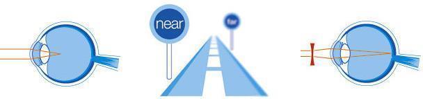
Both Myopia (short-sightedness) and Hypermetropia (long-sightedness) are regular eye conditions that mean light does not focus on the retina of the eye. The two conditions can be effortlessly redressed utilizing medicine glasses or contact lenses or in mild cases by laser eye surgery.2
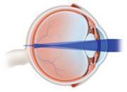
2.8.1 Myopia - Short Sighted
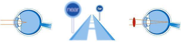
Myopia is the term used to define short sightedness. Light from a distant object forms an image before it reaches the retina. This could be because the eye is too long, or the cornea or crystalline lens is too strong.9
A myopic person has clear vision when looking at objects close to them, but distant objects will appear blurred.9Myopia is easily corrected at your local Vision Express optician using prescription glasses or contact lenses specifically designed to counteract the effect. A concave lens (minus powered) is placed in front of a myopic eye, moving the image back to the retina and clarifying the image.9 9
Figure 3: In a myopia eye, light from a distant object comes to a focal point before reaching the retina and then diverges again
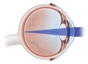
2.8.2 Hypermetropia (Hyperopia) - Long Sighted
Hypermetropia means long sight and is where the image of a nearby object is formed behind the retina. This could be because the eye is too short, or the cornea or crystalline lens does not refract the light enough.9
A hypermetropic person may have blurred vision when looking at objects close to them, and clearer vision when looking at objects in the distance. By placing a convex (plus powered) lens in front of a hypermetropic eye, the image is moved forward and focuses correctly on the retina.9
Page | 41 Research Publish Journals
(Online)
Vol. 8, Issue 2, pp: (37-51), Month: October 2020 - March 2021, Available at: www.researchpublish.com
Figure 4: In a hypermyopic eye, light from a near object comes to a focal point behind (past) the retina.
2.9 PROGNOSIS
Normally, gentle to direct astigmatism can be remedied with eyeglasses or contact lenses. While it used to be the situation that contact lens for astigmatism must be unbending contact focal points (Rigid Gas Permeables, likewise called GP lens), this is never again genuine. Presently, soft contact lens called toric contact focal points can rectify astigmatism. Yet, while delicate toric focal points might be suitable for a few, on the off chance that you have serious astigmatism, rigid contacts or glasses might be a superior alternative. 6,4,8
2.10 TREATMENT
2.10.1 NON SURGICAL PROCEDURES
CONTACT LENS
Before contact lens fitting, a visual history including past contact lens experience had to be acquired and an exhaustive restorative eye assessment must be performed . Patients should be aware that contact lens can be related with the improvement of visual issues, including microbial corneal ulcers that might be vision harm, and that overnight wear contact focal points is related with an expanded danger of ulcerative keratitis . 2,5,8
Abnormal astigmatism happens when by retinoscopy or keratometry, the primary meridians of the cornea, in general, are not opposite to each other. Although everyone's eyes have no less than a little measure of irregular astigmatism, this term is clinically utilized just for terribly irregular corneas, for example, those happening with keratoconus or corneal scars. Round and hollow display focal points can do little to enhance vision in these cases, thus for best optical adjustment, unbending contact focal points are needed. High astigmatic errors can be corrected adequately with inflexible gaspermeable and hybrid contact lenses. For instances of more better measures of corneal astigmatism, back surface toric contact lens design to limit corneal aspect and enhance centration. 2,5,8
Hand crafted delicate toric contact lens give another way to remedy high astigmatic refractive mistakes. These contact lens offer great centration when legitimately fitted, an adaptable wear plan, and enhanced relief in a few patients. Despite, suitable contact lens development is basic for agreeable wear and repair of corneal attachment 2,5,8
EYEGLASSES
Eyeglasses are the easiest and most secure methods for remedying a refractive error (astigmatism),therefore eyeglasses ought to be considered before contact lens or refractive surgery. A patient's eyeglasses and refraction should be assessed at whatever point visual side effects create. Patients with low refractive errors (low astigmatism) may not require adjustment, little changes in astigmatism revisions in asymptomatic patients are for the most part not prescribed. Full modification may not be required for people with customary astigmatism. Adults with astigmatism may not acknowledge full round and hollow modification in their first match of eyeglasses or in resulting eyeglasses if their astigmatism has been just mostly adjusted. Generally, generous changes in axis or power are not very much encountered 2,4,6
2.10.2 SURGICAL PROCEDURES
LASER
Laser is a surgical strategy that uses a laser to rectify myopia, farsightedness, and additionally astigmatism. In laser, a thin fold in the cornea is made utilizing either a microkeratome sharp edge or a femtosecond laser. The specialist overlap back the fold, at that point expels some corneal tissue underneath utilizing an excimer laser. The fold is then laid back set up, covering the range where the corneal tissue was evacuated. 10
With myopic individuals, the objective of laser is to level the as well soak cornea; with farsighted individuals, a more extreme cornea is wanted. Laser can likewise amend astigmatism by smoothing an unpredictable cornea into a more ordinary shape.10
International Journal of Healthcare Sciences ISSN 2348-5728
Page | 42 Research Publish Journals
9
International Journal of Healthcare Sciences ISSN 2348-5728 (Online)
Vol. 8, Issue 2, pp: (37-51), Month: October 2020 - March 2021, Available at: www.researchpublish.com
Initial step is to pick a decent laser specialist who can assess whether laser is appropriate for a person. Laser specialist will inspect a persons eyes to decide their well being, what sort of vision revision a person need, and how much laser removal (corneal tissue evacuation) is required. The specialist will likewise get some information about any well being conditions that may exclude you inside and out for laser surgery. 10
In the event that a person is not a possibility for laser, they may meet all requirements for another laser eye surgery, for example, PRK (like laser yet without the fold) or Laser. There are additionally non-laser vision rectification techniques. The medicine and eye structure will be considered to help figure out which method is best for you. 10
Laser is an outpatient methodology, so a person don't need to remain at the surgery focus overnight. The Laser specialist utilizes a PC to modify the laser for your specific solution. The person will be made a request to take a gander at an objective light for a brief span while the laser sends beats of light to easily reshape the cornea. The real laser surgery for the most part takes under five minutes.10
III. RESEARCH FRAMEWORK AND CONCEPT
3.1Research Frame Work
Astigmatism is a refractive error that happens when parallel rays of light entering the non accommoding eye are not centered around the retina. Astigmatism most of the time happens with other vision conditions like nearsightedness which is partial blindness (myopia) and hyperopia which is farsightedness. Astigmatism indications may include blurry vision or areas of distorted vision, eyestrain, headaches, squinting to try to see clearly or eye discomfort. Having these side effects may not really imply that a person has astigmatism, but rather it does show the requirement for a visit to an ophthalmologist for an entire eye exam. Astigmatism has multifactorial etiologies and can emerge from the cornea, the focal point, and even the retina. Both hereditary and ecological components have parts in the advancement of astigmatism. Students especially medical students have a possibility of having astigmatism since most of them already have another condition that get along with astigmatism such as myopia. Eyeglasses are the easiest and most secure methods for remedying a refractive blunder (astigmatism), therefore eyeglasses to be considered before contact lens or refractive surgery. Uncorrected astigmatism can causes poor vision that influence the quality of life. The study is to learn the characteristic of astigmatism in Faculty of Medicine Udayana University could be important to give an appropriate correction earlier and improve their quality of academic live.
3.2Research Concept
This research will be carried out in Faculty of Medicine Udayana University, Bali where medical student from batch 2016-2018 participate to answer a questionnaire to assess the characteristics and perform eye examination towards them. The figure 5 below shows the concept on how this research will be carried out.
Figure 5: Conception of Framework of The Study
Page | 43 Research Publish Journals
4.1
International Journal of Healthcare Sciences ISSN 2348-5728 (Online)
Vol. 8, Issue 2, pp: (37-51), Month: October 2020 - March 2021, Available at: www.researchpublish.com
IV. METHODS OF RESEARCH
Design of Research
This study is a descriptive study with cross-sectional approach where data‟s measurement only be done once. This study aim to determine the cases of astigmatism among medical student of Udayana University.
4.2Location and Duration
4.2.1Location
The location of this research is at the Faculty of Medicine Udayana University, Denpasar- Bali
4.2.2Duration
This research will be conduct for 2 months from 1 April 2019 – 1 May 2019
4.3Population and Sample of Research
4.3.1
Population Variability
Target population in this study is all medical students in Bali. Sample population that were used in this study are Udayana University‟s medical students. The batches involved will be students from batch 2016 - 2018 in Medical faculty. Sample of the study is the medical students from sample population that fulfill inclusion and exclusion criteria.
4.3.2
Criteria of The Subject
4.3.2.1 Inclusion Criteria
All the medical students of Udayana University who registered as batch 2016-2018 and voluntarily participated the study.
4.3.2.2Exclusion Criteria
Student who refused to participated on the study.
4.3.3Sample Size
Minimum sample size needed for the study calculated using a formula for cross-sectional study : N = Zα2 x P x Q (e)2
Explanation :
N = needed sample size
Zα2 = α value is determined. In this study, we used 0.05, so value of Zα2 is 1.96
P = Prevalence of astigmatism based on the epidemiological data described in chapter 1 which was 32.2% (0,32)
Q = 1-P = 1-0,32 = 0,68
e = needed level of absolute accuracy is 0.1. So, estimated population
N = (1.96)2 x 0,32 x 0,68 (0,1)2
N = 87,04
To anticipate drop out, 10% is added to become 87.04 + 10%(87.04) = 95.74 , as a whole number make the value as 96. So, 96 is a minimum sample size needed for this study. Those 96 samples that will be taken for this research from students of the Medical Faculty of Udayana University from batch 2016-2018.
4.3.4
Method of Sampling for Research
The sampling method used in this study is simple random sampling. Its benefit in giving all samples opportunity to participate the research.
Page | 44 Research Publish Journals
International Journal of Healthcare Sciences ISSN 2348-5728 (Online)
Vol. 8, Issue 2, pp: (37-51), Month: October 2020 - March 2021, Available at: www.researchpublish.com
4.4Research Variable
4.4.4Operational Definition
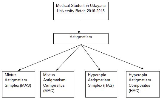
1 Astigmatism is a refractive error that happens when parallel rays of light entering the non accommoding eye are not centered around the retina. Its is classified as :
- Mixtus Astigmatism Simplex (MAS) : One or both principal meridians of the eye are nearsighted. Cylindrical -20D axis 180o
- Mixtus Astigmatism Compositus (MAC): One principal meridian is nearsighted and the other is far sighted. -100< cylindrical< -20D axis 180o
- Hyperopia Astigmatism Simplex (HAS): One or both principal meridians of the eye are farsighted. Cylindrical +20D axis 180o
- Hyperopia Astigmatism Compositus (HAC) One principal meridian is nearsighted and the other is far sighted. +100< cylindrical< -20D axis 180o
2 Demographical Data : Demographic data is the data that cover gender, age, and year of study. The data classified as:
Gender : Gender is divided become male and female
Age : Age is written as they real biological age and will be shown as a mean and standard deviation.
Year of Study : is the period of how long they have been studying in Faculty of Medicine Udayana University
3 Family History :. The history of first degree relative (mom, dad, brothers, sisters) that also experienced astigmatism.
4 Reading habit : is a habit of the student to read the study material everyday. It represent a period of hour they have spent to read them. It is classified as :
< 7 hours
7-14 hours
>14 hours
5 Mobile phone usage : the the amount of time that they have used to interact with their mobile phone screen everyday. It is classified as :
< 7 hours
7-14 hours
>14 hours
6 Television : is the amount of time the student have spent everyday to watch television. It is classified as :
< 7 hours
7-14 hours
>14 hours
4.5Research Instruments and Materials
A self made questionnaire consist of 2 parts. The first part aim to get demographical data which includes name, NIM, gender, age, and year of study. The second part aim to get the risk factor of refractive disorder among student which includes family history, screen time for mobile phone and television and also time for reading.
4.6Research Protocol
Protocol or procedure of this research is divided into the component which is the preparation stage, implementation stage and analysis stage.
1 Preparation Stage
In preparation stage, research proposal is done together with recommendation letter to be sent to Research & Development for collecting data. Letter is sent for approval of ethical clearance.
Page | 45 Research Publish Journals
International Journal of Healthcare Sciences ISSN 2348-5728 (Online)
Vol. 8, Issue 2, pp: (37-51), Month: October 2020 - March 2021, Available at: www.researchpublish.com
2 Implementation Stage
Implementation stage is carried out upon approval from Udayana University to carry out the research. In this stage, the questionnaire will be distributed to the students of medical faculty.
3 Analysis Stage
Analysis stage is carried after collecting the results of questionnaire. After the analysis, final arrangement of the report is done till it‟s finished.
4.7Data Analysis
Data collected from questionnaire will be coded in SPSS 21.0 and through a cleaning and coding. Cross tabulation is used to see a proportion of risk factor among the group. Because it is a descriptive study, chisquare and regression analytic are not necessarily to be done in this study. Data will be shown as tables and graphics.
V. RESULT
5.1 Demographic Characteristics of Sample
The study has been done by distributing the questionnaire consist of 9 questions related to demographic data and astigmatism. A total of 98 respondents are participated on this study, include 32 students from batch 2016, and 33 students from batch 2017 and 2018. All the respondents who participated in this study are wearing glasses and has been diagnosed with astigmatism by their optician. They reported no problem while answering question on the questionnaire.
Female participants are slightly higher than the male participants with the percentage of 59.2% and 40.8% respectively. No respondents aged 26-30 years old. More than half of them aged 20-25 years old, followed by 15-19 years old. The complete data can be seen in Table 5.1.
Table 5.1: Demographic Data of Samples
Demographic Data F (%) Gender
Male Female 40 (40.8) 58 (59.2) Age 15-19 years old 20-25 years old 26-30 years old
Batch 2016 2017 2018
5.2 Family History of Astigmatism
33 (33.7) 65 (66.3) 0 (0.0)
32 (32.6) 33 (33.7) 33 (33.7)
In this study, we found out that the vast majority of respondents have the first degree relatives (can be either parents or siblings) who also diagnosed with astigmatism, which is accounted for 96.9%. On the contrary, only 3.1% of respondents who denied any history of astigmatism among their first degree relatives.
First Degree Relative with Astigmatism
Yes No 0 20 40 60 80 100
Figure 5.2: Family History of Astigmatism
Page | 46 Research Publish Journals
International Journal of Healthcare Sciences ISSN 2348-5728 (Online)
Vol. 8, Issue 2, pp: (37-51), Month: October 2020 - March 2021, Available at: www.researchpublish.com
5.3 Contact Lenses Usage
This study found that about 90.8% of the participants are not wearing any contact lenses, whereas the other 9.2% wearing the contact lenses.
Contact Lenses Usage
0 10
5.4 Hours Spend on Reading in a Day
Figure 5.3: Contact Lenses Usage
In terms of the hours spend to read, majority of the respondent spend around 7-14 hours in a day to read, followed by less than 7 hours to read and more than 14 hours a day, with the percentage of 27.6%, 70.4%, and 2.0% respectively. Complete data can be seen in figure 5.4
Hours Spend on Reading
Figure 5.4: Hours Spend on Reading
5.5 Hours Spend on Using Mobile Phone in a Day
Students in this study spend a long hour on their mobile phone, as we can see in figure 5.4 where more than a half of participants (62.2%) spend more than 14 hours on their mobile phone. We also found that only 2.0% of participant who spend less than 7 hours a day on their phone.
Page | 47 Research Publish Journals
20 30 40 50 60 70 80 90 100 yes no
28% 70% 2%
<7hours 7-14hours >14hours
(Online)
Vol. 8, Issue 2, pp: (37-51), Month: October 2020 - March 2021, Available at: www.researchpublish.com
Hours Spend on Mobile Phones
<7hours 7-14hours >14hours
Figure 5.5 Hours Spend on Mobile Phone
5.6 Hours Spend on Watching TV in a Day
In terms of watching TV, most of the students only spend less than 7 hours to watch TV (52.0%), while there are 34.7% participants who spend 7-14 hours on watching TV. Only 13.3% of participants who spend more than 14 hours on watching TV. Complete data can be seen on Figure 5.6
Hours Spend on Watching TV
<7hours 7-14hours >14hours
Figure 5.6 Hours Spend on Watching TV VI. DISCUSSION
6.1 Discussion
This study found that there are is no significant difference on gender when it comes to astigmatism. The female gender in this study is only slightly higher in proportion compare to the male counterparts. This study result is supported by a study that found no correlation between gender and astigmatism (p=0.24) (Huang et.all, 2014). Another big study involving 26.007 samples also shows that there are no significant difference between the number of patients who suffer astigmatism in terms of the gender (Huang et.all, 2020). A study in 2011 shows that female gender can increase the risk of astigmatism around 1.66 times (95%CI= 1.19-2.31) whereas male gender can only increase the risk as much as 1.37 times (0.96-1.95) (Coudin et.all, 2011) Another study supported this result with the statement that females may have steeper corneas and shorter eyes based on the biometric measurement by keratometry, therefore female is more prone to the astigmatism morphologically (Bernado et.all, 2020). The different result has been found on the newest study that has been done by Wang et.all in 2020 which shows that male gender can increase the risk of getting astigmatism as much as 1.65
International Journal of Healthcare Sciences ISSN 2348-5728
Page | 48 Research Publish Journals
2% 36% 62%
52% 35% 13%
International Journal of Healthcare Sciences ISSN 2348-5728 (Online)
Vol. 8, Issue 2, pp: (37-51), Month: October 2020 - March 2021, Available at: www.researchpublish.com
times compare to the females, also give 2.2 times more risk to suffer high astigmatism. However, unfortunately the mechanism behind this still cannot be explained (Wang et.all, 2020). The studies that has been done 2 years priors shows that total vertical-horizontal axis and anterior cornea were significantly smaller in male eyes than in female eyes in all age groups (P=0269) indicating greater against-the-rule (ATR) astigmatism in male eyes (Hayashi et.all, 2018). Another study also shows similar result where ATR shift was faster for total corneal astigmatism than for anterior corneal astigmatism and it occurred earlier in men than in women (Kim, et.all, 2019). .
This study found that almost all students who suffer from astigmatism have at least one first degree relative who also diagnosed with astigmatism. It is believed that astigmatism can be inherited trough generation, due to the genetics effect to the corneal shape that can affect the astigmatism condition. A study that has been done involving 612 twins shows that there is a higher correlation between corneal astigmatism among monozygotic twins compare to the dizygotic twins (Dirani et.all, 2008). Previous publication also shows that not only astigmatism that can be hereditary, but also myopia. That study found that there was an increase risk of having myopia as much as 2.98 times if the sufferer have two parents with myopia (95%CI 2.47-3.24), which is prove the strong association between parental history of myopia and the myopia incidence (Lim et.all, 2014). That data can also be taken into consideration since various study stated that having myopia is also one of the strong risk factors of having astigmatism (Wang et.all, 2020). Another study also supported the fact that myopia can increase the risk of astigmatism where there is an increase 4.5 times risk to have astigmatism in children with myopia (Huang et.all, 2014). Some genetic study found that a single nucleotide polymorphism on chromosome 2p.13.3 especially in VAX2 gene play a significant role in the hereditary aspect of astigmatism (p=0.000). It is acquainted that VAX2 gene plays a pivotal role in the development of dorsoventral axis of the eye which is important in corneal curvature (Lopes et.all, 2013).
This study found that most students with astigmatism do not wear any contact lenses. Contac lenses are still less preferable to become one of the astigmatism corrector. Previous study shows that soft spehric, soft toric and rigid permeable contact lenses are still become the choice of less than a half astigmatism sufferer. It is caused by the lens insignificant effect towards astigmatism especially the higher degree astigmatism (Kurna et.all, 2010; Young et.all, 2011). One experimental study that has been done towards the astigmatism surferer shows that the spherical lenses failed to mask corneal toriciy durig topography while the thoric lenses can help with central neutralization also decrease corneal cylincer in low to moderate astigmatic eyes significantly (Kurna et.all, 2010). However, although toric lenses is available in quite a useful cylindrical range (powers +6.00 to 29.00 D, three cylinder powers, and 18 axes) which is able to cover 90% of astigmatism lens prescriptions, toric lens fitting requires many more prescriptions than spherical lenses, making it still become the first choice for many astigmatism sufferer (Young et.all, 2011).
This study found that most of the participants spend time on reading for about 7-14 hours per day. There has not been any study that describing the association between hours spend on reading and astigmatism incidence. However there is one study that assessing the correlation of reading posture, gaze angle and distance with the progression of myopia (Parssinen & Kaupinnern, 2016), where we know that myopia is closely related to astigmatism (Huang et.all, 2014; Wang et.all, 2020). That study found that reading while lying down can contribute to the worsen of myopia condition, as well as reading with the eyes more upward angle. Eventough there is no effect on hours spend reading in that study, it is believed that long term reading in an incorrect position could bring a refractive problem towards the eyes. Long term reading with the wrong gazing, especially upward gazing can potentially bring higher tensions in the extraocular muscles, which in turn increases the tensions and pressures on the bulbus oculi. It is also known that eyelid pressure influences the shape of the cornea and that this pressure depends on the angle of gaze (Parsinnen & Kauppinen, 2016).
This study found that most of the participants spend time on their mobile phone more than 14 hours in a day, on the contrary, participant spend time on TV less than 7 hours in a day. Screen exposure may have adverse health efects, particularly on eyes. Studies regarding the impact of excessive screen time on ocular-related health problems, have been largely confined to the ocular symptoms related to eye fatigue, myopia and low vision (Agarwal, et.al, 2013; Huang et.all, 2020) Unfortunately the study that established this relationship is still limited. A previous study found that screen exposure was associated with a higher risk of astigmatism amongst preschoolers and the risk increased as the daily duration and total years of exposure increased (Huang et.all 2020) More study on children shows that watching video or playing game in the mobile phone that require more than 2 continual hours of using smartphone is highly associated with the astigmatism incidence in children (p=0.000), it is caused by over accommodation that can lead to negartive effect towards maturation and emmetropisation of the corneal (Yadav & Suchita, 2018). Another study in adult population shows that smatphone usage more than 2 hours daily continuously is associated with higher likelihood of having myopia
Page | 49 Research Publish Journals
International Journal of Healthcare Sciences ISSN 2348-5728 (Online)
Vol. 8, Issue 2, pp: (37-51), Month: October 2020 - March 2021, Available at: www.researchpublish.com
and astigmatism, where there is an increase 2.18 times risk in every hours spend on using smartphone (95%CI=1.09-4.39) (Kim et.all, 2016). Eventhough the mechanism of screen exposure causing astigmatism is still unknown, there are several mechanism that possibly occurred Excessive screen time at close distances might cause excessive accommodation, thus leading to overworking of the ciliary muscles of the eyes and impacting the natural development of the crystalline lens, impacting its curvature. Alternately, looking at the screens up close may lead to changes in the shape of corneas, due to variations in the palpebral aperture and eye movements performed during the tasks, or the mechanical interactions between the cornea and the eyelids. Also, it may be that staring down at the screen increases pressure from eyelids on the cornea, which can result in increased corneal astigmatism (Agarwal et.all, 2013).
6.2 Study Limitation
This study is a descriptive study, thus it cannot determined the risk factors of astigmatism among participants. This study is also did not do any follow up towards the participants. The current pandemic situation and limited amount of time were made this study have a limited number of participants.
VII. SUMMARY
7.1 Conclusion
From the result of this study it can be concluded that the number of female and male who have astigmatism is almost similar. Most of the astigmatism students have some first degree relatives the diagnosis of astigmatism. Only a small percentage of students with astigmatism wear contact lenses. Most of the students with astigmatism spend more than 14 hours on phone, 7-14 hours on reading and less than 7 hours for watching TV.
7.2 Suggestion
Further longitudinal analytical study should be done to determine the risk factors of astigmatism. Multicenter study that involving more sample number can also help to make the study become more representative. Questionnaire in the electronic form such as google form perhaps can help with this current pandemic situation.
ACKNOWLEDGEMENT
I, Sharleeanna Mecdonah, medical student of Universitas Udayana with NIM no: 1602511233 would like to take this opportunity to express my grateful prayers and thanks to the Lord for aid in successfully completing my Elective Study entitled “Astigmatism”.
I would like to express my heartiest and outmost thanks to the following parties for assisting me in my paper work and also guiding me through this report writing as well as developing a good understanding of the topic with me:-
1. dr. Eka Sutyawan, Sp.M, as my first guidance lecturer, for guiding me throughout the research paper since the day 1.
2. dr. Jayanegara, Sp.M, as my second guidance lecturer, for correcting my mistakes along the way.
3. Dr. dr. Anak Agung Mas Putrawati Triningrat, Sp.M, as my examiner, for your time and consideration.
4. And also my dear beloved friends from Medical Faculty of Udayana University whom helped and supported me on this research paper.
All of outmost, I have gained tremendous amount of benefit through this whole journey. I had a chance to explore more about the field of topic I have researched on. In addition, I have learned how to take full responsibility of my own research paper. Overall I have learnt many wonderful and important facts about this topic.
Thank you.
REFERENCES
[1] “What Is Astigmatism?” (American Academy of OphthalmologySeptember 7, 2018) <https://www.aao.org/eyehealth/diseases/what-is-astigmatism> accessed July 14, 2019
[2] “Management of Astigmatism” (Dieudonne Kaimbo Wa Kaimbo (2013). - Dr. Michael Goggin (Ed.) <https://www.intechopen.com/books/astigmatism-optics-physiology-and-management/astigmatism>accessed March 8, 2018
[3] “Mutiara Medika” [2013] Keluhan Mata Silau pada Penderita Astigmatisma Dibandingkan dengan Miopia 127 <journal.umy.ac.id › index.php › article › download> accessed April 31, 2019
Page | 50 Research Publish Journals
International Journal of Healthcare Sciences ISSN 2348-5728 (Online) Vol. 8, Issue 2, pp: (37-51), Month: October 2020 - March 2021, Available at: www.researchpublish.com
[4] Hashemi H and others, “The Prevalence of Astigmatism and Its Determinants in a Rural Population of Iran: the „Nooravaran Salamat‟ Mobile Eye Clinic Experience” (Middle East African journal of ophthalmology2014) <https://www.ncbi.nlm.nih.gov/pmc/articles/PMC4005184/> accessed January 14, 2019
[5] Vinas MD and others, “Astigmatism Impact on Visual Performance” (2013) 90 Optometry and Vision Science 1430.
[6] Davis A and others, “The Relation Between Convergence Insufficiency and Astigmatism” (Investigative Ophthalmology & Visual ScienceJune 11, 2015) <https://iovs.arvojournals.org/article.aspx?articleid=2335341> accessed April 17, 2019
[7] Cotter SA, “Risk Factors for Astigmatism in Preschool Children” (https://www.aaojournal.org/article/S01616420(11)00610-5/pdf) < https://doi.org/10.1016/j.ophtha.2011.06.031> accessed March 22, 2019
[8] Xiao O and others, “Prevalence of Amblyopia in School-Aged Children and Variations by Age, Gender, and Ethnicity in a Multi-Country Refractive Error Study” (2015) 122 Ophthalmology 1924.
[9] Myopia, Hyperopia and Astigmatism: A Complete Review with View of Differentiation (International Journal of Science and Research (IJSR) )
[10] Marieb EN and Marieb & Hoehn, “Human Anatomy & Physiology, 10th Edition” (Pearson) <https://www.pearson. com/us/higher-education/program/Marieb-Human-Anatomy-Physiology-Plus-Mastering-A-P-with-e-Text-AccessCard-Package-10th-Edition/PGM239608.html> accessed March 31, 2019
[11] L. Huang et al., "Screen Exposure during Early Life and the Increased Risk of Astigmatism among Preschool Children: Findings from Longhua Child Cohort Study", International Journal of Environmental Research and Public Health, vol. 17, no. 7, p. 2216, 2020.
[12] R. McKean-Cowdin et al., "Risk Factors for Astigmatism in Preschool Children", Ophthalmology, vol. 118, no. 10, pp. 1974-1981, 2011.
[13] J. Kim et al., "Association between Exposure to Smartphones and Ocular Health in Adolescents", Ophthalmic Epidemiology, vol. 23, no. 4, pp. 269-276, 2016. Available: 10.3109/09286586.2015.1136652.
[14] L. Lim, Y. Gong, E. Ah-Kee, G. Xiao, X. Zhang and S. Yu, "Impact of Parental History of Myopia on the Development of Myopia in Mainland China School-Aged Children", Ophthalmology and Eye Diseases, vol. 6, p. OED.S16031, 2014.
[15] M. Lopes et al., "Identification of a Candidate Gene for Astigmatism", Investigative Opthalmology & Visual Science, vol. 54, no. 2, p. 1260, 2013.
[16] O. Pärssinen and M. Kauppinen, "Associations of reading posture, gaze angle and reading distance with myopia and myopic progression", Acta Ophthalmologica, vol. 94, no. 8, pp. 775-779, 2016.
[17] J. Wang et al., "Astigmatism in school students of eastern China: prevalence, type, severity and associated risk factors", BMC Ophthalmology, vol. 20, no. 1, 2020.
[18]G. Young, A. Sulley and C. Hunt, "Prevalence of Astigmatism in Relation to Soft Contact Lens Fitting", Eye & Contact Lens: Science & Clinical Practice, vol. 37, no. 1, pp. 20-25, 2011.
Page | 51 Research Publish Journals