International Journal of Healthcare Sciences ISSN 2348-5728 (Online)
Vol. 8, Issue 2, pp: (277-290), Month: October 2020 - March 2021, Available at: www.researchpublish.com

International Journal of Healthcare Sciences ISSN 2348-5728 (Online)
Vol. 8, Issue 2, pp: (277-290), Month: October 2020 - March 2021, Available at: www.researchpublish.com
Faruk Yusuf1, Yousif Safaa Aldeen Subhi Al Daghestani1, Manish Kumar1 , Indu Melkani1 , Dileep Singh Baghel1 , Bimlesh Kumar1*, Linu Dash1, Anupriya1 , Varimadugu Bhanukirankumar Reddy1, Amrik Singh2 , Amarish Kumar Sharma3
1School of Pharmaceutical Sciences, Lovely Professional University, Punjab, India
2School of Hotel Management & Tourism, Lovely Professional University, Punjab, India
3Scool of Bioengineering & Bioscience, Lovely Professional University, Punjab, India
Abstract: Diabetic nephropathy (DN) or diabetic kidney disease means the degeneration of kidney function which can be seen in chronic type 1 and type 2 diabetes mellitus patients. Diabetic nephropathy is the leading cause of chronic renal disease and a major cause of cardiovascular mortality. The development of the disease is known to occur in different type stages and is connected to glycemic and blood pressure control. Although, despite aggressive blood sugar control the prevalence of chronic kidney disease (CKD) I in diabetic people have not been seen in the last two decades: which has now lead to the recognition of new additional factors in the development. The two main risk factors for diabetic nephropathy are hyperglycemia and arterial hypertension, but the genetic susceptibility in both type 1 and type 2 diabetes is of great importance. Other risk factors are smoking, dyslipidemia, proteinuria, glomerular hyperfiltration, and dietary factors. The nutritional station of the patient is a very vital and changeable factor that may influence CKD procedure and the result. It directly comes from the traditional diet choices that the patient makes base on poor nutritional recognition. Dietary management of DN victims is challenging, as the factors of diet overload on the kidney function required to be maintained with malnutrition. The single best evidence-based therapy for diabetic nephropathy is therapy with a RAS-blocking medication. The introduction of antioxidant and anti-inflammatory agents to this field had also added a wealth of knowledge. However, many of these agents are still waiting for well-designed clinical studies to prove their beneficial therapeutic role.
Keywords: Diabetic Nephropathy(DN), renal disease, Nano formulation, Diabetes mellitus(DM)
Kidney disease is associated with increased total mortality and cardiovascular morbimortality in the general population and patients with T2DM (Type 2 diabetes) [47]. The prevalence of DN varies according to ethnicity: it is much higher in African-Americans, Asians, and Native-Americans than in Caucasians. African-Brazilians are very more susceptible to progress to end-stage renal disease(ESRD) than people of European ancestry but they appear to be a similar prevalence of micro or macroalbuminuria [2]. Diabetic nephropathy becomes the main cause of end-stage renal failure in the western part of the world [14]. Part of the most important clinical factor of diabetes is connected to chronic tissue complications. Histological changes in DN are identical in T1DM and T2DM. People with T1DM (Type 1 diabetes) and T2DM have equivalent rates of proteinuria, azotemia and ultimately End-stage kidney disease (ESKD) [44]. This was then estimated to grow to almost 550 million people by the year 2035. [1] The early morphological signs of renal damage include nephromegaly and a modified Doppler, but the degree of damage is best ascertained from proteinuria and GFR (Glomerular filtration rate). The core function of the glomerulus is the GFB (Glomerular Filtration Barrier), which has the unique capacity to selectively filter molecules and proteins by size and charge, thus maintaining the body‘s electrolyte and pH balance and blood homeostasis. [31] Renal disease is a major cause of morbidity and mortality for a patient with insulin-independent diabetes mellitus (IDDM) and is now becoming an increasingly important clinical problem in Non-
International Journal of Healthcare Sciences ISSN 2348-5728 (Online)
Vol. 8, Issue 2, pp: (277-290), Month: October 2020 - March 2021, Available at: www.researchpublish.com
insulin-dependent diabetes mellitus (NIDDM) [14]. A little term increase in hyperglycemia does not lead to serious clinical complications [1] The complication was traditionally thought to result from the interaction between hemodynamic and metabolic factors [44, 48]. The time and severity of the increase in glucose level is the major causative feature in starting organ damage. The average occasion of diabetic nephropathy is higher as of the first 10 to 20 years after diabetic onset. It takes up to almost 15 years for the blood vessels in the organs like the eyes, kidney, and nerves to get influenced. [1] It was examined that almost more than 20 and up to 40% of the diabetic victims will generate Chronic kidney disease (CKD) but which is based on the population, with a very vital number that generates ESKD needing renal replacement therapies like kidney transplantation. Diabetes which has no clinical sign of kidney destruction during the initial 20 to 25 years is very importantly less likely to cause major renal complication in 4/24 the late period of a victim's life. [1] The Joint Committee on Diabetic Nephropathy has revised its Classification of Diabetic Nephropathy (Classification of Diabetic Nephropathy 2014) in line with the widespread use of key concepts, such as the eGFR and CKD. [18]
DN has been didactically categorized into stages according to the UAE values. The values used to characterize these stages are described in the table.1. [2]
Table 1: Diabetic nephropathy stages based on urinary albumin excretion
Stages Urine with marked time (μg/min) 24 hours urine (mg/24h) Random urine sample
Albumin conc (mg/l) Albumin/creatinine Ratio(mg/g)
Normoalbuminuria <20 <30 <17 <30
Microalbuminuria 20 199 30 299 17 173 30 299
Macroalbuminuria > 200 > 300 >174 >300
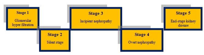
The concept of kidney disease is based on the presence of albuminuria and/or impaired renal function that lasts for more than three months [47]. Moreover, albuminuria predicts an increased risk of myocardial infarction, stroke, cardiovascular (CV) death, total death, and heart failure in patients with T2DM in comparison with non-diabetic patients [47]. The relationship between the mechanisms responsible for the development of albuminuria and renal impairment is not clear [47, 11]. DN is characterized by microalbuminuria and macroalbuminuria, and morphological changes such as glomerular thickening, interstitial fibrosis, the formation of nodular glomerulosclerosis and decreased endothelial cell fenestration [48]. Microalbuminuria is referred to as the risk factor for the generation of macroalbuminuria but not all the victims advance to this particular stage and some may even regress to Normoalbuminuria.[17,2] The first examination suggested that almost about 75 to 80% of type 1 diabetic victims with microalbuminuria would advance to proteinuria for over a period 6 of 14 years and up to date, the examination suggested that 30 to 45 % of the microalbuminuric victims will advance to proteinuria over 10 years follow up [2] Some of them will present regression to normoalbuminuria and this can lead to a result of more intensive sugar and blood pressure (BP) control process employed within the late decades than within the first studies. The regression of the microalbuminuria is more often among patients with a short duration of microalbuminuria, glycohemoglobin A1C (HbA1c) below 8%, systolic BP >115mmHg, and favorable lipid profile. [4] Unrestrained of the role as a prognostic feature for macroalbuminuria, the looks of microalbuminuria, reflecting a stage of generalized endothelial dysfunction, could be a dangerous factor for heart disease and mortality. [2] As seen in the microalbuminuria stage, there is no expectation of GFR to decline. Once the patient has developed macroalbuminuria, the GFR is expected to decline to 1.2 ml/min/month in type 1 DM. But this could be decreased by BP treatment. IN type 2 DM, the rate of the GFR decline is unpredictable or less predictable.[2] A mean decline of almost 0.5 ml/min/month has been examined, but in some victims, GFR may remain stable for a very long period. However, the greater GFR decline is related to many advanced diabetic glomerulopathies and poor metabolic control. Hence the progress of kidney disorder can be explained in different stages which is given in fig.1. Extensive studies in the Western world have demonstrated that diabetic patients with microalbuminuria have an increased risk of progression to overt proteinuria, and after some time, renal failure [11] Among the 1,478 patients with T2DM who had available data for calculating both GFR and albuminuria, patients with albuminuria without renal impairment and nonalbuminuric renal impairment were compared. The prevalence of albuminuria without renal impairment (albuminuria alone) (13.5%) and nonalbuminuric renal impairment (14.7%) was similar [47].
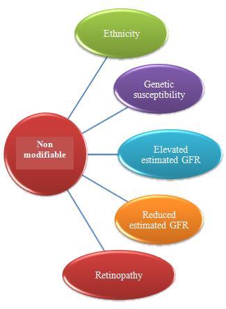
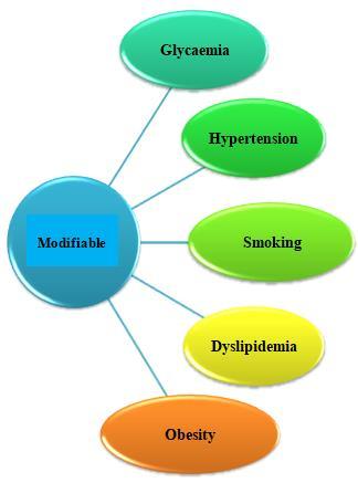
Many epidemiological studies demonstrate that ethnicity, family history, gestational diabetes, elevated blood pressure, dyslipidemia, obesity, and insulin resistance are the major risk factors of diabetic nephropathy [49]. There are two most basic risk factors of DN and which are hyperglycemia and arterial hypertension (which is a pressure that is acting very importantly on the walls of the arteries). [1] After the studies, it was seen that even with the presence of hyperglycemia and also elevated BP for a very long period, DN develops only in about 40% of the patients. So this outcome raised the concept that DN will be generated only in a susceptible subset of victims [2] and based on the family studies, it was confirmed that a genetic factor will also contribute to the development of DN in both type 1 and type 2 DM. Once the DN occurs, the advancement factors may act, which may favor the evolution to more advanced stages. There is proof that some of these factors which are involved in the generation of proteinuria are also similar to the loss of GFR, but others are special to each one of them (Fig.2.). [2]
1. Hyperglycemia: It is known to be the minor reason or risk factor for the development of macroalbuminuria in both type 1 and type 2 DM. In the presence of micro and macroalbuminuria, the role of metabolic control is less defined, although some studies have verified the decrease of blood glucose on GFR. And it has been shown that the transplantation of the pancreas reverses renal damage in type 1 DM victims with mild to progressed DN lesions. A decrease of about 1% HbA1c with a 37% decrease in the microvascular endpoint. [1, 2, 7, 10] Moreover, it was demonstrated that pancreas transplantation reversed renal damage in type 1 DM patients with mild to advanced DN lesions [7]. Recently a large trial also reinforced the importance of intensive treatment of DM to decrease the microvascular complications [10]. Hyperglycemia secondary to DN might induce major structural changes in the glomerulus, such as the thickening of the glomerular basement membrane, the impairment of glomerular cells within glomeruli [31]
2. Arterial Hypertension: It is the major or main reason for the development of DN and is also known to be the most relevant factor related to its progression. According to The United Kingdom Prospective Diabetes Study (UKPDS) analysis, is has shown every 10 mm Hg reduction in systolic BP is related to the reduction factor of microvascular complications with a small risk among patients with systolic BP >120 mm Hg.[2] Systolic and diastolic blood pressure was measured according to current guidelines. Age-specific normal values were obtained from the Task Force on Blood Pressure Control in Children and Adolescents. [49] The risk for cardiovascular events synergistically increases in patients with both diabetes mellitus and hypertension [1, 7, 2, 10]. The frequency of hypertension is elevated with the progression of renal damage in both diabetic and nondiabetic patients [7].
3. Proteinuria: This can advance to DN on its self. >2g/24 h of proteinuria shows a greater risk of EDRD. When the leakage of albumin is increased, it might probably include the glomerular damage but only through the activation of inflammatory cascades. It has been shown that the treatment of DN base on proteinuria is by decreasing urinary albumin secretion. [1, 2, 10] The prevalence of DN is high in patients with T2DM manifesting heavy proteinuria. Renal biopsy should be performed in diabetics in the atypical clinical scenario [42].
4. Smoking: It is another risk features of DN and can lead to advancement. Although some of the research did not confirm the observation. So it is advised that an individual should stop smoking in any of the stages of DN to avoid any kind of heart and cancer risk. [2] Inflammation is associated with increased resting energy expenditure (REE) in patients with chronic kidney disease. Oxidative stress, on the other hand, appears not to increase REE. Smoking is a common mechanism for generating oxidative stress and inflammation. Whether smokers have increased REE and if so, whether it is accounted for by the prooxidant and inflammatory state is not known [2, 6, 10].
International Journal of Healthcare Sciences ISSN 2348-5728 (Online)
Vol. 8, Issue 2, pp: (277-290), Month: October 2020 - March 2021, Available at: www.researchpublish.com
5. Dyslipidemia: High serum cholesterol has been seen to be a great risk factor for GFR loss of macroalbuminuria in type 1 DM. An increase in the serum cholesterol level leads to the development of DN in type 2 DM while in type 1, an increase in patient serum triglycerides, total and LDL cholesterol are all related to macro and microalbuminuria. [1, 3, 10]
HDL and LDL cholesterol were treated as independent, continuous variables, whereas for macroalbuminuria, dyslipidemia was treated as a dichotomous variable. [49]. Increasing evidence suggests that obesity is a risk factor for diabetes and chronic kidney diseases. As a marker of obesity, high body mass index (BMI) has been reported to be related to diabetic nephropathy (DN) and ESRD [8].
Figure 2: Risk factors that contribute to the development of DN [44]
6. Others: Oxalosis is a metabolic disorder characterized by the deposition of oxalate crystals in various organs including the kidney. Whereas primary forms result from genetic defects in oxalate metabolism, secondary forms of oxalosis can result from excessive intestinal oxalate absorption or increased endogenous production, e.g. after intoxication with ethylene glycol. Acute secondary oxalate nephropathy due to excessive dietary intake of oxalate may lead to acute renal failure in patients with a preexisting renal disease like mild diabetic nephropathy. Attention should be paid to special food behaviors when reasons for acute renal failure are explored. [46]
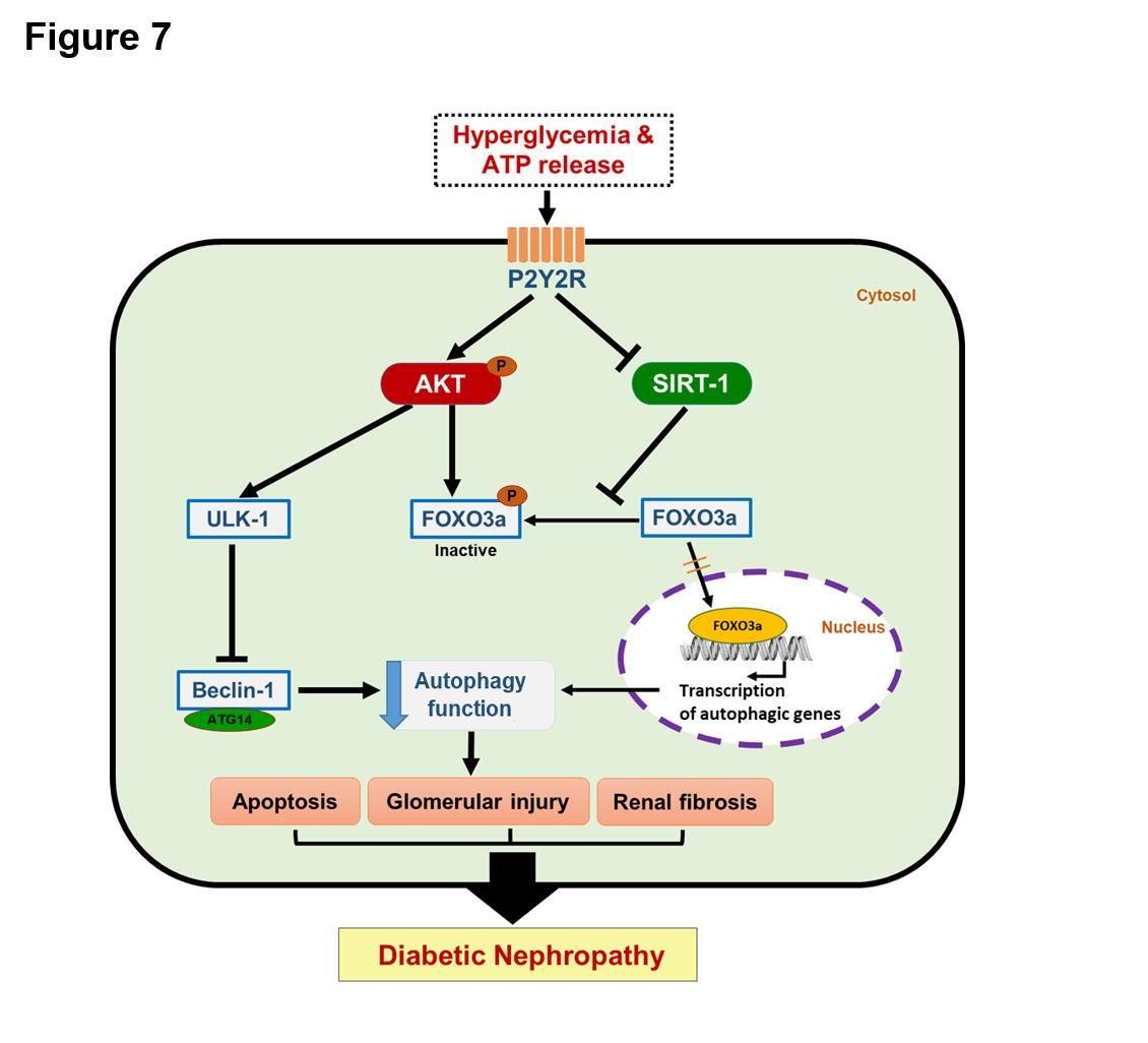
P2Y2R deficiency attenuates diabetic nephropathy [40]. Activation of purinergic receptors including P2Y2R has been associated with the pathogenesis of renal diseases such as polycystic kidney and glomerulonephritis. [52] However, the role of P2Y2R and its precise mechanisms in DN remain unknown. We hypothesized that P2Y2R deficiency may play a protective role in DN by modulating the autophagy signaling pathway. [50] Hyperglycemia and Adenosine triphosphate (ATP) release were induced in wild type DN mice and positively correlated with renal dysfunction. [51] Conversely, P2Y2R knockout markedly attenuates albuminuria, podocytes loss, and the development of glomerulopathy, renal tubular injury, apoptosis, and interstitial fibrosis induced by DN.[53] These protective effects were associated with inhibition of Protein kinase B (AKT)-mediated Forkhead box protein (FOXO3a) phosphorylation and induction of FOXO3a-induced autophagy gene transcription.[59]Furthermore, inhibitory phosphorylation of ULK-1 was decreased, and the downstream Beclin-1 autophagy signaling was activated in P2Y2R deficiency. An increased SIRT-1 and FOXO3a expression in P2Y2R deficiency also enhanced autophagy response, thereby ameliorating renal dysfunction in DN. [58, 60] P2Y2R contributes to the pathogenesis of DN by impairing autophagy and serves as a therapeutic target for treating DN. P2Y1R deficiency protected against renal capillary loss, fibrosis, and apoptosis in a crescentic glomerulonephritis model. [58] Subsequent analysis showed that the decrease of podocyte number is a good predictor of progression of albuminuria. [22]
P2Y2R activation via hyperglycemia and extracellular ATP increases AKT phosphorylation, and the increased AKT activity promotes inhibitory ULK-1 phosphorylation that down-regulates autophagy through inhibition of Beclin-1 and ATG14 complex required for phagophore formation, resulting in renal apoptosis, glomerular injury, and interstitial fibrosis. Also, the increased phosphorylation of FOXO3a by AKT activity and the inhibited SIRT-1(Selective Internal Radiation Therapy) activity reduced autophagy response by downregulating transcription of autophagy genes. Conversely, P2Y2R knockout mice have reduced AKT phosphorylation and increased FOXO3a and SIRT-1 expression to rescue autophagy response and attenuate the progression of DN. [40, 54 – 57]
The candidate anti-diabetic drug, [RuII(H3ucp)Cl(PPh3)](H4ucp=2,6-bis-((6-amino-1,3- dimethyl-uracil imino)methylene)pyridine exhibited Renoprotective effects in HFHC diet-induced pre-diabetic rats by reducing blood glucose, fluid intake, and urinary output while ameliorating oxidative stress and antioxidant defense enzymes, reducing aldosterone and KIM-1 concentrations.[37].
An evolutionarily conserved biologic process that epithelial cells were transdifferentiated into motile mesenchymal cells, is involved with the wound healing and stem cell behavior, and the progression of fibrosis and cancer. In the process of EMT, their epithelial characteristics are disappeared, including the loss of cell-cell junctions and apical-basal polarity, cytoskeleton reorganization, and are with high production of extracellular matrix (ECM), indicating the promoted migratory and invasive ability [72, 73] Additionally, it is widely recognized that EMT is a key process that contributes to DN, resulting to tubulointerstitial fibrosis, and even ESRD. It was uncovered that epithelial cells were in the process of EMT in kidney fibrosis tissues [74], and the increased serum creatinine level and the severity of interstitial damage are associated with the number of mesenchymal marker-positive epithelial cells [75,76]. Furthermore, mesenchymal-like αsmooth muscle actin (α-SMA)-positive epithelial cells were identified in the kidney tissues from DN patients, indicating that epithelial cells are undergoing EMT in the kidney in DN
International Journal of Healthcare Sciences ISSN 2348-5728 (Online)
Vol. 8, Issue 2, pp: (277-290), Month: October 2020 - March 2021, Available at: www.researchpublish.com
After exposure to pathologic stimuli (e.g., high glucose, advanced glycation end-products (AGEs), and Reactive oxygen species. (ROS), the renal cells are triggered by several signaling cascades that facilitate chronic inflammation. AGEs interact with Receptor for AGEs (RAGE) and activates NADPH oxidase (Nox2) at Endoplasmic reticulum (ER) and cell membranes, as well as, Nox4 at the ER membrane. Apart from AGEs, High mobility group box (HMGB1) and S100/calgranulins are other ligands for RAGE. Protein kinase C (PKC) is also upstream of Nox. To exert a proinflammatory effect, Nox induces ROS production and Inhibitory kappa B kinase (IKK)-dependent Jun N-terminal kinase (NF-κB) activation. Meanwhile, Nox activation leads to ER stress. In response to ER stress, UPR proteins, which are composed of PKR-like eukaryotic initiation factor 2α kinase (PERK), activating transcription factor (ATF) 6, and Inositol requiring enzyme (IRE1) enhance the adaptive pathway to maintain ER proteostasis via ATF4, ATF6, and X-box binding protein (XBP1), respectively. In prolonged ER stress, ATF4 induces CHOP (enhancer-binding protein homologous protein) to discard dysfunctional proteins in the apoptotic pathway. UPR transducers interfere with lipogenesis and gluconeogenesis genes. ER stress alters insulin signaling and causes insulin resistance by IRE1/JNK (Jun N-terminal kinase) signaling and JNK-dependent IRS-1(Insulin receptor substrate) serine phosphorylation. Also, IRS-1 can be downregulated by PKC (Protein kinase C). In the end, AGE/RAGE/Nox signaling-mediated ER stress causes kidney inflammation, glomerular hypertrophy, podocytes injury, proteinuria, and eventually renal fibrosis. [3]. AGE/RAGE/Nox signaling triggers ER stress in DN. Targeting at critical molecules of this axis, including RAGE, Nox, and UPRtransduced proteins (i.e., PERK, ATF6, and IRE1), will be beneficial in treating these diseases and other relevant kidney disorders. Specific miRNAs and lncRNAs can rescue those pathological events by interfering at particular targets, highlighting ncRNAs as a new layer of therapeutic strategies against DN. We prospect that ncRNAs listed in this review, with the exosomal delivery system, will pave the way for the ncRNA-based therapeutic approach in kidney diseases related to AGE/ RAGE/Nox/ER stress cascade. [3]
Pathogenesis of DN is a complex and multifactorial process that appears to be a combination of inflammation, oxidative stress, and epigenetic factors [79, 80]. However, the role of glomerular hyperfiltration early in the course of diabetes lies at the heart of the pathophysiology of DN. Vallon et al., established this through the single-nephron GFR studies, which showed that the dysregulated tubuloglomerular feedback (TGF) in diabetic rats leads to a reduced tone in afferent arteriole which causes glomerular hypertension [81]. Hence, it is important to understand that a) juxtaglomerular apparatus fine-tunes the glomerular filtration and is an important homeostatic mechanism that regulates glomerular pressures b) adenosine is an important mediator of TGF; and c) in diabetes, the activity of sodium-glucose transporter (SGLT-2) is increased, leading to enhanced absorption of sodium in the proximal convoluted tubule.
This results in a decreased distal delivery of sodium, which in turn signals the TGF mechanism to decrease the afferent arteriolar tone. Ultimately, this leads to glomerular hyperfiltration, thus begins the pathologic changes of DN [20] The mechanism of decreased distal sodium delivery leading to glomerular hyperfiltration was confirmed by the phenomenon called the ―salt paradox‖. In this phenomenon, when rats in early phases of diabetes were salt-restricted, they absorbed most of the sodium in the proximal convoluted tubule that lowered the delivery of sodium to the distal convoluted tubule. This decreased local adenosine levels resulting in the dilation of the afferent arteriole. We refer readers to Vallon et al. for an excellent review of these mechanisms [81]
Many of the therapeutic modalities are already approved by clinical trials that proved the safety and efficacy of these modalities. Others are still waiting for this approval. These 2 categories will be discussed under ‗‗Approved treatment‖ and ‗‗Potential therapeutic modalities‖ respectively [5] A multidisciplinary approach for management [44] for DN is recent approach. DN and ESKD remain a significant clinical problem. In diabetic individuals in whom no single treatment can halt the progression of DN, a multidisciplinary approach delivered by a multi-professional team remains the most sensible strategy. The main goals are Reno-protection and optimization of treatment using ACE inhibitors or ARBs increasing to the maximum dose, restrict salt intake with a diuretic, RAS blockers, and calcium-channel blocker. In
International Journal of Healthcare Sciences ISSN 2348-5728 (Online)
Vol. 8, Issue 2, pp: (277-290), Month: October 2020 - March 2021, Available at: www.researchpublish.com
addition to this, statins according to requirement, fibrates to lower cholesterol. Apart from this, all the risk factors should be taken care of. There are several methods of treating DN, which include the use of Drugs (table.2.), enzymes/Antibodies, Nano formulations, etc.
Vascular endothelial growth factor B (VEGF-B) is a crucial factor in promoting abnormal lipid metabolism as a part of the vascular endothelial growth factor family[61]VEGF-B can specifically up regulate fatty acid transport proteins (FATPs) and increase ectopic lipid deposition, which ultimately leads to insulin resistance by binding to the membrane receptors neuropilin-1 (NRP-1) or vascular endothelial growth factor receptor (VEGFR)-1[62]. Recent studies have reported that VEGF-B can also promote lipid accumulation in podocytes and directly result in DN by reducing podocytes' insulin sensitivity[33] Therefore, an anti-VEGF-B monoclonal antibody is expected to treat DN by simultaneously regulating lipid metabolism and improving insulin resistance. However, there is no definitive evidence that reducing VEGF-B signaling could alleviate oxidative stress and inflammatory responses, which are equally the leading causes of DN [63 - 65] Antagonism of VEGF-B could efficiently ameliorate DN by reducing renal lipotoxicity [39]. IL-22 (Interleukin) can be produced by CD4 T (cluster of differentiation 4) helper subtypes (such as Th17 cells and Th22 cells) and innate lymphoid cells (ILCs, such as natural killer cells, dendritic cells, and macrophages) as one of the interleukins10 cytokine superfamilies‘[66] IL-22 mainly binds to the receptors IL-22R1 and IL-10R2, and then specifically activates the intracellular JAK/STAT3(Janus kinase/signal transducers and activators of transcription) signaling pathway, thereby mediating anti-inflammatory and other biological effects [67 - 69] IL-22 can significantly reduce inflammatory cytokines (such as IL-1b) and simultaneously protect renal function in DN mice.[70] Moreover, studies have also indicated that IL22 can alleviate renal injury caused by renal ischemia-reperfusion.[71] Thus, we hypothesize that combining VEGF-B antibody and IL-22 can alleviate renal lipotoxicity and insulin resistance as well as ameliorate inflammatory responses.[33]
Table 2. Drugs for the treatment of DN
Drug Name Uses Side Effects Reference Lisinopril ACE Inhibitors Dizziness, hypotension, hyperkalemia, increased blood urea nitrogen
2,89
Enalapril Dizziness Trandolapril Fever, sweating Ramipril Dizziness, Quinapril Fever, sweating Canagliflozin SGLT-2 inhibitor loss of appetite 20 Perindopril ACE Inhibitors Dizziness, sweating 2,89 Moexipril Fever, sweating Benazepril Dizziness, Losartan AR Blockers Fever, sweating Dizziness Irbesartan Fever, sweating Dizziness Fosinopril ACE Inhibitors Dizziness, Captopril Fever, sweating Quinapril Fever, sweating Pyridoxamine nausea, vomiting, diarrhea 90,2 R-147176 Nausea, Vomiting Benfotiamine Upset stomach Nausea Dizziness Alagebrium (ALT-711) None 90,2 Kremezin (AST-120) Retards progression of CKD Appetite loss, Nausea, and Vomiting
Pentoxifylline Prevents the enhanced expression of cytokine and IL Nausea, vomiting, gas, belching Ruboxistaurin Reduces the actions of VEGF Dyspepsia. Aliskiren AR Blockers Nausea, Vomiting, Diarrhea. Sulodexide Decreasing the concentration of glycosaminoglycans Dyspepsia,Heartburn,Dizziness
Naproxen Anti-inflammatory Drug Nausea and Vomiting. 6,90 Ibuprofen Diclofenac Aspirin
International Journal of Healthcare Sciences ISSN 2348-5728 (Online)
Vol. 8, Issue 2, pp: (277-290), Month: October 2020 - March 2021, Available at: www.researchpublish.com
Nanomedicine and nano delivery systems are a relatively new but rapidly developing science where materials in the nanoscale range are employed to serve as means of diagnostic tools or to deliver therapeutic agents to specifically targeted sites in a controlled manner. Nanotechnology offers multiple benefits in treating chronic human diseases by site-specific, and target-oriented delivery of precise medicines. Recently, there are several outstanding applications of nanomedicine (chemotherapeutic agents, biological agents, immunotherapeutic agents, etc.) in the treatment of various diseases. [84] Nanotechnology is the measurement and manipulation of material at the level of 1– 100 nanometres (nm), 1 nm being 10−9 or one billionth of a meter (Nanos, Greek, ‗dwarf‘). When this science is applied specifically to the problems of medicine, it is called ‗nanomedicine‘ [85] The current review, presents an updated summary of recent advances in the field of nanomedicines and nano based drug delivery systems through comprehensive scrutiny of the discovery and application of nanomaterials in improving both the efficacy of novel and old drugs (e.g., natural products) and selective diagnosis through disease marker molecules. The opportunities and challenges of nanomedicines in drug delivery from synthetic/natural sources to their clinical applications are also discussed. In addition, we have included information regarding the trends and perspectives in Nanomedicine area. [84]
Numerous biopolymeric materials are utilized in drug delivery systems. These materials are:
1. Chitosan is a linear polysaccharide composed of randomly distributed β-(1→4)-linked D-glucosamine (deacetylated unit) and N-acetyl-D-glucosamine (acetylated unit). It is made by treating the chitin shells of shrimp and other crustaceans with an alkaline substance, such as sodium hydroxide.
2. Alginate is another biopolymeric material that has been used as drug delivery is alginate. This biopolymer presents final carboxyl groups, being classified as anionic mucoadhesive polymer, and presents greater mucoadhesive strength when compared with cationic and neutral polymers [91, 92].
3. Xanthan gum (XG) is a high molecular weight heteropolysaccharide produced by Xanthomonas campestris. It is a polyanionic polysaccharide and has good bioadhesive properties. Because it is considered non-toxic and nonirritating, xanthan gum is widely used as a pharmaceutical excipient [93].
4. Cellulose and its derivatives are extensively utilized in the drug delivery systems basically for modification of the solubility and gelation of the drugs that resulted in the control of the release profile of the same [94].
5. Liposomes They were discovered by Alec Bangham in 1960. Liposomes are used in the pharmaceutical and cosmetics industry for the transportation of diverse molecules and are among the most studied carrier system for drug delivery. Liposomes are an engrained formulation strategy to improve drug delivery. They are vesicles of spherical form composed of phospholipids and steroids usually in the 50–450 nm size range [95].
6. Polymeric micelles are nanostructures made of amphiphilic block copolymers that gather by themselves to form a core-shell structure in the aqueous solution. The hydrophobic core can be loaded with hydrophobic drugs (e.g. camptothecin, docetaxel, paclitaxel), at the same time the hydrophilic shell makes the whole system soluble in water and stabilizes the core. Polymeric micelles are under 100 nm in size and normally have a narrow distribution to avoid fast renal excretion, thus permitting their accumulation in tumor tissues through the EPR effect. [96]
7. Dendrimers are highly bifurcated monodisperse, well-defined, and three-dimensional structures. They are globularshaped and their surface is functionalized easily in a controlled way, which makes these structures excellent candidates as drug delivery agents[102-104].
8. Inorganic nanoparticles include silver, gold, iron oxide, and silica nanoparticles are included. Studies focused on them are not as many as there are on other nanoparticle types discussed in this section although they show some potential applications.
9. Nanocrystals are pure solid drug particles within the 1000 nm range. These are 100% drugs without any carrier molecule attached to them and are usually stabilized by using polymeric steric stabilizers or surfactants
10. Metallic nanoparticles in recent years, the interest in using metallic nanoparticles has been growing in different medical applications, such as bioimaging, biosensors, target/ sustained drug delivery, hyperthermia, and photoablation therapy [101, 100].
International Journal of Healthcare Sciences ISSN 2348-5728 (Online)
Vol. 8, Issue 2, pp: (277-290), Month: October 2020 - March 2021, Available at: www.researchpublish.com
To mimic native insulin activity, materials have been developed that encapsulate insulin, glucose oxidase, and catalase for glucose-responsive insulin delivery. A major challenge, however, has been achieving the desired kinetics of both rapid and extended-release. Here, we tune insulin release profiles from polymeric nanoparticles by altering the degree of modification of acid-degradable, acetylated-dextran polymers. Nanoparticles synthesized from dextran with a high acyclic acetal content (94% of residues) show rapid release kinetics, while nanoparticles from dextran with a high cyclic acetal content (71% of residues) release insulin more slowly. [82]
Ursodeoxycholic acid (UDCA) is a secondary hydrophilic bile acid, metabolized in the gut, by microbiota. UDCA is currently prescribed for primary biliary cirrhosis and recently has shown βcell-protective effects, which suggests potential antidiabetic effects. Thus, this study aimed to design targeted delivery microcapsules for oral uptake of UDCA and test its effects in type 1 diabetes (T1D). UDCA microcapsules brought about a reduction in elevated blood glucose, reduced inflammation, and altered concentrations of the primary bile acid chenodeoxycholic acid, and the secondary bile acid lithocholic acid, without affecting the survival rate of mice. [86]
3. Oral administration of angiotensin-(1–7) ameliorates
It has been shown that activation of the angiotensin (Ang)-(1–7)/Mas axis of the renin-angiotensin system leads to improved glucose uptake. In the recent study, it was intended to evaluate, whether this effect could be exploited therapeutically. It was first confirmed that Ang-(1–7) improves insulin signaling and glucose uptake in vitro in cultured cardiomyocytes. An oral Ang-(1–7) formulation reverses hyperglycemia and its consequences in an animal model of DM2 and represents a novel therapeutic option for the treatment of DM2 and other cardio-metabolic diseases [1]
Metformin HCl belongs to class biguanides. It is considered the first-line agent for the treatment of patients newly diagnosed with type 2 diabetes as a supplement to diet and exercise, despite its drawbacks of short biological half-life of 1.5–1.6 h and daily requirement of 1.5– 3 g/day with an absolute bioavailability of 50–60% when administered orally. Metformin HCl niosomal gel (applied transdermally every 2 days) has a better sustained anti-diabetic effect as compared to the orally given metformin HCl tablets (given daily) which also helped to alleviate the problems associated with oral metformin HCl tablets. Also, one of the main results achieved from this is the capability of using metformin HCl niosomal patches for helping the enhancement and improvement of wound healing that represent a vital problem for diabetic patients. Hence, the optimized transdermally applied niosomal formulations represent a promising carrier for metformin HCl to provide a better lifestyle for diabetic patients. [87] The combined synergistic effects of Metformin and biopolymers are due to their corresponding mechanism to enhance glucose uptake, minimized the adverse effects during diabetic therapy[88]
DN is a global concern and its treatment is required to consider from the time of initiation of diabetes. Patients must consider all the restrictions of diet and they must take the drug on time. It is also very important that they must be aware of all the complications associated with it. The advancement of technology used in drug formulations and its development indicates that scientists are working very hard to treat this. These nanoformulation aspects are very successful to control diabetes.
The authors are thankful to Lovely Professional University for providing a suitable environment to complete this manuscript.
Conflict of interests
None
International Journal of Healthcare Sciences ISSN 2348-5728 (Online)
Vol. 8, Issue 2, pp: (277-290), Month: October 2020 - March 2021, Available at: www.researchpublish.com
1. Sulaiman MK. Diabetic nephropathy : recent advances in pathophysiology and challenges in dietary management. DiabetolMetab Syndr. Published online 2019:1-5. doi:10.1186/s13098-019-0403-4
2. Zelmanovitz T, Gerchman F, Balthazar APS, Thomazelli FCS, Matos JD, Canani LH. Diabetology & Metabolic Diabetic nephropathy. 2009;17:1-17. doi:10.1186/1758-5996-1-10
3. Pathomthongtaweechai N, Chutipongtanate S. Biomedicine & Pharmacotherapy AGE / RAGE signaling-mediated endoplasmic reticulum stress and future prospects in non-coding RNA therapeutics for diabetic nephropathy. Biomed Pharmacother. 2020;131(August):110655. doi:10.1016/j.biopha.2020.110655
4. Gunzler D, Bleyer AJ, Thomas RL, et al. Diabetic nephropathy in a sibling and albuminuria predict early GFR decline : a prospective cohort study. Published online 2013:1-9.
5. Sharaf UAA, Din E, Salem MM, Abdulazim DO. Diabetic nephropathy : Time to withhold development and progression - A review. JAdvRes. 2017;8(4):363-373. doi:10.1016/j.jare.2017.04.004
6. Agarwal R. Smoking , oxidative stress and inflammation : Impact on resting energy expenditure in diabetic nephropathy. 2005;9:1-9. doi:10.1186/1471-2369-6-13
7. Ito H, Mifune M, Abe M, et al. Hypertension resistant to antihypertensive agents commonly occurs with the progression of diabetic nephropathy in Japanese patients with type 2 diabetes mellitus : a prospective observational study. Published online 2012:1-6.
8. Chen H, Shen W, Ge Y, Zhang Y, Xie H, Liu Z. The relationship between obesity and diabetic nephropathy in China. Published online 2013.
9. Misra PS, Szeto SG, Krizova A, Gilbert RE, Yuen DA. Renal histology in diabetic nephropathy predicts progression to end-stage kidney disease but not the rate of renal function decline. Published online 2020:1-12.
10. Navarro-gonzález JF, Mora-fernández C, Fuentes MM De. in the pathogenesis of diabetic nephropathy. Nat Publ Gr. 2011;7(6):327-340. doi:10.1038/nrneph.2011.51
11. Joy J, Lutale K, Thordarson H, Abbas ZG, Vetvik K. Microalbuminuria among Type 1 and Type 2 diabetic patients of African origin in Dar Es Salaam , Tanzania. 2007;8:1-8. doi:10.1186/1471-2369-8-2
12. Rigalleau V, Garcia M, Lasseur C, et al. Large kidneys predict poor renal outcome in subjects with diabetes and chronic kidney disease. Published online 2010.
13. Bramham K. Diabetic Nephropathy and Pregnancy. Semin Nephrol. 2017;37(4):362-369. doi:10.1016/j.semnephrol.2017.05.008
14. Cooper ME. Pathogenesis , prevention , and treatment of diabetic nephropathy. 1998;352(panel 1).
15. Rubio-guerra AF, Rubio-guerra AF. Diabetic nephropathy and inflammation. 2014;5(3):393-398. doi:10.4239/wjd.v5.i3.393
16. Fineberg D, Jandeleit-dahm KAM, Cooper ME. Diabetic nephropathy : diagnosis and treatment. Nat Publ Gr 2013;9(12):713-723. doi:10.1038/nrendo.2013.184
17. Study PD, Americans A, Americans N, Asso- AD. Diabetic Nephropathy : Diagnosis , Prevention , and Treatment. Published online 2005.
18. Haneda M, Utsunomiya K, Koya D, Babazono T, Moriya T, Makino H. A new Classi fi cation of Diabetic Nephropathy 2014 : a report from Joint Committee on Diabetic Nephropathy. 2015;6(2):242-246. doi:10.1111/jdi.12319
19. Kato M, Natarajan R. Diabetic nephropathy emerging epigenetic mechanisms. Nat Publ Gr. 2014;10(9):517-530. doi:10.1038/nrneph.2014.116
20. Keri KC, Samji NS, Blumenthal S, et al. Diabetic nephropathy : newer therapeutic perspectives Diabetic nephropathy : newer therapeutic perspectives. J Community Hosp Intern Med Perspect. 2018;8(4):200-207. doi:10.1080/20009666.2018.1500423
International Journal of Healthcare Sciences ISSN 2348-5728 (Online)
Vol. 8, Issue 2, pp: (277-290), Month: October 2020 - March 2021, Available at: www.researchpublish.com
21. Lim AKH, Tesch GH. Inflammation in Diabetic Nephropathy. 2012;2012. doi:10.1155/2012/146154
22. Maezawa Y, Takemoto M, Yokote K. Cell biology of diabetic nephropathy : Roles of endothelial cells , tubulointerstitial cells and podocytes. 2015;6(1):3-15. doi:10.1111/jdi.12255
23. Mcknight AJ, Duffy S, Maxwell AP, Maxwell AP, Mcknight AJ. Genetics of Diabetic Nephropathy : a Long Road of Discovery. Published online 2015. doi:10.1007/s11892-015-0610-9
24. Watts RL, Zimmerman JL. Positive Accounting Theory : A Ten Year Perspective. 1990;65(1):131-156.
25. Reutens AT. Epidemiology of Diabetic Nephropathy. 2011;170:1-7.
26. He Z, Hou H, Zhang D, et al. Effects of dialysis modality choice on the survival of end-stage renal disease patients in southern China : a retrospective cohort study. Published online 2020:1-11.
27. Nishizono R, Kogou H, Ishizaki Y, et al. Concurrent minimal change nephrotic syndrome and type 1 diabetes mellitus in an adult Japanese woman : a case report. Published online 2020:1-7.
28. Singh DK, Winocour P, Farrington K. REvIEWS Oxidative stress in early diabetic nephropathy : fueling the fire. NatPublGr. 2010;7(3):176-184. doi:10.1038/nrendo.2010.212
29. Tervaert TWC, Mooyaart AL, Amann K, et al. Pathologic Classification of Diabetic Nephropathy. Published online 2010:556-563. doi:10.1681/ASN.2010010010
30. Wada J, Makino H. Inflammation and the pathogenesis of diabetic nephropathy. 2013;152:139-152. doi:10.1042/CS20120198
31. Manuscript A. Lab on a Chip. Published online 2017. doi:10.1039/C7LC00134G
32. Umanath K, Lewis JB. Update on Diabetic Nephropathy : Core Curriculum 2018. AmJKidneyDis. 2018;71(6):884895. doi:10.1053/j.ajkd.2017.10.026
33. Shen Y, Chen W, Han L, et al. VEGF-B Antibody and Interleukin-22 Fusion Protein Ameliorates Diabetic Nephropathy through Inhibiting Lipid Accumulation and Inflammatory Responses. Acta Pharm Sin B. Published online 2020. doi:10.1016/j.apsb.2020.07.002
34. Huang W, Liu W, Xiao Y, et al. Biomedicine & Pharmacotherapy Tripterygium and its extracts for diabetic nephropathy : E ffi cacy and pharmacological mechanisms. 2020;121(September 2019). doi:10.1016/j.biopha.2019.109599
35. Xu Y, Ouyang C, Lyu D, et al. Biomedicine & Pharmacotherapy Diabetic nephropathy execrates epithelial-tomesenchymal transition ( EMT ) via miR-2467-3p / Twist1 pathway. Biomed Pharmacother. 2020;125(December 2019):109920. doi:10.1016/j.biopha.2020.109920
36. Liu C, Zhao S, Zhu C, et al. Biomedicine & Pharmacotherapy Ergosterol ameliorates renal in fl ammatory responses in mice model of diabetic nephropathy. Biomed Pharmacother. 2020;128(February):110252. doi:10.1016/j.biopha.2020.110252
37. Patience L, Wilkinson M, Maikoo S, Noel I, Siphosethu P, Khathi A. Biomedicine & Pharmacotherapy Amelioration of risk factors associated with diabetic nephropathy in diet- induced pre-diabetic rats by an uracilderived diimine ruthenium ( II ) compound. Biomed Pharmacother. 2020;129(July):110483. doi:10.1016/j.biopha.2020.110483
38. Wang E, Wang L, Ding R, et al. Astragaloside IV acts through multi-scale mechanisms to e ff ectively reduce diabetic nephropathy. PharmacolRes. 2020;157(February):104831. doi:10.1016/j.phrs.2020.104831
39. Shen Y, Chen W, Han L, et al. VEGF-B antibody and interleukin-22 fusion protein ameliorates diabetic nephropathy through inhibiting lipid accumulation and inflammatory responses. Acta Pharm Sin B. 2020;(xxx). doi:10.1016/j.apsb.2020.07.002
40. Dusabimana T, Kim SR, Park EJ, et al. Jo ur l P re of. Mol Metab. Published online 2020:101089. doi:10.1016/j.molmet.2020.101089
41. Wang W, Jiang S, Tang X, Cai L, Paul N. Jo UrNa lP Of. Elsevier B.V; 2019. doi:10.1016/j.bbadis.2019.165589
International Journal of Healthcare Sciences ISSN 2348-5728 (Online)
Vol. 8, Issue 2, pp: (277-290), Month: October 2020 - March 2021, Available at: www.researchpublish.com
42. Wang X, Li J, Huo L, et al. Clinical characteristics of diabetic nephropathy in patients with type 2 diabetic mellitus manifesting heavy proteinuria : a retrospective analysis of 220 cases. Diabetes Res Clin Pract. Published online 2019:107874. doi:10.1016/j.diabres.2019.107874
43. Gupta S, Goyal P, Feinn RS, Mattana J. Role of Vitamin D and Its Analogues in Diabetic Nephropathy: A Metaanalysis. AmJMed Sci. 2019;357(3):223-229. doi:10.1016/j.amjms.2018.12.005
44. Muthuppalaniappan VM, Sheaff M. Identi fi cation and management of diabetic nephropathy Key points. Published online 2019. doi:10.1016/j.mpmed.2019.07.010
45. Sanajou D, Ghorbani A, Argani H, Aslani S. AGE-RAGE axis blockade in diabetic nephropathy : Current status and future directions. EurJPharmacol. 2018;833(April):158-164. doi:10.1016/j.ejphar.2018.06.001
46. Albersmeyer M, Hilge R, Schröttle A, Weiss M, Sitter T, Vielhauer V. Acute kidney injury after ingestion of rhubarb : secondary oxalate nephropathy in a patient with type 1 diabetes. Published online 2012.
47. Coll-de-tuero G, Mata-cases M, Rodriguez-poncelas A, et al. Chronic kidney disease in the type 2 diabetic patients : prevalence and associated variables in a random sample of 2642 patients of a Mediterranean area. Published online 2012.
48. Rao V, Rao ALBV, Hong S, Candasamy M, Kumar S. Diabetes & Metabolic Syndrome : Clinical Research & Reviews Diabetic nephropathy : An update on pathogenesis and drug development. Diabetes Metab Syndr Clin Res Rev. 2019;13(1):754-762. doi:10.1016/j.dsx.2018.11.054
49. Aller ANG. Diabetic Nephropathy in 27,805 Children, Adolescents, and Adults With Type 1 Diabetes. 2007;30(10):2523-2528. doi:10.2337/dc07-0282.Additional
50. Ding Y, Choi ME. Autophagy in diabetic nephropathy. Published online 2015. doi:10.1530/JOE-14-0437
51. Solini A, Usuelli V, Fiorina P. The Dark Side of Extracellular ATP in Kidney Diseases. Published online 2014:1-10. doi:10.1681/ASN.2014070721
52. Menzies RI, Tam FW, Unwin RJ, Bailey MA. Purinergic signaling in kidney disease. Kidney Int. (Figure 1):1-9. doi:10.1016/j.kint.2016.08.029
53. Su J, Li S, Chen Z, et al. and Clinical Practice Evaluation of podocyte lesion in patients with diabetic nephropathy : Wilms ‘ tumor-1 protein used as a podocyte marker. 2010;87:167-175. doi:10.1016/j.diabres.2009.10.022
54. Kraus A, Grampp S, Goppelt-struebe M, et al. P2Y2R is a direct target of HIF-1 α and mediates secretion-dependent cyst growth of renal cyst-forming epithelial cells. Purinergic Signal. Published online 2016. doi:10.1007/s11302016-9532-5
55. Schwiebert EM, Wallace DP, Braunstein GM, et al. Autocrine extracellular purinergic signaling in epithelial cells derived from polycystic kidneys. Published online 2002:763-775.
56. Medicine I, Biology C, Superiore I. Purinergic modulation of mesangial extracellular matrix production : Role in diabetic and other glomerular diseases. 2005;67:875-885.
57. Hohenstein B, Renk S, Lang K, et al. P2Y1 Gene Deficiency Protects from Renal Disease Progression and Capillary Rarefaction during Passive. Published online 1802:494-505. doi:10.1681/ASN.2006050439
58. Murtaza G, Khan AK, Rashid R, et al. Review Article FOXO Transcriptional Factors and Long-Term Living. 2017;2017.
59. Daitoku H, Sakamaki J, Fukamizu A. Biochimica et Biophysica Acta Regulation of FoxO transcription factors by acetylation and protein – protein interactions ☆. BBA - Mol Cell Res. 2011;1813(11):1954-1960. doi:10.1016/j.bbamcr.2011.03.001
60. Wang W, Sun W, Cheng Y, Xu Z, Cai L. Role of sirtuin-1 in diabetic nephropathy. Published online 2019.
61. Bry M, Kivelä R, Leppänen V, Alitalo K. PHYSIOLOGY AND DISEASE. Published online 2014:779-794. doi:10.1152/physrev.00028.2013
International Journal of Healthcare Sciences ISSN 2348-5728 (Online)
Vol. 8, Issue 2, pp: (277-290), Month: October 2020 - March 2021, Available at: www.researchpublish.com
62. Scotney P, Nyqvist D, Same E. Targeting VEGF-B as a novel treatment for insulin resistance and type 2 diabetes. Published online 2012:1-7. doi:10.1038/nature11464
63. Mehrotra D, Wu J, Papangeli I, Chun HJ. Endothelium as a gatekeeper of fatty acid transport. Trends Endocrinol Metab. 2014;25(2):99-106. doi:10.1016/j.tem.2013.11.001
64. Mehlem A, Palombo I, Wang X, Hagberg CE, Eriksson U. PGC-1α coordinates mitochondrial respiratory capacity and muscular fatty acid uptake via regulation of VEGF-B. Published online 2016:1-40.
65. Opazo-r L, Mar G, Mezzano S. Lipotoxicity and Diabetic Nephropathy : Novel Mechanistic Insights and Therapeutic Opportunities. :1-30.
66. Sabat R, Ouyang W, Wolk K. Therapeutic opportunities of the. Nat Publ Gr. 2014;13(1):21-38. doi:10.1038/nrd4176
67. Chen W, Zhang X, Fan J, et al. T h e r a n o s t i c s Tethering Interleukin-22 to Apolipoprotein A-I Ameliorates Mice from Acetaminophen-induced Liver Injury. 2017;7(17). doi:10.7150/thno.20955
68. Virus HB. Drug Delivery Strategies for Antivirals against Hepatitis B Virus. Published online 2018. doi:10.3390/v10050267
69. Wu Y, Min J, Ge C, et al. Interleukin 22 in Liver Injury , Inflammation and Cancer. 2020;16. doi:10.7150/ijbs.38925
70. Wang S, Li Y, Fan J, et al. Interleukin-22 ameliorated renal injury and fibrosis in diabetic nephropathy through inhibition of NLRP3 inflammasome activation. NatPublGr. 2017;(826). doi:10.1038/cddis.2017.292
71. Xu M, Feng D, Wang H, Guan Y, Yan X, Gao B. IL-22 Ameliorates Renal Ischemia-Reperfusion Injury by Targeting Proximal Tubule Epithelium. Published online 2014:967-977. doi:10.1681/ASN.2013060611
72. Kalluri R, Neilson EG. Epithelial-mesenchymal transition and its implications for fibrosis. 2003;112(12). doi:10.1172/JCI200320530.For
73. Thiery JP, Acloque H, Huang RYJ, Nieto MA. Review Epithelial-Mesenchymal Transitions in Development and Disease. Published online 2009:871-890. doi:10.1016/j.cell.2009.11.007
74. Biology C, Liu H, Wang X, et al. The International Journal of Biochemistry Effects and mechanism of miR-23b on glucose-mediated epithelial-to-mesenchymal transition in diabetic nephropathy. Int J Biochem Cell Biol. 2016;70:149-160. doi:10.1016/j.biocel.2015.11.016
75. Astaldi MAPR, Errario FRF, Iardino LAG, Ntonio GIDELLA. Epithelial-mesenchymal transition of tubular epithelial cells in human renal biopsies. 2002;62:137-146.
76. Loeffler I, Wolf G, Jena D-. Epithelial-to-Mesenchymal Transition in Diabetic Nephropathy: Fact or Fiction? Published online 2015:631-652. doi:10.3390/cells4040631
77. Westra H, Peters MJ, Esko T, et al. Systematic identification of trans eQTLs as putative drivers of known disease associations. doi:10.1038/ng.2756
78. Ward LD, Kellis M. HaploReg : a resource for exploring chromatin states , conservation , and regulatory motif alterations within sets of genetically linked variants. 2012;40(November 2011):930-934. doi:10.1093/nar/gkr917
79. Nguyen D V, Shaw LC, Grant MB. Inflammation in the pathogenesis of microvascular complications in diabetes. 2012;3(December):1-8. doi:10.3389/fendo.2012.00170
80. Sharma D, Bhattacharya P, Kalia K, Tiwari V. Diabetic nephropathy : New insights into established therapeutic paradigms and novel molecular targets. Diabetes Res Clin Pract. 2017;128:91-108. doi:10.1016/j.diabres.2017.04.010
81. Vallon V. Tubuloglomerular Feedback and the Control of. Published online 2003.
82. Langer R, Anderson DG. Glucose-Responsive Nanoparticles for Rapid and Extended Self-Regulated Insulin Delivery. Published online 2020. doi:10.1021/acsnano.9b06395
International Journal of Healthcare Sciences ISSN 2348-5728 (Online)
Vol. 8, Issue 2, pp: (277-290), Month: October 2020 - March 2021, Available at: www.researchpublish.com
83. Santos SHS, Giani JF, Burghi V, Miquet JG. Oral administration of angiotensin- ( 1 – 7 ) ameliorates type 2 diabetes in rats. Published online 2014:255-265. doi:10.1007/s00109-013-1087-0
84. Patra JK, Das G, Fraceto LF, et al. Nano based drug delivery systems : recent developments and future prospects. J Nanobiotechnology. Published online 2018:1-33. doi:10.1186/s12951-018-0392-8
85. Zhi Z, Saxl T, Birch DJS. Nanomedicine and its potential in diabetes research and practice. 2008;(July):604-610. doi:10.1002/dmrr
86. Mooranian A, Zamani N, Ionescu CM, Takechi R, Luna G, Mikov M. Oral gavage of nano ‑ encapsulated conjugated acrylic acid ‑ bile acid formulation in type 1 diabetes altered pharmacological profile of bile acids , and improved glycaemia and suppressed inflammation. Pharmacol Reports. 2020;(0123456789). doi:10.1007/s43440019-00030-z
87. Fawzy R, Rahman A, El-gamil MA. ce pt e d cr. J Liposome Res. 2018;0(0):000. doi:10.1080/08982104.2018.1556291
88. Chinnaiyan SK, Karthikeyan D, Gadela VR. PT US CR. Int J Biol Macromol. Published online 2018:#pagerange#. doi:10.1016/j.ijbiomac.2018.12.009
89. Kim Y, Park CW. New therapeutic agents in diabetic nephropathy. Published online 2017:11-25.
90. Dash A, Maiti R, Kumar T, Bandakkanavar A, Pandey BL. Novel Drug Treatment for Diabetic Nephropathy. Hong Kong JNephrol. 2011;13(1):19-26. doi:10.1016/S1561-5413(11)60003-3
91. Yong K, Mooney DJ. Progress in Polymer Science Alginate : Properties and biomedical applications. Prog Polym Sci. 2012;37(1):106-126. doi:10.1016/j.progpolymsci.2011.06.003
92. Sosnik A. Alginate Particles as Platform for Drug Delivery by the Oral Route : State-of-the-Art. 2014;2014.
93. Goswami S, Naik S. Natural gums and its pharmaceutical application. 2014;3(1):112-121.
94. Sun B, Zhang M, Shen J, He Z, Fatehi P, Ni Y. Applications of Cellulose-based Materials in Sustained Drug Delivery Systems. Published online 2019:2485-2501. doi:10.2174/0929867324666170705143308
95. Bozzuto G. Liposomes as nanomedical devices. Published online 2015:975-999.
96. Xu W, Ling P, Zhang T. Polymeric Micelles , a Promising Drug Delivery System to Enhance Bioavailability of Poorly Water-Soluble Drugs. 2013;2013(1).
97. Balaji AB, Pakalapati H, Khalid M, Walvekar R, Siddiqui H. 1 - Natural and Synthetic Biocompatible and BiodegradablePolymers. Elsevier Ltd; 2018. doi:10.1016/B978-0-08-100970-3.00001-8
98. Parveen I, Mahmud I. Biodegradable Natural Polymers for Biomedical Applications 2 . Important Properties of Biodegradable Materials / Polymer 3 . Classification of Natural Biodegradable Polymers 4 . Natural Polymers for Biomedical Application. 2019;5:67-80.
99. No Title
100. Mcnamara K, Tofail SAM, Mcnamara K, Tofail SAM. Advances in Physics : X Nanoparticles in biomedical applications. AdvPhysX. 2017;2(1):1-35. doi:10.1080/23746149.2016.1254570
101. Mcnamara K, Tofail SAM. biomedical applications. Phys Chem Chem Phys. Published online 2015. doi:10.1039/C5CP00831J
102. Madaan K, Kumar S, Poonia N, Lather V, Pandita D. Drug-dendrimer interactions and toxicity. 2014;6(3). doi:10.4103/0975-7406.130965
103. Article F. Dendrimer-based nanodevices for targeted drug. Published online 2013. doi:10.1039/c3tb20724b
104. Kesharwani P, Xie L, Mao G, Padhye S, Iyer AK. Hyaluronic acid-conjugated polyamidoamine dendrimers for targeted delivery of 3,4-difluorobenzylidene curcumin to CD44 overexpressing pancreatic cancer cells. Elsevier BV. Published online 2015. doi:10.1016/j.colsurfb.2015.09.043