ISSN 2348-313X (Print)
International Journal of Life Sciences Research ISSN 2348-3148 (online) Vol. 9, Issue 1, pp: (27-33), Month: January - March 2021, Available at: www.researchpublish.com

ISSN 2348-313X (Print)
International Journal of Life Sciences Research ISSN 2348-3148 (online) Vol. 9, Issue 1, pp: (27-33), Month: January - March 2021, Available at: www.researchpublish.com
1MS.ROOPALISARWA, 2MS.DIVYASHARMA
1(AUTHOR), M.SC. OPTOMETRY STUDENT,DEPARTMENT OF OPTOMETRY. SHRI RAWATPURASARKAR UNIVERSITY , RAIPUR , CHHATTISGARH. ROOPALISARWA@GMAIL.COM.
2(CO- AUTHOR), ASSISTANT PROFESSOR, DEPARTMENT OF OPTOMETRY. SHRI RAWATPURASARKAR UNIVERSITY, RAIPUR CHHATTISGARH. SRU.OPTMDIVYA@GMAIL.COM.
Abstract: To investigate the changes in endothelial cell count [ECC] at 15 days and 2 month in eyes undergoing femtosecond laser assisted cataract surgery [FLACS]. METHODS:-Hundred [100] patients had one eye operated by femtosecond laser assisted cataract surgery [FLACS] [catalase precision laser system]. Endothelial count, hexagonality, central thickness was measured by using non-contact specular microscopy and pachymetry. preoperatively, 15 days and 2 months postoperatively.
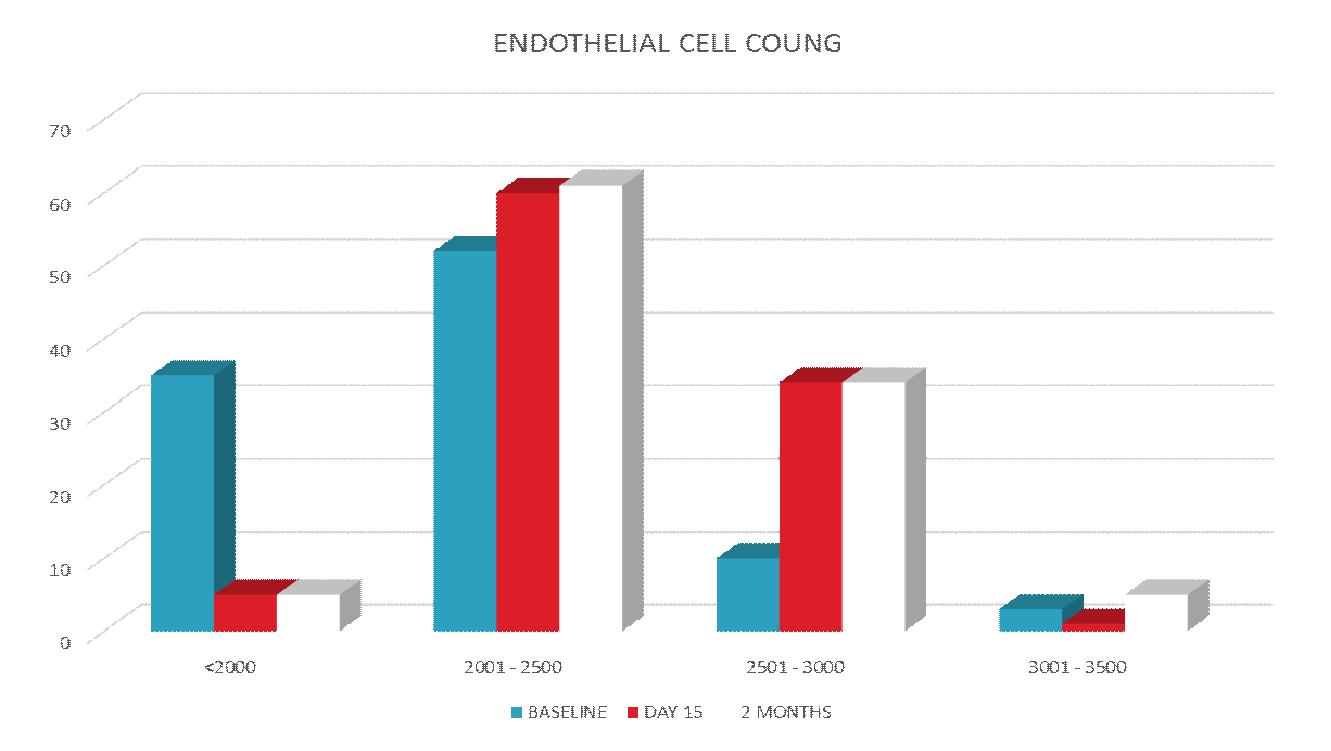
Results: Preoperatively endothelial cell count was 2500 or less in 35 eyes [35%],2501to 3000 in 52 eyes [52%] ,3001to 3500 in 10 eyes [10%],3501 to 4000 in 3 eyes [3%].15days post operatively endothelial cell count was 2000 or less in 5 eyes [5%],2001 to 2500 in 60 eyes [60%],2501 to 3000 in 34 eyes [34%], 3001 to 3500 in 1 eye [1%].2 month operatively endothelial cell count was 2000 or less in 5 eyes [5%], 2001 to 2500 in 61 eyes [61%],2501 to 3000 in 34 eyes [34%].
Conclusion: Femtosecond laser assisted cataract surgery [FLACS] appears to be as safe for corneal endothelial cells postoperatively.
Keywords: femto secand laser, cataract, endothelial cell count [ECC], surgery, cornea, phacoemulsification, preoperatively , postoperatively.
An estimation of 670 million peoples, are visually impaired- 41 million people are blind and 268 million have low vision. Cataract are the most common ocular disease and leading cause of low vision and blindness all around world, accounts for 52% of the global burden of blindness, representing more than 20 million people in world. The cataract is responsible for blur vision. {11}
Cataract can be defined as loss of transparency of the human crystalline lens in the eye. In which the refractive index of the lens significantly changes over distance and near approximating the wavelength of the light. The human crystalline lens consists of three structures: capsule, cortex and nucleus. In which the fetal nucleus is the most centrally located portion and new fiber is led down over time it turns into a hard-central nucleus {18}. The new fibers join each other form a Y-shaped suture line anteriorly and posteriorly. This central portion of the lens over time matures and hardens to form nuclear cataract which are treated with cataract surgeries. In most developing countries, blindness is associated with nutrition, economic and social implications. This is directly affecting on the populations, people who reside in underserved areas. An estimated 85-90% of people who are affected with cataract disease reside in developing countries. In current situation many infrastructure and technology are available to cure for the visually impaired persons. Moreover, So
ISSN 2348-313X (Print)
International Journal of Life Sciences Research ISSN 2348-3148 (online) Vol. 9, Issue 1, pp: (27-33), Month: January - March 2021, Available at: www.researchpublish.com
many areas have limited eye care capabilities and facilities to cope with high demand for cataract surgery and managements. These countries exhibit the largest back lock of IMSC (immature senile cataract) or MSC (mature senile cataract) cataract surgeries. Surgery is the most common or only treatment for cataracts thus performed by ophthalmologists and after surgery prescribed supplements with a pair of spectacles. Normal vision can be restored through the surgical removal of opacity of the lens, facilitated by implantation of an [IOL] intra ocular lens in the eye.
Cornea description;The cornea is the major refractive element of the eye. This requires optical transparency and smooth curve surfaces. The corneal has six layers: the epithelium layer, bowman’s (basement) membrane, the stroma, the duas layer, Descemet’s membrane, and the inner surface the endothelial layer. The epithelium provides the barrier to foreign particles the outside world. The stroma layer provides the refractive shape in the cornea. The endothelium layer maintains the nutrition of the cornea and corneal cells and the hydration in the stroma, all the nutrients comes from the aqueous humor and through the endothelium and nourishes the cornea. {18}
Femtosecond laser has emerged and assisted cataract surgery in recent years, and gaining popularity as the best procedure for management of cataracts in patients in the developing world. {5 } .The treatment with femtosecond laser is safe and gives better visual outcomes for patients such as early visual correction, visual rehabilitation and emmetropia. Major disadvantage of femtosecond laser cataract surgery is cost of the surgery.
Femtosecond laser cataract surgery is the latest technology for the treatment of cataracts. Anterior capsulotomy, corneal incision, and lens fragmentation are the three procedures that are performed with femtosecond laser assisted cataract surgery. In femtosecond laser assisted cataract surgery the phaco time and phaco energy is reduced significantly so this produces the postoperative recovery time, problems due to postoperative patients the IOP will be increases, and postoperative corneal edema and striate keratopathy are causing in the eye. Complication rates are low and recovery is quick and fast. Usually anesthesia will be giving with drops sometimes supplemented with a local injection if needed. {14} The major concern during surgery is corneal endothelial damage during cataract surgery for all ophthalmologists. Old age, small pupil size, large nucleus, high nucleus grade greater infusion volume, type of intraocular lens, greater amount of total emitted used energy, and longer duration of surgery are causing endothelial cell loss.
This was a prospective study trial with a cohort of 100 patients was offered FLACS on contra-lateral eyes. All patients to be informed about trail and included in the trial and consent was taken from-
Inclusion criteria - presence of cataract who are willing for femtosecond laser assisted cataract surgery included in the study, Patient of either sex, Patient of age 40 to 85 years, Patient with no media opacity other than cataract.
Exclusion criteria-Patients with complicated cataract, Patients with traumatic cataract, Patients with retinal pathology. Patients who have preoperative inflammatory ocular diseases, Patient who have media opacifications other than cataract, Patient who does not follow-up, Previous intraocular-surgeries
Preoperative Evaluation will include: -Best corrected visual acuity (BCVA), Intra ocular pressure measurement, Slit lamp examination, Specular cell counts
Outcome measurement; Follow up will be performed after a period of 15 days, 2 months after surgery. The follow up visits will include-Visual acuity testing, Slit lamp examination, Specular microscopy.
Preoperatively endothelial cell count was 2500 or less in 35 eyes [35%],2501to 3000 in 52 eyes [52%] ,3001to 3500 in 10 eyes [10%],3501 to 4000 in 3 eyes [3%]. {Table 6}15days post operatively endothelial cell count was 2000 or less in 5 eyes [5%],2001 to 2500 in 60 eyes [60%],2501 to 3000 in 34 eyes [34%], 3001 to 3500 in 1 eye [1%].2 month operatively endothelial cell count was 2000 or less in 5 eyes [5%], 2001 to 2500 in 61 eyes [61%],2501 to 3000 in 34 eyes [34%]. {Table 7}
ISSN 2348-313X (Print)
International Journal of Life Sciences Research ISSN 2348-3148 (online) Vol. 9, Issue 1, pp: (27-33), Month: January - March 2021, Available at: www.researchpublish.com
TABLE 1: Table showing age distribution among the no. of cases operated
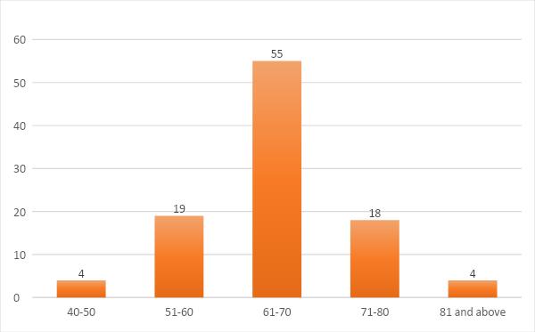
AGE (years) NO. OF CASES(n=100) PERCENTAGE 40-50 4 4% 51-60 19 19% 61-70 55 55% 71-80 18 18% 80 ABOVE 4 4%
TABLE 2: Table showing sex distribution of cases operated
SEX NO. OF CASES(n=100) PERCENTAGE MALE 59 59% FEMALE 41 41% TOTAL 100 100%
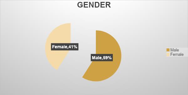
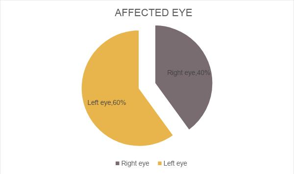
TABLE 3: Distribution of patients according to the eyes being operated
EYE NO. OF CASES(n=100) PERCENTAGE RIGHT EYE 40 40% LEFT EYE 60 60% TOTAL 100 100%
ISSN 2348-313X (Print)
International Journal of Life Sciences Research ISSN 2348-3148 (online) Vol. 9, Issue 1, pp: (27-33), Month: January - March 2021, Available at: www.researchpublish.com
ISSN 2348-313X (Print)
International Journal of Life Sciences Research ISSN 2348-3148 (online) Vol. 9, Issue 1, pp: (27-33), Month: January - March 2021, Available at: www.researchpublish.com
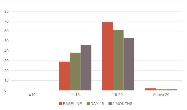
Table 8: Comparison of preoperative and postoperative Intraocular Pressure INTRAOCULAR PRESSURE BASELINE DAY 15 2 MONTHS ≤10 0 0 0 11-15 29 38 46 16-20 69 61 53 Above 21 2 1 1
Femtosecond lasers is the way for cataract surgery will be performed, with promising preliminary results showing selfsealing corneal incisions and faster recovery rate. Consistently accurate capsulorrhexis, which optimizes adequate centration and positioning of an intraocular lens [IOL], {11} during surgery they decrease phaco-emulsification energy, so there’ll be decrease endothelial cell loss The potential benefit of femtosecond laser assisted cataract surgery is from
ISSN 2348-313X (Print)
International Journal of Life Sciences Research ISSN 2348-3148 (online) Vol. 9, Issue 1, pp: (27-33), Month: January - March 2021, Available at: www.researchpublish.com
the subgroup of patients requiring no phaco-emulsification energy. There is a decreased incidence of corneal endothelial cell loss in post operative patients. We focused on the effect of femtosecond laser on the postoperative corneal endothelial count. In postoperative patients, there was a significant cell count decreases in central corneal endothelial cell count compared with preoperative values. The corneal endothelial cell count was higher in the femtosecond laser group at 15 days, 2 months in follow-up. {2} Patients in the femtosecond laser showed better vision on first postoperative day. Takács et al. demonstrated less corneal swelling and endothelial cell damage in patients undergoing femtosecond laserassisted cataract surgery than in those undergoing a conventional phacoemulsification technique at all visits. {1} Limitations of our study were as follows: first, there was a lack of analysis of endothelial cell morphology, patients were not matched according to lens density.
To conclude, femtosecond laser-assisted cataract surgeries appear to be as safe management for cataracts, with reduced the ultrasonic energy and lower effective phaco-emulsification time and after surgery having less corneal endothelial cell loss. {1}, {11}, {14}.
The author would like to acknowledge participant for their patience and coordination.
[1] Nagy ZZ, Kránitz K, Takacs AI, Miháltz K, Kovács I, Knorz MC. Comparison of intraocular lens decentration parameters after femtosecond and manual capsulotomies. J Refract Surg 2011; 27:564–569.
[2] Kránitz K, Takacs A, Miháltz K, Kovács I, Knorz MC, Nagy ZZ. Femtosecond laser capsulotomy and manual continuous curvilinear capsulorrhexis parameters and their effects on intraocular lens centration. J Refract Surg 2011; 27:558–563.
[3] Cho YK, Chang HS, Kim MS. Risk factors for endothelial cell loss after phacoemulsification: comparison in different anterior chamber depth groups. Korean J Ophthalmol 2010; 24:10–15.
[4] Grewal DS, Brar GS, Grewal SP. Correlation of nuclear cataract lens density using Scheimpflug images with Lens Opacities Classification System III and visual function. Ophthalmology 2009; 116:1436–1443.
[5] Musket S, Sarayba M, Ignacio T, Fram N. Femtosecond laser-assisted cataract incisions: architectural stability and reproducibility. J Cataract Refract Surg 2010; 36:1048–1049.
[6] Plankter DV, Blumenkranz MS, Andersen D, Wiltberger M, Marcellino G, Gooding P et al. Femtosecond laserassisted cataract surgery with integrated optical coherence tomography. Sci Transl Med 2010; 2:58ra85.
[7] Gain P, Thuret G, Kodjikian L, et al. Automated tri-image analysis of stored corneal endothelium. Br J Ophthalmol 2002; 86:801–8. [PMC free article] [PubMed] [Google Scholar]
[8] Gundersen HJ. Estimators of the number of objects per area unbiased by edge effects. Microsc Acta 1978; 81:107–17. [PubMed] [Google Scholar]
[9] Doughty MJ, Muller A, Zaman ML. Assessment of the reliability of human corneal endothelial cell-density estimates using a noncontact specular microscope. Cornea 2000; 19:148–58. [PubMed] [Google Scholar]
[10] Schimmelpfennig BH. Direct and indirect determination of nonuniform cell density distribution in human corneal endothelium. Invest Ophthalmol Vis Sci 1984; 25:223–9. [PubMed] [Google Scholar]
[11] Nagy ZZ, Ecsedy M, Kovács I, Takács Á, Tátrai E, Somfai GM, Cabrera DeBuc D. Macular morphology assessed by optical coherence tomography image segmentation after femtosecond laser-assisted and standard cataract surgery. J Cataract Refract Surg 2012; 38:941–946.
[12] S. J. Tuft and D. J. Coster, “The corneal endothelium,” Eye, vol. 4, no. 3, pp. 389–424, 1990.View at: Publisher Site | Google Scholar
ISSN 2348-313X (Print) International Journal of Life Sciences Research ISSN 2348-3148 (online) Vol. 9, Issue 1, pp: (27-33), Month: January - March 2021, Available at: www.researchpublish.com
[13] F. W. Stocker, “The endothelium of the cornea and its clinical implications,” Transactions of the American Ophthalmological Society, vol. 51, pp. 669–786, 1953.View at: Google Scholar
[14] R. A. Laing, M. M. Sandstrom, A. R. Berrospi, and H. M. Leibowitz, “Changes in the corneal endothelium as a function of age,” Experimental Eye Research, vol. 22, no. 6, pp. 587–594, 1976.View at: Publisher Site | Google Scholar
[15] S. J. Tuft and D. J. Coster, “The corneal endothelium,” Eye, vol. 4, no. 3, pp. 389–424, 1990.View at: Publisher Site | Google Scholar
[16] F. W. Stocker, “The endothelium of the cornea and its clinical implications,” Transactions of the American Ophthalmological Society, vol. 51, pp. 669–786, 1953.View at: Google Scholar
[17] R. A. Laing, M. M. Sandstrom, A. R. Berrospi, and H. M. Leibowitz, “Changes in the corneal endothelium as a function of age,” Experimental Eye Research, vol. 22, no. 6, pp. 587–594, 1976.View at: Publisher Site | Google Scholar
[18] A.K Khurana book “Diseases of eye’, diseases of lens {179}.