ISSN 2348-313X (Print)
International Journal of Life Sciences Research ISSN 2348-3148 (online)
Vol. 9, Issue 3, pp: (20-26), Month: July - September 2021, Available at: www.researchpublish.com

ISSN 2348-313X (Print)
International Journal of Life Sciences Research ISSN 2348-3148 (online)
Vol. 9, Issue 3, pp: (20-26), Month: July - September 2021, Available at: www.researchpublish.com
Department of Microbiology
Department of Microbiology Sant Gadge Baba Amravati University, Amravati (Maharashtra), India
Email: nirajghanwate@gmail.com
Abstract: The power of Staphylococcus aureus to make biofilm is considered to be a serious virulence factor influencing its survival and persistence in both the environment and host. In Staphylococcus aureus biofilms, a number one explanation for persistence infections is because it is highly immune to immune defences and antimicrobial therapies. This study was aimed to gauge the antibiofilm effect of varied enzymes against biofilm formation by clinical isolates of Staphylococcus aureus. Within the present study total of 120 clinical specimens of pus samples were collected from hospitals and processed for isolation and identification of S. aureus. Growth was found in 96 specimens. Out of 96, 64 were found to be Gram-positive cocci and 32 Gram-negative bacilli. From 64 Gram-positive cocci 52 were Coagulase positive i.e Staphylococcus aureus and 12 Coagulase-negative. All Staphylococcus aureus isolates were further investigated for biofilm formation by the Tissue Culture Plate method. From 52 isolates 32 (62%) moderate biofilm forming, 16 (30%) weak biofilm forming and 04 (08%) strong biofilm forming. Biofilm formed by Staphylococcus aureus isolates was treated by 1% solution Amylase and Lysozyme enzymes to gauge the antibiofilm activity of enzymes for the detachment of biofilm. It was observed that Amylase and Lysozyme showed considerable antibiofilm activity against Staphylococcus aureus biofilm. All the strong and moderate biofilms produced by strains of Staphylococcus aureus were rendered non biofilm. Thus, these enzymes are useful as antibiofilm agents against biofilm formed by pathogenic Staphylococcus aureus.
Keywords: Biofilm, TCP method, enzymes, Staphylococcus aureus.
A survey on Nosocomial Infection concluded that at any time, over 1.4 million people worldwide are affected by infections acquired in treatment booth, with an accounted 80,000 deaths annually [1,2] Biofilm is an important character of nosocomial pathogens. Biofilm is an association of microorganism that adhere to solid surfaces and are embedded in a matrix of extracellular polymeric substances (EPS) consisting of carbohydrates, proteins, and nucleic acids, in an environment containing liquids [3-5]. Biofilm associated cells stay irreversibly on various kinds of surfaces, including living tissues and indwelling medical devices as catheters valves, prosthesis then forth [6-8] and acts as endless source of contamination and infection [9]. According to National institute of health (NIH) about 65% of all microbial infections and 80% of all chronic infections are associated with biofilms [10]
Staphylococcus aureus is a major cause of severe infections in both developed and developing countries. The most common S. aureus diseases are skin and soft tissue infection, ranging from mild and superficial conditions such as impetigo and folliculitis, to potentially fatal and deep-seated infections such as cellulitis, pyomyositis and fasciitis [11]. The ability of Staphylococcus aureus to form biofilm is considered to be a major virulence factor influencing its survival and persistence in both the environment and the host. It is also important nosocomial pathogen [12,13]. Biofilms forming on the surface of indwelling medical devices by organisms such as Staphylococcus epidermidis and Staphylococcus aureus constitute a leading cause of infections [14].
Bacteria protected within biofilm exopolysaccharides are up to 1,000 times more resistant to antibiotics than planktonic cells (free floating), which generates serious consequences for therapy and severely complicates treatment options. The increased biofilm resistance to conventional treatments enhances the need to develop new control strategies. In recent years, several green nonlethal strategies for biofilm control have been developed, because the mode of action of these of these novel antibiofilm agents is much less susceptible to the emergence of resistance [13,15-17]. The use of substances to
ISSN 2348-313X (Print)
International Journal of Life Sciences Research ISSN 2348-3148 (online)
Vol. 9, Issue 3, pp: (20-26), Month: July - September 2021, Available at: www.researchpublish.com
induce biofilm removal directly by destroying the physical integrity of the biofilm matrix would be an attractive alternative for both medical and industrial applications where complete biofilm removal is essential [14]. Biofilm disruption based on enzymatic activity is thought to serve as a plausible strategy to combat persistent infections associated with biofilms, as enzymatic treatment improves the antibiotic susceptibility of microbial biofilms.
Considering the above and based on previous results, the present study is aimed to evaluate the antibiofilm effect of amylase and lysozyme against clinical isolates of Staphylococcus aureus
Pus from wound was collected using sterile cotton swabs packed in sterile screw capped bottle and transferred to laboratory. Isolation and identification of S. aureus isolates was done by Standard methods. Confirmation was done by Coagulase (slide test). Prompt clumping of the organism indicated the presence of bound Coagulase [18]
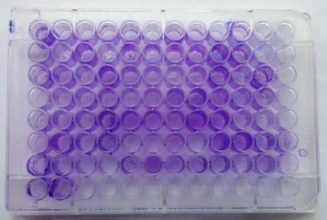
Biofilm formation by isolated strains of Staphylococcus aureus was done by tissue culture plate method. This quantitative test [19] is taken into account the gold-standard method for biofilm detection. Organisms isolated from fresh agar plates were inoculated in 10ml trypticase soy broth with 1% glucose. Broths were incubated at 370C for 24 h. The cultures were then diluted 1:100 with fresh medium. Individuals well of sterile 96 well-flat bottom polystyrene tissue culture plates (Tarsons, made in Korea) were filled with 200μl of the diluted cultures. The plates were incubated at 370C for 24 h. After incubation, contents of each well were removed by gentle tapping. The wells were washed with 0.2 ml of phosphate buffer saline (pH 7.2) fourfold to remove free floating bacteria. Biofilm formed by bacteria adherent to the wells were fixed by 2 % sodium acetate and stained gentian violet (0.1%). Excess stain was removed by using deionized water and plates were kept for drying (Fig 2). Optical density (OD) of stained adherent biofilm was obtained by using micro-ELISA auto reader at wavelength 570 nm. OD values below 0.120 were considered non biofilm forming, 0.120-0.240 was considered moderate and greater than 0.240 as strong biofilm producing.
The procedure for testing the antibiofilm activity of enzymes was similar as described above till the growing and washing of the biofilm. After washing 200μl of 1% solution of enzymes was added in each well and again incubated overnight. After incubation wells were washed with Phosphate buffer saline. Biofilm was fixed by 2 % sodium acetate and stained with crystal violet (0.1%). Excess stain was removed by using deionized water and plates were kept for drying. Optical density (OD) of stained adherent biofilm was obtained by using micro-ELISA auto reader at wavelength 570 nm.
A total 120 clinical specimens of pus from wound were collected aseptically from hospitals of Amravati District, Maharashtra and processed by standard procedure for the isolation and identification of Staphylococcus aureus.
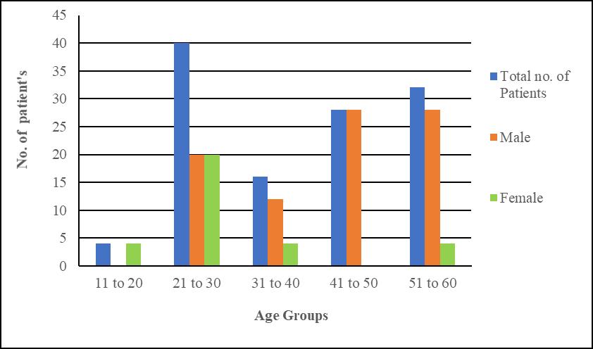
Fig. 1: Distributions of patient’s data
ISSN 2348-313X (Print)
International Journal of Life Sciences Research ISSN 2348-3148 (online) Vol. 9, Issue 3, pp: (20-26), Month: July - September 2021, Available at: www.researchpublish.com
Figure 1, depicts that the patients at the age of 21-30 are most infected with Staphylococcus aureus infection. The percentage of infection in the age group of 21-30 yrs is 33%. Males are more infected with Staphylococcus aureus infections as compared to female with the percentage of 73% and 27% respectively
B. Characterization of bacterial isolates from pus samples by standard method:
Pus from wound samples was collected aseptically from hospitals. The specimens were processed using standard methods for isolation and identification of Staphylococcus aureus
Out of 120 specimens of pus, the growth was observed in 96 specimens from which 64 were found to be Gram positive cocci and 32 Gram negative rods. From 64 Gram positive cocci, 52 were Coagulase positive Staphylococci and 12 Coagulase negative Staphylococci (Table 1).
Table 1: Numbers of Coagulase positive and Coagulase negative Staphylococci isolated from pus samples
Total specimens (pus) Growth No growth 120 96 24
Total isolates Gram positive cocci Gram negative 96 64 32
Total Staphylococci Coagulase positive Coagulase negative 64 52 12
C. Biofilm formation by the S. aureus
A total of 52 S. aureus were isolated from 120 pus samples. These were further investigated for biofilm formation by TCP method (Table 2). Almost 69 % strains of S aureus showed formation of biofilm.
Table 2: Biofilm formation by the S. aureus isolates
Sr. No. Biofilm formation Number
1 Moderate biofilm 32
2 Strong biofilm 04
3 Weak/non biofilm 16
Biofilm formed by Staphylococcus aureus isolates was treated by Amylase and Lysozyme to evaluate the antibiofilm activity of enzymes. Isolates were tested for presence of biofilm before and after enzymatic exposure and their O.D. was compared
ISSN 2348-313X (Print)
International Journal of Life Sciences Research ISSN 2348-3148 (online) Vol. 9, Issue 3, pp: (20-26), Month: July - September 2021, Available at: www.researchpublish.com
Optical density of biofilm after treatment of Amylase was significantly decreased indicating decrease in the biofilm integrity Figure 3 depicts that 4 isolates PS 13, PS 36, PS 49 and PS 80 with strong biofilm was decreased after the treatment of amylase to non-biofilm. Similarly, the optical density of 16 isolates of S. aureus PS 1, PS 11, PS 17, PS 23, PS 27, PS 33, PS 40, PS 45, PS 48, PS 53, PS 62, PS 66, PS 75, PS 82, PS 93 and PS 110 was also decreased from moderate to non-biofilm and the OD of remaining isolates (PS 9, PS 12, PS 21, PS 25, PS 32, PS 46, PS 51, PS 55, PS 61, PS 64, PS 68, PS 70, PS 78, PS 88, PS 97 and PS 105) also changed significantly after treatment with Amylase
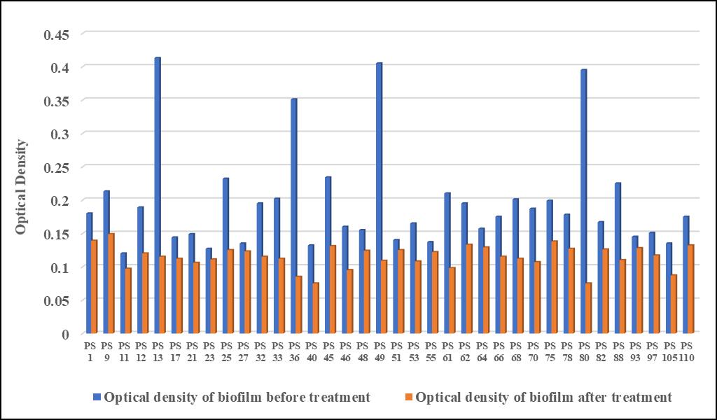
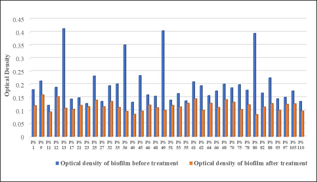
Fig. 3: Antibiofilm activity of Amylase against S. aureus biofilm
Similar results were seen with the optical densities of Staphylococcus aureus biofilm before and after treatment with lysozyme Again PS 13, PS 36, PS 49 and PS 80 changed from strong biofilm to non-biofilm PS 11, PS 17, PS 21, PS 23, PS 32, PS 33, PS 40, PS 46, PS 53, PS 61, PS 66, PS 68, PS 70, PS 88, PS 97 and PS 105 isolates biofilm decreased from moderate to non-biofilm and PS 1, PS 9, PS 12, PS 25, PS 27, PS 45, PS 48, PS 51, PS 55, PS 62, PS 64, PS 75, PS 78, PS 82, PS 93 and PS 110 also decreased their optical density significantly after treatment with Lysozyme. (Fig 4).
Fig. 4: Antibiofilm activity of Lysozyme against S. aureus biofilm
ISSN 2348-313X (Print)
International Journal of Life Sciences Research ISSN 2348-3148 (online) Vol. 9, Issue 3, pp: (20-26), Month: July - September 2021, Available at: www.researchpublish.com
It was clear that the Amylase and Lysozyme showed antibiofilm activity which helps for the detachment of Staphylococcus aureus biofilm Lysozyme showed great results against Staphylococcus aureus biofilm on basis of their optical density value compared to before and after treatment. Amylase too exhibited good antibiofilm activity against Staphylococcus aureus biofilm. Both enzymes are useful as antibiofilm agent against biofilm formed by Staphylococcus aureus.
In the present study, we discussed the enzymatic detachment of Staphylococcus aureus biofilm. Staphylococcus aureus is an opportunistic human pathogen found within the microbiota of mucosa and skin, capable of causing diverse healthcareassociated infections in altogether age groups. Besides the skin and soft tissue infections, Staphylococcus aureus has also increasingly been recognized as an evidence for severe invasive diseases like osteomyelitis, septic arthritis, and pneumonia. Staphylococcus aureus was isolated from clinical specimens of pus to detect the biofilm formation by Sanchez et al in 2016 [15]. They discussed that biofilm may be a complex microbial community highly immune to antimicrobials. Similarly, Lister and Harswill in 2018 [20] demonstrated the Staphylococcus aureus may be a major explanation for nosocomial and community-acquired infections and represents a big burden on the healthcare system. Staphylococcus aureus attachment to medical implants, and host tissue and therefore the establishment of a mature biofilm, play a crucial role within the persistence of chronic infections.
During this study, Staphylococcus aureus strains isolated from wound exudates were characterized for biofilm production. Total of 52 isolates of which 32 (62%) moderate biofilm, 16 (30%) weak biofilm, and 04 (08%) strong biofilm identified were from 120 clinical specimens of pus. Torlak et al in 2017 [12] discussed on the isolation of biofilm forming S. aureus from dental pus They concluded that dental clinic environments should be considered a possible reservoir for biofilmproducing S. aureus and thus cross-contamination.
Our findings demonstrate that Lysozyme and Amylase exhibit biofilm releasing activity against Staphylococcus aureus biofilm. These enzymes were active at physiologically achievable concentrations and prevented biofilm formation. Thus, these enzymes might be used as an agent to stop or treat Staphylococcus aureus infections of catheters and other medical devices. Similar results were observed by Kaplan et al [21] for S. epidermidis biofilm. They demonstrated that the Nacetyl-glucosaminidase enzyme was active at physiologically achievable concentrations and prevented biofilm formation
Chen et al [22] discussed novel therapeutic solutions aside from the traditional antibiotic therapies as an urgent need to stop or treat current biofilm infections They discussed the biofilm-forming process of bacteria supported their understanding of the molecular mechanism of biofilm formation. Small molecules and enzymes are developed to inhibit or disrupt biofilm formation.
In the present investigation, it had been observed that biofilm detachment of Staphylococcus aureus was possible by the treatment of Amylase and Lysozyme As the major component of the EPS is polysaccharide, amylase has inhibited EPS by preventing the adherence of the microbial cells, thus making amylase a suitable antimicrobial agent. The work of Molobela et al. (2010) showed the successful use of α-amylase from Bacillus amyloliquefaciens and glucoamylase from Aspergillus niger on the Gram-negative biofilm-forming bacteria Pseudomonas fluorescence. EPS was reduced by 42.5% in the presence of the enzyme in a challenge of 90 minutes. Microscopic studies were performed to assess the reduction of biofilm and the ability of the enzyme to degrade the EPS and disperse the cells, which resulted in reduction of the biofilm Ghanwate et al [24, 25] discussed on prevention of biofilm formation within the urinary catheter by Pseudomonas aeruginosa. It was determined by coating the catheter with some amylase and lysozyme. It was concluded that it prevented biofilm production by Pseudomonas for 7 days in Amylase and 6 days in Lysozyme treated catheter. Present study also noted that the results of antibiofilm activity of Amylase and Lysozyme against Staphylococcus aureus biofilm and showed considerable antibiofilm activity Application of suitable enzymes for degrading the structural components of the biofilm matrix will weaken it so that it can be more easily removed by mechanical processes. Since the sugar backbone of the biofilm matrix is composed mainly of carbohydrate residues, carbohydrate-based enzymes, such as amylase, might be used to hydrolyze and thereby denature the biofilm matrix (Lembre et al., 2012). Lysozyme had the utmost detachment power against Staphylococcus aureus biofilm. Antibiofilm activity of lysozyme is associated with the protective function of the innate immune system against infections with biofilms [27] and relies on the hydrolytic activity against peptidoglycan. In addition, lysozyme has an on-lytic mechanism related to its cationic and hydrophobic characteristics, which lead to bacterial autolysis [28] It also has a direct antibacterial effect on Staphylococcus aureus.
ISSN 2348-313X (Print)
International Journal of Life Sciences Research ISSN 2348-3148 (online) Vol. 9, Issue 3, pp: (20-26), Month: July - September 2021, Available at: www.researchpublish.com
Present study evaluated the antibiofilm effect of Lysozyme and Amylase enzymes against clinical isolates of Staphylococcus aureus biofilm. It was observed that S. aureus was the cause of almost 43% of wound infections. Thus, concluded that Staphylococcus aureus was the common isolate from pus samples. Isolates of Staphylococcus aureus when tested for biofilm formation by tissue culture plate method showed 62% with Moderate biofilm, 8% with Strong biofilm and 30% with weak biofilm. Biofilm formed by Staphylococcus aureus isolates was treated by Amylase and Lysozyme. Lysozyme exhibits great results on the basis of Optical density as compared to Amylase against Staphylococcus aureus biofilm before and after treatment. Amylase also showed significant antibiofilm activity. Both enzymes can be used as antibiofilm agents against biofilm formed by pathogenic Staphylococcus aureus. This will be an alternative antimicrobial therapy to regulate the formation of microbial biofilms. Thus, from this study, it is concluded that the biofilm formed by Staphylococcus aureus was degraded or detached in presence of Lysozyme and Amylase enzymes. Enzymes can be effective anti-biofilm agents, and they are environmentally friendly and easily biodegradable.
[1] Nazir A, kadriz SM (2014) An overview of hospital acquired infections and the role of the microbiology laboratory. International Journal Research Medical Science 2(1):21-27.
[2] Reddy PS, John MS, Devi PV, Kumar SS (2015) Nosocomial infections among patients admitted in general ICU: study from a tertiary care hospital in South India. International Journal of Medical Science and Public health 5(8):1523-1527.
[3] Souza PR, Andrade D, Cabral DB, Watanabe E (2014) Endotracheal tube biofilm and ventilator associated Pneumonia with mechanical ventilation. Microbiology Scholar Research technology 77(4):305-312.
[4] Mukharji R, Patil A and Prabhune A (2015) Role of extracellular protease in biofilm disruption of gram-positive bacteria with special emphasis on Staphylococcus aureus biofilms. Enzyme engineering 4(1):1-7.
[5] Vasudevan R (2014) Biofilms: microbial cities of scientific significance. Journal of Microbiology and Experimentation 171(3):1-16.
[6] Parsek MR, Singh PK (2003) Bacterial biofilms an emerging link to disease pathogenesis. Annual Review of microbiology 57:677-701.
[7] Olsen I (2015) Biofilm-specific antibiotic tolerance and resistance. European Journal of Clinical Microbial Infectious Diseases 34(5):877-886.
[8] Wu H, Moser C, Wang HZ et al (2015) Strategies for combating bacterial biofilm infections. International Journal of Oral Science 7(1):1-7.
[9] Hall CW, Mah TF (2017) Molecular mechanisms of biofilm-based antibiotic resistance and tolerance in pathogenic bacteria. FEMS Microbial Review 41(3):276-301.
[10] Jamal M, Tasneem U, Hussain T et al (2015) Bacterial Biofilm, it’s composition, formation and role in human infections. Research and Reviews Journal of microbiology and biotechnology 4(3):1-14.
[11] Kwiecinski J, Jacobsson G, Karlsson M et al. (2013) Staphylokinase promotes the establishment of Staphylococcus aureus skin infections while decreasing disease severity. Journal of infectious diseases 208:990-999.
[12] Torlak E, Korkut E, Uncu AT, et al (2017) Biofilm formation by Staphylococcus aureus isolates from a dental in Konya, Turkey. Journal of Infection and public health 10:809-813.
[13] Kwiecinsky J, Peetermans M, Liesenbarghs L et al [2016] Staphylokinase control of Staphylococcus aureus biofilm formation and detachment through host plasminogen activation. The journal of infectious Diseases 213:139-148.
[14] Xavier JB, Picioreanu C, Rani SA et al (2005) Biofilm control strategies based on enzymatic disruption of the extracellular polymeric substance matrix-amodelling study. Microbiol 151:3817-3832.
[15] Sanchez E, Morales CR, Castillo S, et al (2016) Antibacterial and anti-biofilm activity of methanolic plant extracts against nosocomial microorganisms. Evidence-Based Complementary and Alternative Medicine :1-8.
ISSN 2348-313X (Print) International Journal of Life Sciences Research ISSN 2348-3148 (online) Vol. 9, Issue 3, pp: (20-26), Month: July - September 2021, Available at: www.researchpublish.com
[16] Katharina R, Driessche FV, Coenye T (2017) Innovative approaches to treat Staphylococcus aureus biofilm related infections. Essay in Biochemistry 61:67-70.
[17] Verma PA (2012) Study on isolation of different type of bacteria from pus. International Journal of Pharmacy and Life sciences 3 (11):2107-2110.
[18] Yasmeen F, Sarwar MI, Hakeem A, et al (2014) Identification of Staphylococcus aureus in pus samples and it’s antimicrobial susceptibility against Imipenem, Tobramycin and Linzolid. International Journal of Basic Medical Sciences and Pharmacy 4(1):2049-4963.
[19] Christensen GD, Simpson WA, Younger JA et al (1995) Adherence of Coagulase negative staphylococci to plastic tissue culture: a quantitative model for the adherence of Staphylococci to medical devices. Journal of Clinical Microbiol 22:996-1006.
[20] Lister JL, Harswill AR (2014) Staphylococcus aureus biofilms recent developments in biofilm dispersal. Frontiers in Cellular and Infection Microbiology 4:1-9.
[21] Kaplan JB, Ragunath C, Velliyagounder K et al (2004) Enzymatic detachment of Staphylococcus epidermidis biofilms. Antimicrobial Agents and Chemotherapy 48(7): 2633-2636.
[22] Chen M, Yu Q, Sun H (2013) Novel strategies for the prevention and treatment of biofilm related infections. International Journal of Molecular Science 14:18488-18501.
[23] Molobela IP, Cloete TE, Beukes M (2010) Protease and amylase enzymes for biofilm removal and degradation of extracellular polymeric substances (EPS) produced by Pseudomonas fluorescence bacteria. African Journal of microbiology research 4(14):1515-1524.
[24] Ghanwate NA, Thakare PV, Bhise PR, et al (2012) Prevention of biofilm formation in urinary catheter by coating enzymes/gentamycin/EDTA. International journal of Biotechnology and Bioengineering 6(10):899-901.
[25] Ghanwate NA, Thakare PV, Bhise PR et al (2014) Prevention of biofilm formation in urinary catheters by treatment with antibiofilm agents. International Journal of Science and Research 3(4):714-717.
[26] Lembre, P., Lorentz, C., Di, P. (2012). “Exopolysaccharides of the Biofilm Matrix: A Complex Biophysical World,” in The Complex World of Polysaccharides. (London, UK: IntechOpen). Available at: https://www.intechopen.com/ books/the-complex-world-of-polysaccharides/exopolysaccharides-of-the-biofilm-matrix-a-complex-biophysicalworld.
[27] M. Hukic, D. Seljmo, A. Ramovic et al. (2018), “The effect of lysozyme on reducing biofilms by Staphylococcus aureus, Pseudomonas aeruginosa, and Gardnerella vaginalis: an in vitro examination,” Microbial Drug Resistance, vol. 24, no. 4, pp. 353–358,.View at: Publisher Site | Google Scholar
[28] J. Humann and L. L. Lenz, (2009) “Bacterial peptidoglycan-degrading enzymes and their impact on host muropeptide detection,” Journal of Innate Immunity, vol. 1, no. 2, pp. 88–97,.View at: Publisher Site | Google Scholar