6 minute read
How is NSCLC diagnosed?
How is NSCLC diagnosed?
Most patients with NSCLC are diagnosed after seeing their doctor to report symptoms such as a persistent cough, a chest infection that won’t go away, dyspnoea, wheezing, coughing blood, chest or shoulder pain that won’t go away, hoarseness or lowering of the voice, unexplained weight loss, loss of appetite or extreme fatigue. A diagnosis of lung cancer is based on the results of the following examinations and tests:
Advertisement
Clinical examination
Your doctor will carry out a clinical examination. He/she will examine your chest and check the lymph nodes in your neck. If there is a suspicion of lung cancer, he/she may arrange for a chest x-ray, or possibly a CT scan, and refer you to a specialist for further testing.
Imaging
Imaging is used to confirm a suspected diagnosis of lung cancer, and to investigate how far the cancer has progressed

Different imaging techniques include: • Chest x-ray: A chest x-ray will enable the specialist to check your lungs for anything that looks abnormal. This is usually the first test that is carried out, based on your symptoms and the clinical examination. • CT scan of chest and upper abdomen: A series of images are taken, which build up a three-dimensional picture of the inside of your body. This allows the specialist to gather more information about the cancer such as the exact location of the tumour in your lungs, whether nearby lymph nodes are affected, and whether the cancer has spread to other areas of the lungs and/or parts of your body. It is a painless procedure and usually takes about 10–30 minutes. • CT scan or magnetic resonance imaging (MRI) scan of the brain: This test allows doctors to rule out or confirm whether the cancer has spread to your brain. An MRI scan uses powerful magnetism to build up detailed images. You may be given an injection of dye into a vein in your arm to help the images show up more clearly. The scan won’t hurt but may be slightly uncomfortable as you will need to lie still inside the scanning tube for about 30 minutes. You will be able to hear and speak to the person doing the scan.
Positron emission tomography (PET)/CT scan: A combination of a CT scan and a PET scan. PET uses low-dose radiation to measure the activity of cells in different parts of the body, so a PET/CT scan gives more detailed information about the part of the body being scanned. A mildly radioactive drug will be injected into a vein in the back of your hand or arm and then you will need to rest for about an hour while it spreads throughout your body. The scan itself will take 30–60 minutes and, although you will need to lie still, you will be able to speak to the person operating the scanner. A PET/CT scan is often carried out to detect whether the cancer has spread to the bones.
Histopathology
Examination of a biopsy is recommended for all patients with NSCLC as it helps to determine the best treatment approach
Histopathology is the study of diseased cells and tissues using a microscope; a biopsy of the tumour allows a sample of cells to be closely examined. Examination of a biopsy is recommended for all patients as it is used to confirm a diagnosis of NSCLC, to identify the histological subtype of NSCLC, and to identify any abnormal proteins within the tumour cells that could help to determine the best treatment for you (Planchard et al., 2018).


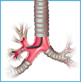
Techniques for obtaining a biopsy include: • Bronchoscopy: A doctor or specially-trained nurse examines the insides of the airways and lungs using a tube called a bronchoscope. It is carried out under local anaesthetic. During a bronchoscopy, the doctor or nurse will take samples of cells (biopsies) from the airways or lungs. • CT-guided needle lung biopsy: If a biopsy is difficult to obtain with a bronchoscopy, your doctor may choose to obtain a biopsy during a CT scan. In this procedure, you will have a local anaesthetic to numb the area. A thin needle is then inserted through your skin into your lung so that the doctor can remove a sample of cells from the tumour. This should only take a few minutes. • Endobronchial ultrasound-guided sampling (EBUS): This technique is used to confirm whether the cancer has spread to nearby lymph nodes, after radiological examinations have suggested that this might be the case. A bronchoscope, containing a small ultrasound probe, is passed through the trachea to see whether any nearby lymph nodes are larger than
normal. The doctor can pass a needle along the bronchoscope to take biopsies from the tumour or the lymph nodes. This test can be uncomfortable but shouldn’t be painful. It takes less than an hour and you should be able to go home the same day after it is finished. Oesophageal ultrasound-guided sampling (EUS): Similar to EBUS, this technique is used to confirm whether the cancer has spread to nearby lymph nodes, after radiological examinations have suggested that this might be the case. However, unlike EBUS, the ultrasound probe is inserted through the oesophagus. Mediastinoscopy: This procedure is more invasive than EBUS/EUS but is recommended as an extra test if EBUS/EUS does not confirm that the cancer has spread to nearby lymph nodes or if the lymph nodes requiring investigation cannot be reached by EBUS. A mediastinoscopy is carried out under general anaesthetic and requires a short stay in hospital. A small cut is made in the skin at the front of the base of your neck and a tube passed through the cut into your chest. A light and a camera attached to the tube allow the doctor to closely look at the middle of your chest – the mediastinum – for any abnormal lymph nodes, as these are the first areas that the cancer may spread to. Samples of tissue and lymph nodes can be taken for further examination.
Ask your doctor for details if you have any questions about these procedures
Cyto(patho)logy
Whereas histopathology is the laboratory examination of tissue or cells, cytology (or cytopathology) is the examination of cancerous cells spontaneously detached from the tumour. Common methods for obtaining samples for cytological examination include: • Bronchoscopy: Bronchial washings (in which a mild salt solution is washed over the surface of the airways) and the collection of secretions can be carried out during a bronchoscopy to look for the presence of cancerous cells. • Thoracentesis/pleural drainage: Pleural effusion is an abnormal collection of fluid between the thin layers of tissue (pleura) that line the lung and the wall of the chest cavity. This fluid can be taken from the pleural cavity by thoracentesis or pleural drainage and examined in the laboratory for the presence of cancerous cells. • Pericardiocentesis/pericardial drainage: Pericardial effusion is an abnormal collection of fluid between the heart and the sac that surrounds the heart (pericardium). This fluid can be taken from the pericardial cavity by pericardiocentesis or pericardial drainage and examined in the laboratory for the presence of cancerous cells. These techniques are carried out in the hospital, usually with the aid of ultrasound to help position the needle. You will be given a local anaesthetic and monitored closely for any complications afterwards. Because of the location of your lungs in your body, obtaining samples of cells/tissue can be difficult and it may be necessary to repeat some of these tests if results are found to be inconclusive.


