

The Journal of Sport and Exercise Science, Vol. 7, Issue 3, 1-9 (2023)
www.jses.net
JSES
ISSN: 2703-240X
No effect of facial expression on running economy in collegiate soccer players
Brian C. Rider1* , Grace L. Ditzenberger1 , Haven L. Westra2 , Thomas J. Sprys-Tellner1, Brooke T. Hedglen1 , Alexander H.K. Montoye2
1Department of Kinesiology, Hope College, USA
2Department of Integrative Physiology and Health Science, Alma College, USA
Received: 02.08.2022
Accepted: 12.10.2022
Online: 27.10.2023
Keywords: Smile Affect Ergogenic aid
The facial feedback hypothesis (FFH) states that activation of facial muscles (i.e., smiling and frowning) can elicit emotional experiences within an individual. A positive emotional experience could result in a more “relaxed state” and result in improved running economy (RE). The purpose of this study was to determine if smiling while running would lead to an improvement in RE among a group of collegiate soccer players. Twenty-four Division III collegiatesoccerplayers(femalesn=14,malesn=10)completedfour,six-minuterunning blocks at 70% of velocity atVO2 max. The order ofbouts was randomised with participants serving astheirown controls.Participants completedrunning blockswhilesmiling (Smile), frowning (Frown), consciously relaxing their hands and upper bodies (Relax), and running as they “normally” would (Control). Each block was separated by two minutes of passive rest. Cardiorespiratory responses were recorded continuously, and participants reported perceived exertion (RPE) after each condition. A repeated measures analysis of variance (ANOVA) was run on all primary variables with a significance level set a priori at 0.05. There were no significant differences in RE across conditions (p > 0.05; Smile: mean = 33.7 mL∙kg-1∙min-1, SD = 4.4; Frown: mean = 34.2 mL∙kg-1∙min-1, SD = 4.1; Relax: mean = 34.2 mL∙kg-1 min-1, SD = 4.1; Control: mean = 34.2 mL∙kg-1 min-1, SD = 3.9). Our findings suggest smiling does not significantly improve RE among a group of collegiate soccer players. Further studies should examine this topic in other athlete groups and at various running intensities.
1. Introduction
Running economy (RE) is a multifaceted concept that is impacted by numerous physiological and biomechanical variables (Barnes & Kilding, 2015b). It is most commonly expressed as an individual’s steady-state oxygen consumption while running at a submaximal speed (Barnes & Kilding, 2015b; Saunders et al.,
2004).RunnerswithahighREutiliselessoxygenandthusrequire less energy to maintain a given pace. As such, RE is frequently cited as a stronger determinant of running performance than maximal oxygen consumption (VO2 max; Saunders et al., 2004). Additionally, RE can account for the difference observed in elite endurance athletes’ performance with otherwise similar VO2 max values, with some studies indicating a difference in RE between
*Corresponding Author: Brian Rider, Department of Kinesiology, Hope College, USA, rider@hope.edu
runners of up to 30% (Conley & Krahenbuhl, 1980; Daniels, 1985). Because of its importance in race performance, training methods to improve RE have been widely researched in the fields of sport and exercise science (Barnes & Kilding, 2015a, 2015b)
Training to improve RE frequently focuses on altering runners’ biomechanics. Spatiotemporal modifications (e.g., minimising vertical oscillations and self-selecting stride length; Cavanagh & Williams, 1982), kinetic and kinematic factors (e.g., leg stiffness and alignment of ground reaction forces, leg axis, stride angle, arm swing; Kerdok et al., 2002; Morin et al., 2007), and neuromuscular factors (e.g., muscle activation during propulsion, agonist–antagonist coactivation; Kyröläinen et al., 2001) are variables which can be modified to improve RE. Additionally, improving leg strength (via resistance and plyometric training) can reduce the amount of work a runner needs to maintain a submaximal speed, resulting in an improved RE (Barnes & Kilding, 2015a)
Previous research has also demonstrated the efficacy of psychological strategies for enhancing RE. Hill et al. (2020) examined the effect of mindfulness training on 31 moderately trained runners. Running economy was significantly improved among the experimental group who completed an eight-week mindfulness training program. Psychological strategies to improve RE often focus on inducing a “relaxed state” while running. Such a “relaxed state” may improve RE by reducing the runner’s overall effort and subsequently the body’s oxygen demand. As such, techniques that can induce this “relaxed” (i.e., parasympathetic) response among participants during running have shown positive associations between a relaxed state and improved RE (Hatfield et al., 1992; Smith et al., 1995; Williams et al., 1991). Smith et al. (1995) assessed the RE of 36 trained runners and found the most economical runners relied more heavily on relaxation techniques compared to the least economical runners. Moreover, Caird et al. (1999) enrolled seven trained runners into a 6-week intervention that combined both biofeedback instruction and relaxation techniques. The authors discovered that the runners were 7.3% more economical following the intervention when utilizing these strategies.
An intriguing studyby Brick et al. (2018) investigated a novel, simple strategy to improve RE that required no equipment or week-long training programs. The authors examined whether creating different facial expressions while running could affect RE. The authors recruited 24 recreational runners (mean VO2 max = 44.8 mL∙kg-1 min-1, SD = 5.7) who completed a treadmill run at 70% of velocity at VO2 max. During four separate six-minute stages, participants were instructed to either “smile,” “frown,” use a relaxation technique involving their thumb and pointer finger (relax),orrun“normally”(control).Theauthorshypothesisedthat smiling would improve the runners’ economy, a belief grounded in the facial feedback hypothesis (FFH) which states that activation of facial muscles (i.e., smiling and frowning) can elicit emotional experiences within the individual (Tourangeau & Ellsworth, 1979). Brick et al. (2018) found that smiling improved the runners’ RE by approximately 2.8% when compared to a frowning (Cohen’s d = -0.23) and control condition (Cohen’s d = -0.19). There was no difference when smiling was compared to a relaxed condition, in which participants were instructed to act as if they were carrying a potato chip between their finger and thumb. Additionally, perceived exertion (RPE) was greater when participants were instructed to frown (mean = 12.29, SD = 1.88)
whilerunningcompared tobothsmiling(mean=11.25,SD=1.94) and relaxing (mean = 11.38, SD = 1.76) conditions. The authors hypothesisedthatsmilingstimulated the“relaxedemotionalstate” within the runners, leading to lowered sympathetic activation and muscle tension and thus resulting in reduced VO2 among the runners.
The FFH, a well-known theory in psychology, postulates that facial movements can provide sensorimotor feedback that can elicit an emotional response (Tourangeau & Ellsworth, 1979). This is incorporated into the larger theoretical concept of “embodied cognition.” Embodied cognition asserts that the mind and body influence one another and that an individual’s emotions are closely related and are, in fact, often affected by physical movement,manipulationandexpression (Glenberg,2010; Shapiro, 2014) This body-mind relationship has been demonstrated in subsequent work (Chang et al., 2014; Strack et al., 1988) and suggests that both smiling and frowning could directly affect how the body responds to an exercise bout. Whereas smiling has the potential to positively impact affective response and emotional state (and thus RE), frowning could result in the opposite effect (Coles et al., 2019). However, influencing one’s emotional state via manipulation of facial muscles leading to a subsequent improvement in RE, though theoretically sound, (Coles et al., 2019), requires additional research. Despite their novelty, the findings of Brick et al. (2018) have not, to our knowledge, been reproduced. In light of the current “replication crisis” in both natural and social sciences, it is essential to examine if previous results can apply to a variety of populations to ensure the robustness of scientific findings (Aarts et al., 2015; Baker, 2015) With a clearer understanding of the impact facial expressions may have on RE, then coaches and athletes of various sports can determine whether to utilise this technique in their training.
Therefore, the purpose of this study was to determine the generalisability of the findings of Brick et al. (2018) via examining the effect of smiling on RE in a group of nonrecreational runners. Additionally, we aimed to address some of the self-reported limitations in Brick et al. (2018) Specifically, we included the measurement of blood lactate (BLa) during each stage in an effort to elucidate apotential physiological mechanism to explain the potential benefit of smiling on RE. We also matched thesexof participantand theresearch staff administering the cues to mitigate any confounding influences on self-report measures due to researcher-participant interactions. Lastly, we chose soccer players because of their similar aerobic fitness levels to recreational runners in the previous study (Brick et al., 2018), their reliance on and familiarity with running as a training/conditioning tool, and our desire to determine whether smiling’s effect on RE was transferable across different athlete populations and age groups. Based on the findings of Brick et al. (2018), we hypothesised that the smiling condition would result in a modest improvement in RE when compared to the frowning and control conditions.
2. Methods
The current study employed a repeated-measures design that consisted of two sessions. Sessions were completed at the same time of day ( two hours) and separated by 3 – 10 days to minimise effects of circadian patterns or risk of residual fatigue,
respectively. Participants were instructed to refrain from caffeine and food two hours prior to testing and strenuous exercise 24 hours prior to testing. Session one consisted of an incremental, treadmill-based VO2 max test, which was used to determine each participant’s running speed during session two. Demographic measurements were alsorecorded during session one. Session two consisted of four, six-minute running blocks during which three separate relaxation-cues and one control block were administered. Each block was separated by two minutes of passive rest. The order in which the cues were administered (“smiling”, “frowning”, “conscious relaxation”, “control”) was balanced using a Latin square design with each participant serving as his or her own control.
2.1. Participants
Twenty-four Division III collegiate soccer players (females n = 14, males n = 10) participated in this study. All participants reported no underlying medical conditions and were accustomed to treadmill running. Participants completed a health history questionnaire (Bredin et al., 2013) and were briefed on all experimental procedures and possible study risks before providing written informed consent. At no point were the participants informed as to the previous study’s hypothesis or results (Brick et al., 2018). The study was approved by the college’s Human Subjects Review Board Committee and adhered to the ethical standards of the Helsinki Declaration. The sample size was identical to that of Brick et al. (2018), who established the sample size based on a moderate effect size (f = 0.25), with a power of 0.80, an alpha level of 0.05, a modest correlation between repeated measures (r = 0.50), and four conditions.
2.2. Apparatus
Height was assessed to the nearest 0.1 centimeter using a wall stadiometer (Detecto, Webb City, MO). Weight and body composition were assessed via bioelectrical impedance using a Tanita Body Composition Analyzer (Tanita BF-350, Arlington Heights,IL).Restingbloodpressure(BP)wasmeasured manually via a standard adult cuff sphygmomanometer and stethoscope. Resting heart rate (HR) was measured via manual palpation of the radial artery. Blood lactate (BLa) was taken using Standard Universal Precautions and a Lactate Plus Analyzer (Nova Biomedical, Walthman MA, USA). Respiratory variables (VO2 max, maximal carbon dioxide production [VCO2], respiratory frequency, tidal volume, minute ventilation [VE], and respiratory exchange ratio [RER, ratio of VCO2:VO2]) were measured using the TrueOne 2400 Parvo Medics metabolic cart (Parvo Medics; Salt Lake City, UT) throughout both exercise tests. A Hans Rudolph (Shawness, KS) mask which fully covered the nose and mouth was used to allow participants to create the required facial expressions. Heart rate was assessed continuously using the Polar RS400 (Polar Electro Oy, Kempele, Finland) chest strap monitor, which was fitted just below the xiphoid process of the sternum. Immediately following the completion of each running block, participants were asked to rate their perceived effort using the Borg Rating of Perceived Exertion (RPE) scale (Borg, 1982). As a recalled in-task measure of affective valence, participants were asked to report how good or bad they felt during each block using
Hardy and Rejeski 11-point Feeling Scale (FS; Hardy & Rejeski, 1989). For perceived activation (a k a arousal), participants were asked to report how aroused or ‘worked up’ they felt during the block of running using the six-point Felt Arousal Scale (FAS; Svebak & Murgatroyd, 1985). Each scale was presented to the participant from a researcher of the same sex. For a measure of focus on each cue provided, participants reported their attentional focus during the block on a Likert-type scale with verbal anchors 0 (none ofthe time), +5 (halfof the time), and +10 (all of the time) (Brick et al., 2018)
2.3. Procedure
Session one. The participant’s height, weight, body composition, resting heart rate, and resting blood pressure were measured. The scales used for assessing RPE, affective valence (FS), arousal (FAS),and attentionalfocus(AF) wereexplained priortoexercise testing. The participants completed a VO2 max test to volitional exhaustion on a treadmill with continuous measurement of respiratorygasexchangeusingametaboliccart.Thetestconsisted of two-minute stages with a 1.9 km/h increment increase after each of the first three stages, followed by an increase of 0.96 km/h increments to volitional exhaustion. The final stage in which participants completed at least one minute was considered their maximal speed achieved during the test. Participants began the test at a self-reported “comfortable pace.” Successful achievementof VO2 max wasbasedonachievingaplateauinVO2 (the final two stages of the test were within 2.0 mL∙kg-1 min-1). Absent an observable plateau, VO2 max was based on the following two criteria: HR within 10 beats of age-predicted max (220 ‒ age) and respiratory exchange ratio (RER) ≥ 1.10 (Balady et al., 2010) The treadmill incline was maintained at 0% throughout the test. During the last 30 seconds in each of the first three stages, participants were asked to report their RPE, affective valence (FS), and arousal (FAS), familiarizing them with the scales prior to session two. Following the VO2 max test, the participants were asked to recount their attentional focus (AF) during the first 3 stages of the test to record their “normal” thoughts during treadmill running in a laboratory environment. Thoughts were categorised into the previous study’s attentional focus categories of active self-regulation, involuntary distraction, internal sensory monitoring and active distraction (Brick et al., 2018). The participant’s speed for session two was determined by calculating70%ofthefinalspeedatwhichthe participantreached their VO2 max, which we hereafter denote as 70% of velocity at VO2 max.
Session two Participants warmed up by completing three minutes at 50% of the speed of their maximal speed on the VO2 max test followed by two minutes at 70% of velocity at VO2 max. Following the five-minute warm-up, participants had two minutes of passive rest and began the six-minute running blocks performed at 70% of velocity at VO2 max at 0% grade. Each sixminute block was separated with a two-minute passive rest interval. Instructions were read by an investigator, of the same sex as the participant, from a script during the two-minute passive rest intervals and a reminder statement of the given cue was read to the participant at the end of each minute during the six-minute running block. Participants were asked to hold the attentional cue for as much of the six-minute running block as possible.
2.4. Facial expression cues
The attentional cues were read from a script taken directly from Brick et al. (2018). Prior to the smiling condition, participants were instructed: “For this running block, please focus on smiling. While several different types of smile exist, please focus on producing what you would consider a ‘real’ smile. Real smiles involve both one’s mouth and one’s eyes. Please monitor your facial expression and keep smiling.”
Prior to the frowning condition, participants were instructed: “For this running block, please focus on frowning. A frown is produced when one brings the eyebrows together and down, and the eyes are narrowed to a slit. During running, you might consider this a face of intense effort. Please focus on producing what you would consider a ‘real’ frown or face of intense effort. Please monitor your facial expression and keep frowning.”
For the conscious relaxation condition, the following instructions were read to the participants prior to running: “For this running block, please focus on your hands and upper-body, keeping your hands and upper-body as relaxed as possible while running with your normal gait. One cue might be to focus on touching your thumb and index finger together as lightly as possible as if you were holding a crisp and trying not to break it, or to hold your fingers in a relaxed position. Please monitor your hands and upper-body and keep them relaxed.”
Participants received the following instructions prior to the control condition: “For this running block, please focus on those thoughts you would normally focus on during running. For example, during your VO2 max test you said you focused on(each participant’s most frequent thoughts during session one) during thestartandmiddlepartsofthatrun.Pleasemonitoryourthoughts and focus on your normal thoughts during running.”
The final sentence of each instruction was read to the participants after every minute of the running condition by the same investigator of the same sex (Brick et al., 2018) Immediately following each block, affective measures (RPE, FS, and FAS) were taken. Then, as a manipulation check, participants reported how long they were able to maintain each facial expression/attentional cue. They responded to a Likert-type scale from 0 – 10 with zero meaning “none of the time”, 5 meaning “half of the time”, and 10 meaning “all of the time.” This scale was adopted directly from Brick et al. (2018). The same male and female investigator who delivered the instructions prior to each running block administered the affective measure scales and conducted the post session interview. This was done in an effort to ensure consistency in approach by the research team (the same female investigator was responsible for the female participants and the male investigator male participants).
Next, BLa was measured within 30 seconds of the completion of thestageviafingerstick. The tipofthe index fingerofonehand was first sterilised with a 70% isopropyl alcohol prep pad (Medline Industries, Inc., Mundelein, IL). The finger was then lanced and approximately 0.5 – 0.7 μl of blood was applied to the edge of the test strip where it was analyzed Lactate Plus Analyzer (Nova Biomedical, Walthman MA, USA).
Lastly, the cue instructions for the upcoming stage were read during the final minute of the rest interval. Upon completion of session two, participants were asked to recount their specific thoughts during each of the four running blocks. These thoughts were further categorised into the attentional focus categories
Following the completion of this session, participants were informed of the study purpose and hypothesis.
2.5. Data analysis
Physiologic data (relative and absolute VO2, VCO2, respiratory frequency, tidal volume, VE, RER, HR) from minutes 4 – 6 of each six-minute block were extracted and averaged to ensure steady-state values. All variables of interest (relative and absolute VO2, VCO2, respiratory frequency, tidal volume, VE, RER, HR, BLa, RPE, FS, FAS, and AF) were compared across the four conditions (Smile, Frown, Relax, Control) using repeatedmeasures analysis of variance and a p-value of p ≤ 0.05 to indicate statistical significance. In the case of a significant F-statistic, post hoc analyses were conducted with Holm-Bonferroni sequential pvalue adjustment (Holm, 1979). Additionally, Cohen’s d effect sizes were calculated for between-group comparisons to understand the magnitude of differences among groups, and were interpreted asminimal(<0.20),small(0.20 –0.49),medium(0.50 – 0.79), large (≥ 0.80; Cohen, 1992). All analyses were conducted in SPSS version 24.0 (IBM Corp., Armonk, NY).
3. Results
Participant demographics can be found in Table 1. The sample had above average fitness, with mean VO2 max values in the 75th percentile for women and the 70th percentile for men; body composition values were similar, with mean body fat percentage in the 50th percentile for women and 65th percentile for men (Kaminsky et al., 2015)
Note: All data are presented as means (standard deviation)
Whilestatisticalanalysesontheminute-by-minuteVO2 kinetics within each stage were performed, we developed Figure 1 for visualization of minute-by-minute relative VO2 across all four attentional cue conditions. Mean steady-state relative VO2 in minutes 4 – 6 was similar across the four conditions indicating no differences in RE across groups, F(3, 69) = 0.935, p = 0.429.

Figure 1: Oxygen consumption for each condition (data represent mean values for each minute). Mean steady-state data for minutes 4 – 6 of each stage were included in the statistical analyses. No significant differences were observed between conditions.
Table 2 presents an overall comparison of physiologic and psychological data across the four attentional focus conditions. The ANOVA test statistic was significant for relative VCO2, F(3, 69)= 3.232, p = 0.028, but post hoc analyses revealed no pairwise differences across conditions (p’s > 0.05). Conversely, no differences across conditions were found for respiratory frequency, F(3, 69) = 1.335, p = 0.270; tidal volume, F(3, 69) = 2.233, p = 0.092; VE, F(3, 69) = 0.224, p = 0.879; RER, F(3, 69) = 1.528, p = 0.215; HR, F(3, 69) = 0.264, p = 0.851; BLa, F(3, 69) = 0.219, p = 0.883; RPE, F(3, 69) = 0.463, p = 0.709; FS, F(3, 69) = 0.934, p = 0.429; FAS, F(3, 69) = 0.740, p = 0.532; or AF, F(3, 69)= 0.527, p = 0.665.
Theeffectsizes(Table3)wereall smallorminimal,indicating little clinical difference among any of the conditions for any of the variables. The manipulation check test revealed that participants reported maintaining a smile for 67.1% of that running block. While this point estimate was lower than adherence to the other conditions (Frown 72.9%, Relaxed 70.4%, Control 70.8%), there were no significant differences between conditions in instructional adherence, F(3, 69) = 0.527, p = 0.665. The post-session interviews provided insight into the attentional focus of the participants during each running block.
During the smile condition block, 10 participants (42%) focused on “happy thoughts.” Of these, three (30%) were most economical when smiling. Eight participants (33%) reported focusing on making a smile with their face and of those only three (38%) were most economical in this condition. Four participants (17%) reported being distracted by their mask and of those, one participant (25%) was equally economical during this condition
and also during the frowning condition. One (25%) participant focused on “school work” they had yet to complete and was most economical during this condition. One (25%) participant focused on their breathing during the smiling condition, but this was not their most economical running block.
When frowning, 12 participants (50%) reported focusing on “making their face frown.” Yet, only two (16%) were most economical during this condition. Six participants (25%) focused on negative and unpleasant thoughts (people they don’t like, bad memories), but only one (16%) was most economical doing this. Ofthefiverunnerswhowerenotmosteconomicalwhilefrowning, one reported thinking of their summer plans, another said they were “relaxed” when frowning, another reported being unsure how to frown, and the final two focused on creating a frowning expression with their face (80% and 100% adherence).
Note: All data are presented as means (standard deviation).
4. Discussion
Table 3: Effect sizes for all participants during each condition.
Note: Smile vs. Frown (SvF), Smile vs. Relax (SvR), Smile vs. Control (SvC), Frown vs. Relax (FvR), Frown vs. Control (FvC), Relax vs. Control (RvC).
Of the six participants (25%) who were most economical during the relax condition, four (66%) focused on their hands and/or upper body being “relaxed” during the running block. Of the remaining two participants (33%) who were most economical during this block, one (17%) reported focusing on breathing and the other reported emptying their mind and thinking about “nothing.” Lastly, during the control condition, seven participants (29%) focused on their breathing. Of these, four (57%) were the most economical during this period. Six (25%) reported singing songs in their head or planning out the rest of their schedule for the day, but none of them were most economical during this period.
The null hypothesis was retained as the results were unable to demonstrate improvements in RE across facial conditions, thus failing to reproduce the results of Brick et al. (2018) within a group of collegiate soccer players. Additionally, all effect sizes were small or minimal, indicating a weak, if any, relationship of our measured variables with facial expression.
Our study closely replicated the methods used by Brick et al. (2018) yet also sought to address several of the self-reported limitations. Notably, we added BLa measures in each of the conditions. If BLa had demonstrated a delayed or minimised increase from the baseline measurement, it may have served as a potential physiologic mechanism explaining why smiling improved RE. Specifically, a reduction in BLa during a given running condition would suggest that the physiological demand of the running bout was lower and the participant experienced a less intense and more aerobic running bout. However, BLa variables were unchanged across facial expression conditions. This along with no significant differences in other relevant markers (HR and RPE) across conditions suggests that the facial expression didnot inducea “relaxed state” amongour participants Our study also purposely matched the sex of participant and the research staff providing the cues. This change acted to mitigate any confounding influences on self-report measures due to researcher-participant interactions.
One theory to explain the findings by Brick et al. (2018) is that focusing on creating and maintaining facial expressions distracts from the running task. In other words, the facial expression itself may not have mattered as much as focusing on creating the facial expression. These types of attentional cues can provide a distraction from an unpleasant activity and have been shown to improve affective valence and lower RPE (Philippen et al., 2012), whereas attentional cues that bring focus to the activity (e.g., “lengthen your stride”, “relax your arms”) results in poorer RE (Hill et al., 2019; Schücker & Parrington, 2019). In the previous study by Brick et al. (2018), five runners reported focusing solely on smilingand,of those five,only three werethemost economical while smiling; in contrast, 17 participants reported focusing on pleasantthoughts(active/involuntarydistraction)and,ofthose,11 were most economical. In the current study, participants’ focus ranged across all attentional cues, without any apparent relationship between the reported attentional cues and RE across conditions. There was also no clear trend of the influence of participants’ focus on RE, so this study does not provide groupor individual-level evidence of how focus on certain attentional cues may affect RE.
Another possibility is the differences among our participants’ self-reported ability to focus on the cue (see manipulation check Table 2). To determine how reliably the participants were creating the facial cue, following the treadmill test participants were asked to report how focused they were during each stage. Brick et al. (2018) participants reported being able to focus on the facial cue 82% – 85% of the time whereas our participants reported focusing for 67% – 72% of the running bout. This difference in attentional focus between study participants could possibly explain the difference in RE. Perhaps maintaining a smile for 80% of a running bout (or longer) is necessary to see an improvement in RE. However, as a practical matter, encouraging runners to “smile” for
a long period of time (nearly the entirety of a race that could be far longer than six minutes) regardless of any potential change to RE, appears impractical. Brick et al. (2018) admits this, stating that “…periodic or occasional smiling (as opposed to continuous smiling) may be most appropriate during sustained endurance activity” (p. 18)
An important difference between participants’ perception of running could explain our lack of significant findings. Theoretically, people will engage in activity they find pleasurable and avoid those they find unpleasurable (Higgins, 2006). Hedonic motivation theory holds that the more enjoyable an exercise task is, the more positively mood is impacted post-exercise (Raedeke, 2007). While we did not directly assess weekly running volume or motivation for running, it is reasonable to think that the participants in the previous study likely had both higher running volumes and more positive experience in running than our soccer players. Brick et al (2018) participants reported running on average 39.4 15.6 km/week. The soccer coaches estimated our players were only running an average of 16.1 – 32.2 km/week as part of their normal off-season conditioning. Feeling Scale (FS) scores demonstrate a difference in positive affect. Indeed, when examining recalled in-task affective valence between the two groups, Brick et al. (2018) participants reported greater FS scores compared to ours (Smile: 2.58 1.77 vs. 1.75 1.87; Frown: 1.96 1.83 vs. 1.29 1.60; Relax: 2.50 1.50 vs. 1.63 1.91; Control: 2.54 1.25 vs. 1.75 1.62). Even a small difference in FS scores may result in differences in affect and the standard deviations do not suggest a greater variability in our participants’ scores compared to those in the previous study (Brick et al., 2018). These FS scores speakto“experientialdifferences”andsoitwouldappearasthough the effect of smiling on how the previous study’s participants “experienced” running was more positive compared to our soccer players(Hoggetal.,2010).Williamsetal.(1991)foundthatamong agroupoftrainedmalerunners,acorrelationexistedbetweenlower reported negative affect and improved RE. Therefore, perhaps the participants who already had a greater affinity towards running (recreational runners) would more likely be impacted by smiling while running. The reported affective valence scores reflect a more positive experience among the previous study’s runners compared to our participants, suggesting that despite the running test being a similar experience physiologically, emotionally the pleasant vs. unpleasant nature of the task was different, potentially contributing to the difference in findings between studies (Brick et al., 2018) This is supported by some past literature that has found a relationship between positive affect and endurance performance (Blanchfield et al., 2014; Philippen et al., 2012)
Future studies should examine more practical applications of this technique. For example, field testing where running performance (time) is measured in place of RE would more directly assess the practical application of smiling or other facial expressions while running. Also, designing a protocol using electromyography (EMG) to measure changes in muscle activation when smiling and frowning could elucidate possible physiological causes behind any differences in RE. It is worth noting the possibility that smiling will not positively affect RE in someone who doesn’t already have a positive perception of running or whose training is not focused on improving running performance. Soccer performance relies on fine motor skills developed over time (passing, dribbling, shooting) and is unlikely
to be significantly impacted by improvements in a player’s RE. Though improvements in RE could indirectly affect soccer performance (improved training and conditioning) any direct impact on soccer performance is unlikely. Therefore, coaches, trainers, and players may need to be considered when evaluating theREresultsandiftheyareimportantforthatsport’sperformance.
While our study did build upon previous research (Brick et al., 2018) and closely followed their protocol notable limitations to our study exist. First, the sample size did not allow for robust subanalysesbyageortrainingstatus,whichmayhaveimpactedresults. Additionally,onlyonerunningintensitywaschosen,soitisunclear if facial expression may affect RE at higher or lower running intensities. An additional limitation was the inability of the investigators to directly observe whether participants were compliant with the instructions during each facial condition. The manipulation check was meant to account for this, however due to the use of a facemask to collect expired gases, it was not possible to know for certain the duration that participants spent modulating their face into a smile or frown during each condition. Yet, it is important to note that the ability to accurately capture EMG while wearing the sensors under the mask necessary to obtain inspired/expired gasses would prove challenging. Likely, any amount of noise the mask would add to the EMG recording would render the data difficult to interpret. Lastly, an important point to consideristhatwedidnotaccountforparticipants’emotionalstatus prior to testing. High levels of stress or emotional distress could have influenced the participants’ response to the facial expression manipulation. Though we aimed to mimic their study design, not accounting for emotional state prior to testing precluded us from using this as a potential explanation for our disparate findings.
The theory that a more relaxed runner will have an economical advantage has experimental support. The established training methods to improve RE require considerable time and consistent application by the athlete. Thus, new techniques/tools that require lesseffort,time,andcost areofinteresttocoachesandrunners. The concept of modifying one’s facial expression during a run to improve RE is enticing due to its simplicity and is not easily dismissed because of its grounding in previously established psychological principles (i.e., FFH) and the findings of Brick et al (2018). However, our results do not support the theory that smiling duringasubmaximalrunningboutwillnecessarilyinducearelaxed state nor improve physiologic measures among collegiate soccer players. Nevertheless, that does not mean it could not still be beneficial for the type of recreational running population that Brick et al. (2018) studied. Therefore, relying on smiling to improve RE should be considered cautiously until future studies can affirm if, when, and in whom it may be most effective.
Conflict of Interest
The authors declare no conflict of interests.
Acknowledgment
We wish to thank our participants who gave their time and energy to make this study possible. We also would like to thank Dr. Kelley Strohacker (University of Tennessee) and Dr. Scott Conger (Boise State University) for assistance in manuscript review prior to final submission.
References
Aarts, A. A., Anderson, J. E., Anderson, C. J., Attridge, P. R., Attwood, A., Axt, J., et al. (2015). Estimating the reproducibility of psychological science. Science, 349(6251), 1–7
https://doi.org/10.1126/science.aac4716
Baker, M. (2015). Reproducibility crisis: Blame it on the antibodies. Nature, 521(7552), 274–276. https://doi.org/10.1038/521274a
Balady, G. J., Arena, R., Sietsema, K., Myers, J., Coke, L., Fletcher, G. F., Forman, D., Franklin, B., Guazzi, M., Gulati, M., Keteyian, S. J., Lavie, C. J., Macko, R., Mancini, D., & Milani, R. V. (2010). Clinician’s guide to cardiopulmonary exercise testing in adults. Circulation, 122(2), 191–225.
https://doi.org/10.1161/CIR.0b013e3181e52e69
Barnes, K. R., & Kilding, A. E. (2015a). Strategies to improve running economy. Sports Medicine, 45(1), 37–56. https://doi.org/10.1007/s40279-014-0246-y
Barnes, K. R., & Kilding, A. E. (2015b). Running economy: Measurement, norms, and determining factors. Sports Medicine – Open, 1, 1–15. https://doi.org/10.1186/s40798015-0007-y
Blanchfield, A., Hardy, J., & Marcora, S. (2014). Non-conscious visual cues related to affect and action alter perception of effort and endurance performance. Frontiers in Human Neuroscience, 8, 1–16
https://doi.org/10.3389/fnhum.2014.00967
Borg, G. A. (1982). Psychophysical bases of perceived exertion. Medicine andScience inSportsand Exercise, 14(5), 377–381.
Brick, N., McElhinney, M., & Metcalfe, R. (2018). The effects of facial expression and relaxation cues on movement economy, physiological, and perceptual responses during running. Psychology of Sport and Exercise, 34, 20–28
https://doi.org/10.1016/j.psychsport.2017.09.009
Caird, S. J., McKenzie, A. D., & Sleivert, G. G. (1999). Biofeedback and relaxation techniques improves running economy in sub-elite long distance runners. Medicine and Science in Sports and Exercise, 31(5), 717–722.
https://doi.org/10.1097/00005768-199905000-00015
Cavanagh, P. R., & Williams, K. R. (1982). The effect of stride length variation on oxygen uptake during distance running. Medicine and Science in Sports and Exercise, 14(1), 30–35.
https://doi.org/10.1249/00005768-198201000-00006
Chang, J., Zhang, M., Hitchman, G., Qiu, J., & Liu, Y. (2014). When you smile, you become happy: Evidence from resting state task-based fMRI. Biological Psychology, 103, 100–106. https://doi.org/10.1016/j.biopsycho.2014.08.003
Cohen, J. (1992). Statistical Power Analysis. Current Directions in Psychological Science, 1(3), 98–101.
Coles, N. A., Larsen, J. T., & Lench, H. C. (2019). A metaanalysis of the facial feedback literature: Effects of facial feedback on emotional experience are small and variable. Psychological Bulletin, 145(6), 610–651.
https://doi.org/10.1037/bul0000194
Conley, D. L., & Krahenbuhl, G. S. (1980). Running economy and distance running performance of highly trained athletes. Medicine andScience inSportsand Exercise, 12(5), 357–360.
Daniels, J. T. (1985). A physiologist’s view of running economy. Medicine andScience inSportsand Exercise, 17(3), 332–338.
Glenberg, A. M. (2010). Embodiment as a unifying perspective for psychology. Wiley Interdisciplinary Reviews. Cognitive Science, 1(4), 586–596. https://doi.org/10.1002/wcs.55
Guglielmo, L. G. A., Greco, C. C., & Denadai, B. S. (2009). Effects ofstrengthtrainingonrunning economy. International Journal of Sports Medicine, 30(1), 27–32.
https://doi.org/10.1055/s-2008-1038792
Hardy,C.J.,&Rejeski,W.J.(1989).Notwhat,buthowonefeels: The measurement of affect during exercise. Journal of Sport & Exercise Psychology, 11(3), 304–317.
Hatfield, B. D., Spalding, T. W., Mahon, A. D., Slater, B. A., Brody, E. B., & Vaccaro, P. (1992). The effect of psychological strategies upon cardiorespiratory and muscular activity during treadmill running. Medicine and Science in Sports and Exercise, 24(2), 218–225.
Higgins, E. T. (2006). Value from hedonic experience and engagement. Psychological Review, 113(3), 439–460.
https://doi.org/10.1037/0033-295X.113.3.439
Hill, A., Schücker, L., Hagemann, N., Babel, S. A., MacMahon, C., & Strauß, B. (2019). Attentional focusing in running: Implicit focus manipulations reflect the effects for explicit instructions. International Journal of Sport and Exercise Psychology, 19(2), 203–214.
https://doi.org/10.1080/1612197X.2019.1674898
Hill, A., Schücker, L., Wiese, M., Hagemann, N., & Strauß, B. (2020). The influence of mindfulness training on running economy and perceived flow under different attentional focus conditions – an intervention study. International Journal of Sport and Exercise Psychology, 19(4), 564–583
https://doi.org/10.1080/1612197X.2020.1739110
Hogg,M.A.,Abrams,D.,&Martin,G.N.(2010).Socialcognition andattitudes.InN.Carlson,G.N.Martin,&W.Buskist(Eds.), Psychology (pp 646–677) Harlow: Pearson Education Limited.
Holm, S. (1979). A simple sequentially rejective multiple test procedure. Scandinavian Journal of Statistics, 6(2), 65–70
Kaminsky, L. A., Arena, R., & Myers, J. (2015). Reference standards for cardiorespiratory fitness measured with cardiopulmonary exercise testing: Data from the fitness registry and the importance of exercise National Database. Mayo Clinic Proceedings, 90(11), 1515–1523.
https://doi.org/10.1016/j.mayocp.2015.07.026
Kerdok, A. E., Biewener, A. A., McMahon, T. A., Weyand, P. G., & Herr, H. M. (2002). Energetics and mechanics of human running on surfacesofdifferent stiffnesses. JournalofApplied Physiology, 92(2), 469–478.
https://doi.org/10.1152/japplphysiol.01164.2000
Kyröläinen, H., Belli, A., & Komi, P. V. (2001). Biomechanical factors affecting running economy. Medicine and Science in Sports and Exercise, 33(8), 1330–1337.
https://doi.org/10.1097/00005768-200108000-00014
Morin, J. B., Samozino, P., Zameziati, K., & Belli, A. (2007). Effects of altered stride frequency and contact time on legspring behavior in human running. Journal of Biomechanics, 40(15), 3341–3348.
https://doi.org/10.1016/j.jbiomech.2007.05.001
Philippen, P. B., Bakker, F. C., Oudejans, R. R. D., & CanalBruland, R. (2012). The effects of smiling and frowning on perceived affect and exertion while physically active. Journal of Sport Behavior, 35(3), 337–353.
Raedeke, T. D. (2007). The relationship between enjoyment and affective responses to exercise. Journal of Applied Sport Psychology, 19(1), 105–115.
https://doi.org/10.1080/10413200601113638
Saunders, P. U., Pyne, D. B., Telford, R. D., & Hawley, J. A. (2004). Factors affecting running economy in trained distance runners. Sports Medicine, 34(7), 465–485. https://doi.org/10.2165/00007256-200434070-00005
Saunders, P. U., Telford, R. D., Pyne, D. B., Peltola, E. M., Cunningham, R. B., Gore, C. J., & Hawley, J. A. (2006). Short-term plyometric training improves running economy in highly trained middle and long distance runners. Journal of Strength and Conditioning Research, 20(4), 947–954. https://doi.org/10.1519/R-18235.1
Schücker, L., & Parrington, L. (2019). Thinking about your running movement makes you less efficient: Attentional focus effects on running economy and kinematics. Journal ofSports Sciences, 37(6), 638–646.
https://doi.org/10.1080/02640414.2018.1522697
Shapiro, L. A. (Ed.). (2014). The Routledge handbook of embodied cognition (1st Ed.). Routledge.
Smith, A. L., Gill, D. L., Crews, D. J., Hopewell, R., & Morgan, D.W. (1995). Attentional strategy use by experienced distance runners: Physiological and psychological effects. Research Quarterly for Exercise and Sport, 66(2), 142–150.
https://doi.org/10.1080/02701367.1995.10762221
Spurrs, R. W., Murphy, A. J., & Watsford, M. L. (2003). The effect of plyometric training on distance running performance. European Journal of Applied Physiology, 89(1), 1–7.
https://doi.org/10.1007/s00421-002-0741-y
Strack, F., Martin, L. L., & Stepper, S. (1988). Inhibiting and facilitating conditions of the human smile: A nonobtrusive test of the facial feedback hypothesis. Journal of Personality and Social Psychology, 54(5), 768–777.
https://doi.org/10.1037//0022-3514.54.5.768
Svebak, S., & Murgatroyd, S. (1985). Metamotivational dominance: A multimethod validation of reversal theory constructs. Journal of Personality and Social Psychology, 48, 107–116. https://doi.org/10.1037/0022-3514.48.1.107
Tourangeau, R., & Ellsworth, P. C. (1979). The role of facial response in the experience of emotion. Journal of Personality and Social Psychology, 37(9), 1519–1531.
https://doi.org/10.1037//0022-3514.37.9.1519
Turner, A. M., Owings, M., & Schwane, J. A. (2003). Improvement in running economy after 6 weeks of plyometric training. Journal of Strength and Conditioning Research, 17(1), 60–67.
Williams, T. J., Krahenbuhl, G. S., & Morgan, D. W. (1991). Mood state and running economy in moderately trained male runners. Medicine and Science in Sports and Exercise, 23(6), 727–731.

The Journal of Sport and Exercise Science, Vol. 7, Issue 3, 10-18 (2023)
www.jses.net
JSES
ISSN: 2703-240X
Does the leap-for-distance test correlate with short sprint performance in young soccer players? A between- and within-player analysis
Mihkel Laas1* , Matthew Wright1 , Shaun McLaren2 , Matthew Portas1,3 , Guy Parkin4 , Daniel Eaves5
1School of Health and Life Sciences, Teesside University, UK
2Newcastle Falcons Rugby Club, UK
3Technical Directorate, The Football Association, St. George’s Park, UK
4Pro Sport Support Ltd, UK
5School of Biomedical, Nutritional & Sport Sciences, Newcastle University, UK
Received: 30.01.2023
Accepted: 23.08.2023
Online: 31.12.2023
Keywords:
Sprint
Leap-for-distance
Individual performance change
Young athletes
Fundamental movement skills
Soccer
1. Introduction
This study analysed the longitudinal relationship between short sprint time and the leapfor-distance test using novel motion tracking in young soccer players. Players (n = 144, age 14.8 ± 1.8 years) from six English Elite Player Performance Plan category three clubs completed two linear sprints (10 m, 20 m) and a leap-for-distance test (cm), on three to seven occasions across three seasons. Within-player (repeated measures) and betweenplayer (mean of the repeated measures) correlation coefficients were calculated and stratified by pre- and post-peak heightvelocity (PHV).Verylarge, negativebetween-player correlations were found for leap-for-distance vs. sprint time (10 m: r = -0.70, 95% CI [0.77, -0.61]; 20 m: r = -0.77, 95% CI [-0.83, -0.70]). Correlations were large for pre-PHV (10 m: r = -0.52, 95% CI [-0.71, -0.26]; 20 m: r = -0.62, 95% CI [-0.78, -0.39]) and moderate-to-large for post-PHV (10 m: r = -0.43, 95% CI [-0.60, -0.24]; 20 m: r = -0.54, 95% CI [-0.68, -0.36], respectively). Within-player correlations were trivial-to-small for all players (10 m: r = -0.14, 95% CI [-0.24, -0.06]; 20 m: r= -0.24, 95% CI [-0.34, -0.16]) and for pre-and post-PHV subgroups (r’s < -0.30). Leap-for-distance is a useful discriminator of sprint performance but should not be used for tracking intra-player sprint changes in young soccer players, irrespective of maturation.
Sprint speed is a key component of performance in modern soccer (Haugen et al., 2014), and straight sprinting is crucial in decisive match situations (Faude et al., 2012). Sprint distances in soccer usually range from 10 to 20 m, with most sprints being shorter than 20 m (Di Salvo et al., 2010). Sprint distances at ≤ 20 m are referred to as short sprint in different football codes (Nicholson et al., 2021). Sprint speed testing is common in boys’ soccer
academies, as it is used for talent identification (Unnithan et al., 2012) and monitoring physical performance (Williams et al., 2011). Regular monitoring of sprint capabilities in youth players can provide important information about neuromuscular fitness or fatigue status to inform training prescription. However, regular sprint testing may not always be feasible in an academy training environment, while excessive exposure to maximal velocities may place an undue risk of muscle injury, particularly if a player is fatigued (Small et al., 2009).
*Corresponding Author: Dr Mihkel Laas, School of Health and Life Sciences, Teesside University, UK, mihkel.laas@gmail.com
Speed plays an important role in a young athlete’s physical development and improvements occur naturally in children and adolescents due to anatomical growth and an increased stride length (Oliver et al., 2013). Increased force and power capacities, which are associated with increased stride length, are also trainable throughout childhood and adolescence (Oliver et al., 2013). The rate of speed development can change at different maturationstagesinboys(Philippaertsetal.,2006;Towlsonetal., 2018). Tracking changes in young soccer players’ sprint performance alongside maturation status is therefore important for monitoring the physical development process, and for guiding trainingprogrammingoverextended periods(Wright&Atkinson, 2019; Wrigley et al., 2014). Despite the importance of speed, however, relatively few studies have examined long term changes in sprint performance in young male academy players (Williams et al., 2011; Wrigley et al., 2014).
Research has previously shown that jump and hop test scores are related to sprint performance (Holm et al., 2008; McFarland et al., 2016). These tests are easily administered and pose a reduced risk of adverse outcomes compared to sprinting. Adverse outcomes could mean longer recovery time, longer neuromuscular fatigue of sprints compared to jumps (Gathercole et al., 2015). Single leg horizontal hop distances are also related to acceleration performance inkinetics, includinghorizontal force and impulse (Morin et al., 2015), flight time, and contact time (Lockieetal.,2013),andkinematics,such as stridelength(Lockie et al., 2013). However, accelerations (part of short sprint distance) insoccerarecyclicalandconsistofreciprocalmovementpatterns, which are more related to striding or leaping motion (i.e., left to right leg) than same-to-same leg horizontal hopping. A leap consists of applying horizontal force by taking off from one leg and contacting the ground with the other leg with the aim of achieving maximal horizontal distance as used with testing (Juris et al., 1997). Essentially, the leap is a locomotor foundational movement, which children and adolescents should be able to perform (Hulteen et al., 2018). Despite this, there are relatively few studies that have monitored leap in comparison to hop or jump tests. Previous research has used the leap in anterior cruciate ligament (ACL) rehabilitation but used subjective criteria (Juris et al., 1997). Juris et al. (1997) studied maximal-hop-for-distance and a maximal controlled leap. The maximal horizontal force production of a leaping skill as a pure performance measure without the need to worry about controlling the landing against short sprint performance has not been studied yet.
More recently, an objective and technologically advanced motion tracking movement screening tool has been developed to assess movement skills in academy soccer players (Athletic Movement Analysis Tool [AMAT] Performance). This system has shown high validity against manual measurements for dynamic movements (jumping, hopping, leaping) and technical static marker reliability (Wijnbergen, 2019). Using AMAT in academy soccer players has also demonstrated high (Malcata et al., 2014) between-session reliability for the leap-for-distance test (Intraclass Correlation Coefficient [ICC] = 0.76, 90% CI [0.46, 0.90]; Laas et al., 2021). However, there is a lack of longitudinal data tracking fundamental movements such as the leap-fordistance over time in youth athletes. Furthermore, understanding the one off and longitudinal association between leap-for-distance and sprint performance provides insight into the leap-for-distance
test’s feasibility as a validity and pragmatic predictor of sprint testing both within- and between-players.
The objectives of the current study were therefore to evaluate longitudinal changes, both between- and within-players, in short sprint performance (10 m, 20 m) in relation to the leap-fordistance test in young male academy soccer players, including stratification by maturation. It was hypothesised that longer leap distance would be correlated with faster sprints, and changes in both measures would be related.
2. Methods
2.1. Design
A longitudinal cross-sectional design was used to assess the relationship between short sprint time (10 m, 20 m) and leap-fordistance (assessed via motion tracking) in young soccer players. Players performed the sprint and leap-for-distance tests across three seasons (2017/18, 2018/19, 2019/20). Measurements took place during the players’ regular physical performance testing at their club throughout each season. The players were tested up to seven times throughout the course of this study.
2.2. Participants
Three-hundred and five young soccer players from six category three clubs (U12 to U18 age groups) within the English Elite Player Performance Plan (EPPP) system participated in the study. A number of players had to be withdrawn from the study due to various reasons (injury, absence, release), as a minimum criterion of three data points was required for both the sprint tests and leapfor-distance. This rendered a final sample size of n = 144 (age = 14.8 ± 1.8 years; height = 167.6 ± 11.9 cm; body mass = 56.3 ± 13 kg; years from peak heightvelocity [YPHV] = 0.6 ± 1.6 years). There were three to seven paired (sprint and leap-for-distance) data points per player and the average test-to-test time period was 4.4 ± 1.7 (within-player 4.2 ± 2.4) months. Not all players were assessed at all time-points due to various reasons, including absence and injury, etc. The players’ biological age was estimated via the maturity offset (in years) from YPHV (Mirwald et al., 2002) and categorised as pre-PHV or post-PHV (Portas et al., 2016). This equation was chosen because it is a non-invasive and practical method to estimate biological age in soccer academies (Portas et al., 2016; Towlson et al., 2018). The same formula has been used previously to detect the maturity offset of young soccer players in the ages as in this study (Mendez-Villanueva et al., 2011; Murtagh et al., 2018; Portas et al., 2016). The anthropometric measurements (height, sitting height, weight) were assessed by qualified sport scientists with minimum of three years of experience in recording anthropometric data who followed the International Society for the Advancement of Kinanthropometry (ISAK) guidelines when conducting the assessments. All players were injury-free and medically cleared to participate in training by the club’s medical staff. Ethics release was obtained from Teesside University’s School of Health and Life Sciences Research Ethics Committee to use anonymised data provided by Pro Sport Support Ltd. The study was conducted in accordance with the Declaration of Helsinki.
2.3. Procedures
Two attempts of each sprint and each leap-for-distance test (left to right, right to left) were recorded in each session, with sprints separated by 2 min and leap-for-distance test by 30 s recovery. The leap-for-distance test was undertaken indoors (e.g., gym, sports hall) and sprints were mostly performed outside on a 3G pitch, with one club using an indoor sports hall. The players wore soccer boots in outdoor sprint testing and their normal indoor training footwear during indoor leap-for-distance testing. The players in one club where sprints were done in an indoor hall used their normal indoor training footwear for both leap-for-distance and sprint testing.
Linear sprint speed was assessed via 20 m sprint tests with split times taken at 10 m and 20 m using single beam timing gates (Brower Timing Gate Systems, USA). The height of the timing gates was standardized at approximately the players’ hip height (Haugen & Buchheit, 2016): 75 cm from the ground to the top of the camera lens for the U12 to U14 age groups and 95 cm for the U15 age-groups and older. This is the standard procedure for EPPP academy performance testing (Taylor et al., 2019). Players started 1m behind the first timing gate in a 2-point split stance position and were instructed to set off at a self-selected time and sprint maximally past the 20 m timing gate. The players had a 10minute dynamic warm-up delivered by an accredited strength and conditioning coach prior to each testing session. The standardized warm-up included jogging, activation (squats, lunges, side lunges), mobility (leg kicks, open/close the gate, hamstring sweeps, etc ), shuffling, bounding, and sprinting movements. After their warm-up, the players had one practice sprint prior to the recorded trials.
Leap-for-distance testing took place in the gym or sports hall at each club. Players wore their normal indoor training footwear and reflective markers were attached to the middle of their shoelaces and above the patella, as per the manufacturer’s user guidelines. This process was carried out from valid and reliable procedures(Laasetal.,2021;Wijnbergen,2019)whichhavebeen successfully used to assess movements in soccer academies (Laas et al., 2020; Laas et al., 2021). The tests started and finished with an auditory cue from the AMAT system, which was explained to the players beforehand. The maximum movement performance outcome score was tracked by the 30 Hz depth sensor camera (Kinect™ V2, Microsoft, USA) throughout movement testing, indicating the distance where the players landed following their leap. The leap-for-distance performance was measured in mm (later converted to cm) as the front position of the landing foot fromthe startposition in theanterior-posterior plane. For theleapfor-distance left to right leg test, the players started with their toes behind the start line and both feet flat on the floor. They performed the test on the audio cue and were instructed to “bend [their] left leg, push off with the left foot, hop as far as possible and land on the right foot”, these instructions were replicated vice versa for the right to left side. No restrictions were placed on the arm movement and stabilizing the landing was not necessary. The players performed two trials of the leap-for-distance left to right leg followed by two trials on the opposite side, in that order (according to the manufacturer guidelines, previously found high reliability with youth soccer players; Laas et al., 2021).
2.4. Statistical analysis
Statistical analyses were performed in R (version 3.6.1, R Foundation for Statistical Computing, Vienna, Austria). The mean of the left and right side was used to create a ‘leap-for-distance score’ for each trial. The best (furthest) score of two trials and the best (fastest) sprint times were retained for the analyses. Baseline player characteristics and outcome measures are presented as mean ± standard deviation (SD)
Calculating the overall correlation between two variables with repeated measures can lead to erroneous conclusions as a result of pseudoreplication. Therefore, to determine if players who leaped further also had faster sprint times, the between-player means of each outcome measure were correlated, which is appropriate to remove within-subject differences (Bland & Altman, 1995b). To determine whether a change in leap-for-distance performance was associated with a change in sprint performance in an individual, the within-player correlations via parallel slopes general linear models (ANCOVA) (Bland & Altman, 1995a) were calculated, using the rmcorr package (Bakdash & Marusich, 2017). This is an appropriate method to remove between-subject differences (Bland & Altman, 1995a). All correlation coefficients were presented with 95% confidence intervals (CI) as markers of uncertainty in the estimates, using a bias corrected accelerated bootstrapping technique of 2000 samples with replacement from the original data (Bakdash & Marusich, 2017). Initial plot visual inspection revealed that the leap-for-distance scores and sprint times both had a higher correlation with biological age compared to all other (potentially confounding) growth-related variables (i.e., body mass, height, leg length). Given the high correlation with the tests and the potential influence of biological age on the young soccer players’ physical performance (speed and power) (Mendez-Villanueva et al., 2011; Murtaghetal.,2018),thegroupswerelaterstratifiedusingmaturity offset, either pre-Peak Height Velocity (PHV) or post-PHV (Portas et al., 2016), before the between-player and within-player correlationanalyseswererunonthestratifieddata.Therewasabias towards more mature players (pre-PHV, n = 43 vs. post-PHV, n = 81). Twenty players belonged to a ‘mixed’ group, who started out in the pre-PHV group but changed to the post-PHV group in the course of the data collection; they were excluded from the correlation analyses.
The magnitude of the correlation coefficients was interpreted using the following thresholds: less than 0.1 as trivial, 0.1 to 0.3 as small, 0.3 to 0.5 as moderate, 0.5 to 0.7 as large, 0.7 to 0.9 as very large, 0.9 to 1.0 as nearly perfect, and equivalent to 1.0 as perfect (Hopkins et al., 2009). To compare the two maturation groups, the 95% confidence interval was calculated for the difference between the mean correlation coefficients (Hopkins, 2006). The minimal thresholdforsmallestpracticallyimportantdifferenceincorrelation coefficient between the two groups (rdif) was selected as ± 0.10 (Hopkins, 2006).
3. Results
Fortheleap-for-distanceandsprinttesting,sixplayersweretestedon all seven occasions, 14 players on six occasions, 25 players on five occasions, 76 players on four occasions, and 144 players on three occasions(Table1).Table1showstheplayers’leap-for-distanceand sprint outcome measures at each of the data collection time points (T1 – T7).
Note: Values are represented as mean standard deviations. T = time points; Leap = leap-for-distance; Leap L-R = leap-for-distance left to right leg; Leap R-L = leap-for-distance right to left leg.
The between-player mean of repeated measures correlation coefficients for leap-for-distance were very large with 10 m (r =0.70, 95% CI [-0.77, -0.61]) and 20 m linear sprint times (r =0.77, 95% CI [-0.83, -0.70]). The noticeable negative correlations are visible from the negative slope with both distances in Figure 1A and 1B. The within-player correlation coefficients for leap-
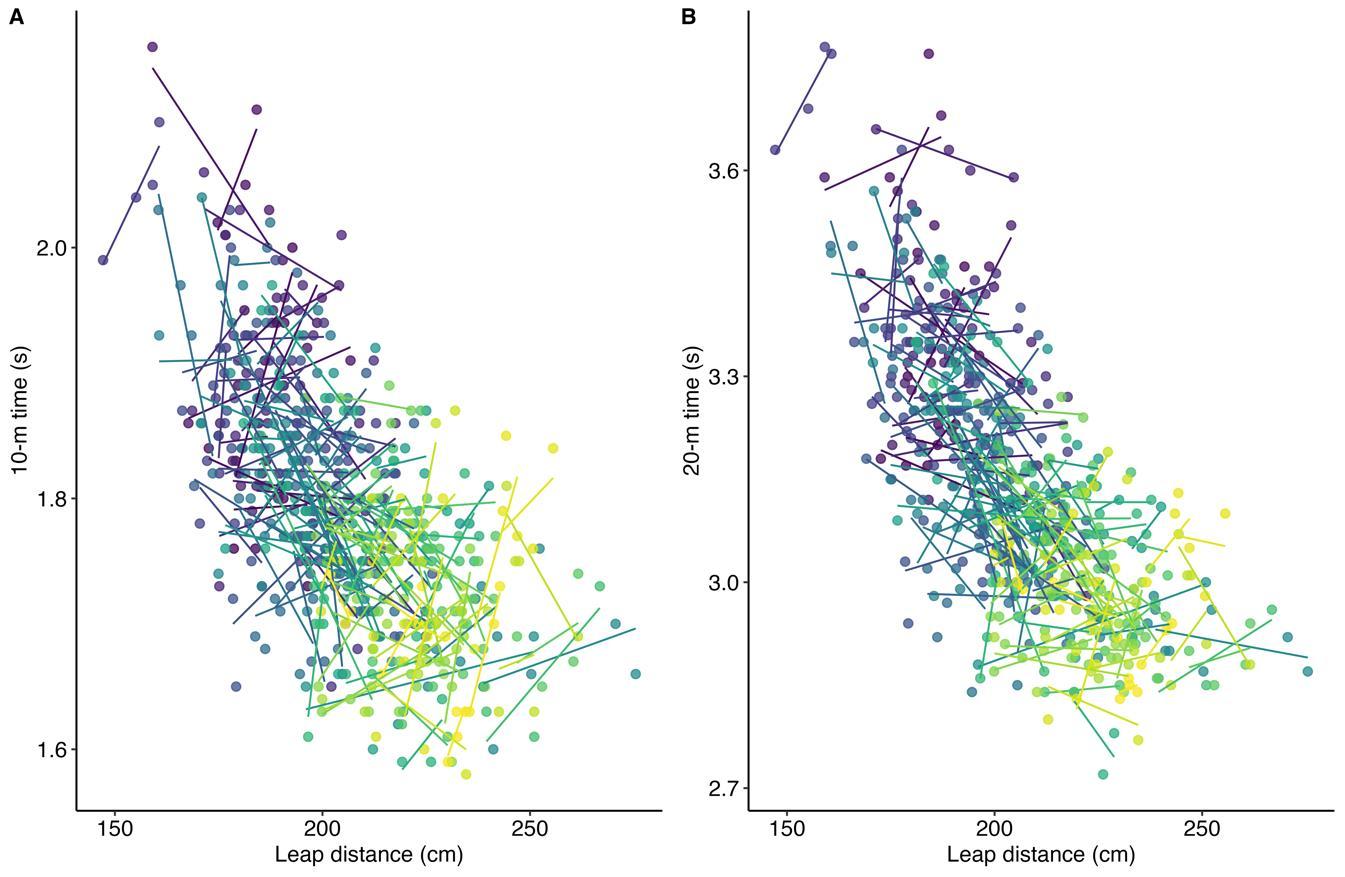
for-distance against 10 m (r = -0.14, 95% CI [-0.24, -0.06]) and 20 m (r = -0.24, 95% CI [-0.34, -0.16]) were small. The lack of within-player relationships is visible from the different directions of the individual slopes in Figure 2. Individual within-player correlations between leap-for-distance and sprint performance ranged between -1.0 and 1.0 for both sprint distances.
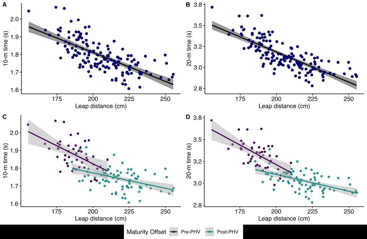
Figure 1: Between-player leap-for-distance and sprint 10 m (A) and 20 m (B) relationships (mean of repeated measures), including via maturity offset stratification (C, D). The grey area in the graph represents the 95% confidence bands of the regression line. For clarity, x and y standard deviation bars about each data point are not shown.
The between-player mean of repeated measures correlation coefficients for leap-for-distance against 10 m sprint time were large and moderate for pre-PHV and post-PHV respectively (r = -0.52, 95% CI [-0.71, -0.26]; r = -0.43, 95% CI [-0.60, -0.24], respectively),andlargefor leap-for-distanceagainst20mforboth pre-PHV and post-PHV (r = -0.62, 95% CI [-0.78, -0.39]; r =0.54, 95% CI [-0.68, -0.36]). The negative correlations with both distances and maturation groups are visible in Figure 1C and 1D. The within-player correlation coefficients of leap-for-distance for pre-PHV against 10 m and 20 m times were small (r = -0.19, 95% CI [-0.35, -0.04]; r = -0.23, 95% CI [-0.38, -0.10]). The withinplayer correlation coefficients of leap-for-distance for post-PHV against 10 m and 20 m were trivial to small (r = -0.08, 95% CI [0.22, 0.02]; r = -0.20, 95% CI [-0.34, -0.08]). The lack of substantial within-player relationships by maturation is visible from the diverse directions of the individual slopes in Figure 3 Similar to the total sample, individual players had both negative
as well as positive correlations between the leap-for-distance and sprint values (10 m, 20 m): ranging across the entire correlation spectrum in both the pre-PHV and post-PHV group (i.e., r range between -1.0 to 1.0).
The differences (pre-PHV minus post-PHV) in mean correlation coefficients between maturation groups were as follows: between-players; leap-for-distance vs. 10 m = rdif -0.09 (-0.38 to 0.20), leap-for-distance vs 20 m = -0.08 (-0.33 to 0.17), within-players; leap-for-distance vs. 10 m = -0.11 (-0.48 to 0.26), leap-for-distance vs 20 m = -0.03 (-0.39 to 0.33). The majority of the point estimates for the difference between groups were below the pre-defined smallest practically important difference (± 0.1). However, the 95% CIs included meaningful small-moderate differences in both between-player and within-player relationships, demonstrating that the groups are not practically equivalent.
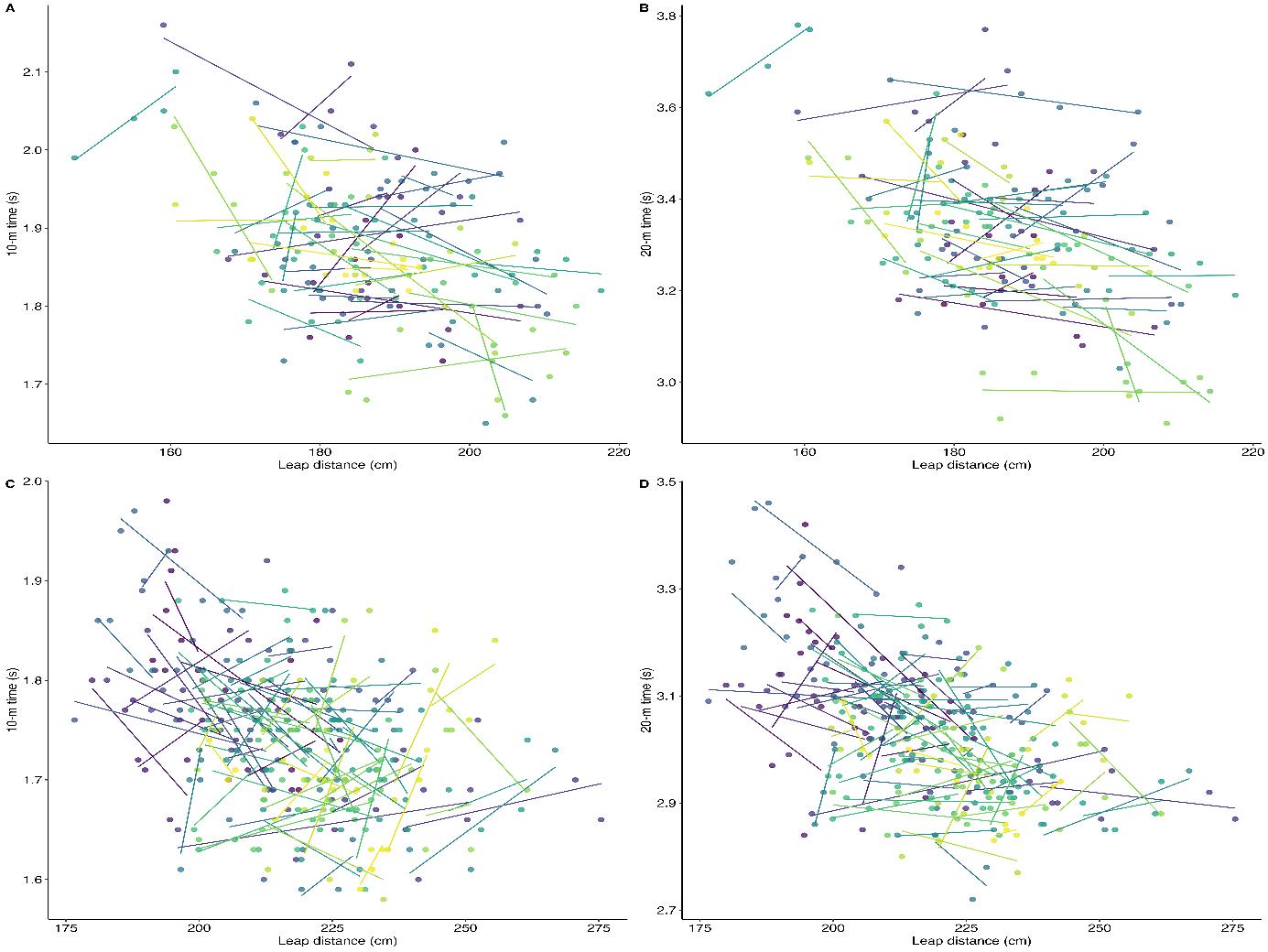
Figure 3: Within-player leap-for-distance and sprint relationships stratified via maturity offset from peak height velocity (PHV) with 10 m pre-PHV (A), post-PHV (C) and 20 m pre-PHV (B) and post-PHV (D). Repeated measures within-player correlation; observations from the same player are given the same colour, with corresponding lines to show the slope for each player.
4. Discussion
This is the first study to investigate longitudinal, within-player sprint performance changes in relation to a leap-for-distance measure in young male soccer players. The results of this study support the first hypothesis by showing clear associations between leap-for-distance and short sprint performance, both pre- and postPHV. This suggests that in similar young male soccer cohorts, the leap-for-distance test would be useful for discriminating sprint ability between players. However, the second hypothesis was not acceptedaswithin-player changesin leap-for-distancedidnot track changes in sprint performance over the duration of testing, irrespectiveofmaturation.Thesefindingsthereforeconfirmthatthe leap-for-distance test is not a good proxy measure for monitoring changes in an individual’s sprint performance over time.
Overall, very large negative associations (between-players) were observed for mean distance in the leap-for-distance test and mean sprint time (Figure 1A, 1B). This indicated that players who had greater distance covered during the leap-for-distance test also had faster sprint times. These results can be explained by the biomechanical similarities between the leap-for-distance and short sprint speed (Lockie et al., 2013; Morin et al., 2015). Similarities also exist between the movement patterns of the leap-for-distance and short sprint speed (i.e., cyclical, reciprocal movement). The findings are consistent with previous research demonstrating that players with high dynamic movement scores (jump) also have faster sprint times (McFarland et al., 2016). The study results demonstrated that this relationship holds over a long time period.
From the findings it was clear that the leap-for-distance length was not appropriate for predicting changes in the sprint times within young players, irrespective of the short sprint distance. Whilst small mean within-player negative relationships existed between the leap-for-distance and sprints, there was a wide range in the within-player individual relationships. A strong positive relationship (greater leap-for-distance length associated with increased [slower] sprint time) was observed in some players, a strong negative relationship in others and in some the relationship was trivial. This finding confirms that changes in the withinplayer leap-for-distance score are not associated with a change in thesprinttimes. Originally,it wasplanned toapplya linearmixed model with repeated sprint times as the outcome variable, and leap-for-distance scores as the predictor. However, the withinplayer correlation analysis revealed no substantial overall relationship, and so further analysis of the type conducted by Goosey-Tolfrey et al. (2020) was not considered instructive.
There might be several reasons why the leap-for-distance test was not a good predictor of sprint times within players over time in this study. One of the reasons could be the prevalence of longterm random error or ’noise’ from the leap-for-distance and sprint testing. The reported short-term testing ’noise’ in the leap-fordistance test was 2.1% (Laas et al., 2021),1.7% in 10 m sprintand 1.4% in 20 m sprint in academy soccer players (Enright et al., 2018). Longitudinal ’noise’ in the tests among young male soccer players has not been established yet on either of these variables. Given the high inter-player variability in the within-player correlation across different time points in this study, it is conceivable that the longitudinal ’noise’ was bigger than the biological change in the sprints. Sprinting is a complex movement and the success in early phase sprint performance (< 20 m) is
dependent on several aspects: horizontal and vertical force application (Lockie et al., 2013; Morin et al., 2015), increased stride frequency (Murphy et al., 2003), shorter contact time (Lockie et al., 2013; Murphy et al., 2003), increased stride length, and longer flight time (Lockie et al., 2013). Inconsistency in any of these elements over repeated measures could have influenced the players` sprint times, potentially adding more ’noise’ to the test. Other factors that could have added to the noise associated with the tests were the environmental changes for the sprint (Haugen & Buchheit, 2016), which were a limitation and not possible to control in this study.
The comparison with maturation revealed large and moderate negative between-player correlations for the pre-PHV and postPHV group between the leap-for-distance and sprint tests. The between-player mean correlation values were compared between maturation groups against a small practically important difference (rdif < 0.10). The 95% CIs for the difference between maturation groups included values beyond this threshold with both distances (10 m, 20 m), indicating that the groups were not practically equivalent and that meaningful differences could not be ruled out statistically. The maturation groups trivial between-player correlation point estimate differences (-0.09 and -0.08) were unexpected as more mature players have outperformed their less mature counterparts in previous research with power and speed activities (Mendez-Villanueva et al., 2011; Murtagh et al., 2018), which would have predicted a stronger negative relationship between the leap-for-distance and sprints in the post-PHV group. The within-player relationships between maturation groups were trivial tosmall(r <-0.30)andtheCIsforthedifferencerangedover the set small important difference threshold with both distances, indicating no clear equivalence between the groups. These findings confirm that maturation did not have a significant impact on the leap-for-distance score and sprint times relationship, although low magnitude correlation between-group differences might exist.
The limitations of the study include the wide CIs of the leapfor-distance test using the AMAT system despite the high reliability (Laas et al., 2021), increasing the potential imprecision of the test with repeated measures. Secondly, the applied nature of the study meant that it was not possible to stringently control the test-to-test time periods due to practical factors. These included a lack of availabilityof the motion tracking system in the clubs or the players being unavailable for testing. Despite this, the test-to-test period was quite consistent (mean 4.4 ± 1.7 months). Lastly, the method of determining biological age via maturity offset has been associated with an error of six months in boys (Mirwald et al., 2002). The latter method is a non-invasive way of assessing maturation in applied settings, which has been successfully used to differentiate maturation status with large groups of young male soccer players (Portas et al., 2016; Towlson et al., 2018).
The study findings suggest that practitioners should not use the leap-for-distance test as a proxy for sprint testing when tracking players over time. Instead, practitioners should consider the individual relationships between the leap-for-distance and sprint scores, which could determine the strength and conditioning strategies that would be suitable for the players` physical development. To optimize individual physical development, the faster players with low leap-for-distance scores could undertake more high force (strength training) and higher leap-for-distance scores with slow sprint times more high velocity based (ballistic)
training (Turner et al., 2021). These assumptions are based on previous recommendations to use jump and short sprint test scores to individualise training and enhance the players` physical profiles (Taylor et al., 2022). Furthermore, if practitioners do not have access to technology for accurately assessing sprint times, a leapfor-distance test might be a useful alternative for identifying quicker and slower players, but this relationship will be weaker when comparing within maturity groups.
5. Conclusion
The results from this study showed that the leap-for-distance test was a good discriminator of short sprint performance between young soccer players. However, the leap-for-distance test was not a good predictor of within-player changes in sprint performance over a longer time period in young soccer players and should not be used as a proxy measure to make intra-athlete speed inferences over short distances. The findings also indicated that sprint distance and maturation did not make a substantial difference in the within-player leap-for-distance and sprint relationships.
Conflict of Interest
The authors declare no conflict of interests.
Acknowledgment
TheauthorswouldliketoexpresstheirgratitudetoProfessorAlan M.Batterham for his invaluable statistical advice throughout the completion of this research. The authors acknowledge the help of Pro Sport Support Ltd for the provision of the anonymised data which was used for this study. At the time of this research, ML and GP were employees of Pro Sport Support Ltd a company seeking the development and commercial sale of practical, marker-based tracking systems for athletic movement screening.
Funding
The project received government funding from a Knowledge Transfer Partnership (Innovate UK) to Pro Sport Support Ltd and Teesside University (KTP 009965. Project title: To develop a specialist technology enhanced Adolescent Movement Analysis Tool and associated training intervention curriculum, exergaming and CPD offers underpinned by leading biomechanical research to improve the physicality of elite youth athletes).
References
Bakdash, J. Z., & Marusich, L. R. (2017). Repeated measures correlation. Frontiers in Psychology, 8, 456–456. https://doi.org/10.3389/fpsyg.2017.00456
Bland, J. M., & Altman, D. G. (1995a). Statistics notes: Calculating correlation coefficients with repeated observations: Part 1 correlation within subjects. BMJ: British Medical Journal, 310(6977), 446–446. https://doi.org/10.1136/bmj.310.6977.446
Bland, J. M., & Altman, D. G. (1995b). Statistics notes:
Calculating correlation coefficients with repeated observations: Part 2 correlation between subjects. BMJ:
British Medical Journal, 310(6980), 633–633.
https://doi.org/10.1136/bmj.310.6980.633
DiSalvo,V.,Baron,R.,Gonzalez-Haro,C.,Gormasz,C.,Pigozzi, F., & Bachl, N. (2010). Sprinting analysis of elite soccer players during European Champions League and UEFA cup matches. Journal of Sports Sciences, 28(14), 1489–1494.
https://doi.org/10.1080/02640414.2010.521166
Enright, K., Morton, J., Iga, J., Lothian, D., Roberts, S., & Drust, B.(2018). Reliability of “in-season” fitness assessments in youth elite soccer players: A working model for practitioners and coaches. ScienceandMedicineinFootball, 2(3), 177–183.
https://doi.org/10.1080/24733938.2017.1411603
Faude, O., Koch, T., & Meyer, T. (2012). Straight sprinting is the most frequent actionin goal situations in professional football. Journal of Sports Sciences, 30(7), 625–631.
https://doi.org/10.1080/02640414.2012.665940
Gathercole, R. J., Sporer, B. C., Stellingwerff, T., & Sleivert, G. G.(2015). Comparison of the capacity of different jump and sprint field tests to detect neuromuscular fatigue. Journal of Strength and Conditioning Research, 29(9), 2522–2531.
https://doi.org/10.1519/JSC.0000000000000912
Goosey-Tolfrey, V., Totosy de Zepetnek, J., Keil, M., BrookeWavell, K., & Batterham, A. M. (2020). Tracking withinathlete changes in whole body fat percentage in wheelchair athletes. International Journal of Sports Physiology and Performance, 16(1), 1–6. https://doi.org/10.1123/ijspp.20190867
Haugen, T., & Buchheit, M. (2016). Sprint running performance monitoring: Methodological and practical considerations. Sports Medicine (Auckland), 46(5), 641–656.
https://doi.org/10.1007/s40279-015-0446-0
Haugen, T., Tønnessen, E., Hisdal, J., & Seiler, S. (2014). The role and development of sprinting speed in soccer. International Journal of Sports Physiology and Performance, 9(3), 432–441. https://doi.org/10.1123/IJSPP.2013-0121
Holm, D. J., Stålbom, M., Keogh, J. W., & Cronin, J. (2008). Relationship between the kinetics and kinematics of a unilateral horizontal drop jump to sprint performance. Journal of Strength and Conditioning Research, 22(5), 1589–1596. https://doi.org/10.1519/JSC.0b013e318181a297
Hopkins, W. G. (2006). A spreadsheet for combining outcomes from several subject groups. Sportscience, 10, 51–53.
Hopkins, W. G., Marshall, S. W., Batterham, A. M., & Hanin, J. (2009). Progressive statistics for studies in sports medicine and exercise science. Medicine and Science in Sports and Exercise, 41(1), 3–12.
https://doi.org/10.1249/MSS.0b013e31818cb278
Hulteen, R. M., Morgan, P. J., Barnett, L. M., Stodden, D. F., & Lubans, D. R. (2018). Development of foundational movement skills: A conceptual model for physical activity across the lifespan. Sports Medicine (Auckland), 48(7), 1533–1540.
https://doi.org/10.1007/s40279-018-0892-6
Juris, P. M., Phillips, E. M., Dalpe, C., Edwards, C., Gotlin, R. S., & Kane, D. J. (1997). A dynamic test of lower extremity function following anterior cruciate ligament reconstruction and rehabilitation. The Journal of Orthopaedic and Sports Physical Therapy, 26(4), 184–191.
https://doi.org/10.2519/jospt.1997.26.4.184
Laas, M. M., Wright, M. D., McLaren, S. J., Eaves, D. L., Parkin, G., & Portas, M. D. (2020). Motion tracking in young male
football players: A preliminary study of within-session movement reliability. Science and Medicine in Football, 4(6), 203–210.
https://doi.org/10.1080/24733938.2020.1737329
Laas, M. M., Wright, M.D., McLaren, S. J., Portas, M. D., Parkin, G., & Eaves, D. L. (2021). Between-week reliability of motion tracking screening: A preliminary study with youth male football players. European Journalof Human Movement, 46, 59–65.
https://doi.org/10.21134/eurjhm.2021.46.11
Lockie, R. G., Murphy, A. J., Schultz, A. B., Jeffriess, M. D., & Callaghan, S. J. (2013). Influence of sprint acceleration stance kinetics on velocity and step kinematics in field sport athletes. Journal of Strength and Conditioning Research, 27(9), 2494–2503. https://doi.org/10.1519/JSC.0b013e31827f5103
Malcata, R. M., Vandenbogaerde, T. J., & Hopkins, W. G. (2014). Using athletes' world rankings to assess countries' performance. International Journal of Sports Physiology and Performance, 9(1), 133–138. https://doi.org/10.1123/ijspp.2013-0014
McFarland,I.T.,Dawes,J.J.,Elder,C.L., &Lockie,R.G.(2016). Relationship of two vertical jumping tests to sprint and change of direction speed among male and female collegiate soccer players. Sports (Basel, Switzerland), 4(1), 11. https://doi.org/10.3390/sports4010011
Mendez-Villanueva, A., Buchheit, M., Kuitunen, S., Douglas, A., Peltola, E., & Bourdon, P. (2011). Age-related differences in acceleration, maximum running speed, and repeated-sprint performance in young soccer players. Journal of Sports Sciences, 29(5), 477–484. https://doi.org/10.1080/02640414.2010.536248
Mirwald, R. L., Baxter-Jones, A. D. G., Bailey, D. A., & Beunen, G. P. (2002). An assessment of maturity from anthropometric measurements. Medicine and Science in Sports and Exercise, 34(4), 689–694. https://doi.org/10.1097/00005768200204000-00020
Morin, J. B., Slawinski, J., Dorel, S., de villareal, E. S., Couturier, A., Samozino, P., Brughelli, M., & Rabita, G. (2015). Acceleration capability in elite sprinters and ground impulse: Push more, brake less? Journal of Biomechanics, 48(12), 3149–3154. https://doi.org/10.1016/j.jbiomech.2015.07.009
Murphy, A. J., Lockie, R. G., & Coutts, A. J. (2003). Kinematic determinants of early acceleration in field sport athletes. Journal of Sports Science and Medicine, 2(4), 144–150.
Murtagh,C. F.,Brownlee,T. E., O’Boyle,A.,Morgans, R., Drust, B., & Erskine, R. M. (2018). Importance of speed and power in elite youth soccer depends on maturation status. Journal of Strength and Conditioning Research, 32(2), 297–303. https://doi.org/10.1519/JSC.0000000000002367
Nicholson, B., Dinsdale, A., Jones, B., & Till, K. (2021). The training of short distance sprint performance in football code athletes: A systematic review and meta-analysis. Sports Medicine (Auckland), 51(6), 1179–1207. https://doi.org/10.1007/s40279-020-01372-y
Oliver, J. L., Lloyd, R. S., & Rumpf, M. C. (2013). Developing speed throughout childhood and adolescence: The role of growth, maturation and training. Strength and Conditioning Journal, 35(3), 42–48.
https://doi.org/10.1519/SSC.0b013e3182919d32
Philippaerts, R. M., Vaeyens, R., Janssens, M., Van Renterghem, B., Matthys, D., Craen, R., & Malina, R. M. (2006). The relationship between peak height velocity and physical
performance in youth soccer players. Journal of Sports Sciences, 24(3), 221–230.
https://doi.org/10.1080/02640410500189371
Portas, M. D., Parkin, G., Roberts, J., & Batterham, A. M. (2016). Maturational effect on Functional Movement Screen™ score in adolescent soccer players. Journal of Science and Medicine in Sport, 19(10), 854–858.
https://doi.org/10.1016/j.jsams.2015.12.001
Small, K., McNaughton,L.R.,Greig,M.,Lohkamp,M., &Lovell, R.(2009). Soccer fatigue, sprinting and hamstring injury risk
International Journal of Sports Medicine, 30(8), 573–578.
https://doi.org/10.1055/s-0029-1202822
Taylor, J. M., Cunningham, L., Hood, P., Thorne, B., Irvin, G., & Weston, M. (2019). The reliability of a modified 505 test and change-of-direction deficit time in elite youth football players. Science and Medicine in Football, 3(2), 157–162.
https://doi.org/10.1080/24733938.2018.1526402
Taylor, J. M., Madden, J. L., Cunningham, L. P., & Wright, M. (2022). Fitness testing in soccer revisited: Developing a contemporary testing battery. Strength and Conditioning Journal, 44(5), 10–21.
https://doi.org/10.1519/SSC.0000000000000702
Towlson, C., Cobley, S., Parkin, G., & Lovell, R. (2018). When does the influence of maturation on anthropometric and physical fitness characteristics increase and subside?
Scandinavian Journal of Medicine & Science in Sports, 28(8), 1946–1955. https://doi.org/10.1111/sms.13198
Turner, A. N., Comfort, P., McMahon, J., Bishop, C., Chavda, S., Read, P., Mundy, P., & Lake, J. (2021). Developing powerful athletes part 2: Practical applications. Strength and Conditioning Journal, 43(1), 23–31.
https://doi.org/10.1519/SSC.0000000000000544
Unnithan, V., White, J., Georgiou, A., Iga, J., & Drust, B. (2012). Talent identification in youth soccer. Journal of Sports Sciences, 30(15), 1719–1726.
https://doi.org/10.1080/02640414.2012.731515
Wijnbergen, M. (2019). Novel algorithms to capture kinematic variables with depth-sensing technology. The development of a reliable, valid and practical movement assessment tool [Doctoral dissertation, Teesside University] Teesside University's Research Portal.
https://research.tees.ac.uk/ws/portalfiles/portal/25374496/Th esis_MarkWijnbergen.pdf
Williams, C. A., Oliver, J. L., & Faulkner, J. (2011). Seasonal monitoring of sprint and jump performance in a soccer youth academy. International Journal of Sports Physiology and Performance, 6(2), 264–275.
https://doi.org/10.1123/ijspp.6.2.264
Wright, M. D., & Atkinson, G. (2019). Changes in sprint-related outcomes during a period of systematic training in a girls’ soccer academy. Journal of Strength and Conditioning Research, 33(3), 793–800.
https://doi.org/10.1519/JSC.0000000000002055
Wrigley, R. D., Drust, B., Stratton, G., Atkinson, G., & Gregson, W.(2014). Long-term soccer-specific training enhances the rate of physical development of academy soccer players independent of maturation status. International Journal of Sports Medicine, 35(13), 1090–1094.
https://doi.org/10.1055/s-0034-1375616

The Journal of Sport and Exercise Science, Vol. 7, Issue 3, 19-30 (2023)
www.jses.net
JSES
ISSN: 2703-240X
Acceleration versus maximum speed sprinting:Apilot study assessing sprint performance, eccentric knee flexor strength, and muscle architecture
Brock W. Freeman1,2*, Scott W. Talpey1, Lachlan P. James3 , Warren B. Young1
1Institute of Health and Wellbeing, Federation University Australia, Ballarat, Australia
2School of Health Sciences and Physiotherapy, The University of Notre Dame Australia, Fremantle, Australia
3School of Allied Health, La Trobe University, Bundoora, Australia
Received: 13.04.2023
Accepted: 12.07.2023
Online: 31.12.2023
Keywords:
Muscle imaging
Eccentric strength
Performance
Speed
Hamstring
1. Introduction
The objective of this study was to implement a pilot protocol to investigate the effects of acceleration and maximum speed sprint training on sprint performance, eccentric knee flexor strength, and knee flexor muscle architecture. A pre- and post-intervention study including a controlgroup was employed. Twelve male field sport athletes (age =23.1 ± 3.2 years, height = 180.1 ± 8.3 cm, body mass = 80.1 ± 6.4 kg) were recruited to participate in this study. Participants were allocated to either an acceleration, maximum speed, or a controlgroup. Participantscompleted pre-andpost-testing consisting ofa 40-metresprint, eccentric knee flexor strength assessment, and an ultrasound of the Biceps Femoris Long Head(BFLH).Traininginterventionswerecompletedover6weekswith2sessionsperweek. Knee flexormuscle sorenessdata wascollectedusinga10-pointscale. BFLH fascicle length increased by 1.95 cm in the acceleration group and 1.67cm in the maximum speed group. Trivial changes were reported in eccentric knee flexor strength for all three groups. Both acceleration and maximum speed training improved sprint performance. Both forms of sprinttrainingwereeffectiveforincreasingBFLH fascicle length.Maximumspeedsprinting mayofferaprophylacticbenefitformodifiablehamstringinjuryriskfactors.Thesefindings should be confirmed with studies on a larger scale.
Hamstring strain injury (HSI) is consistently one of the most common injuries experienced by athletes competing in field sports with a high-speed running (HSR) component (AFL, 2021; Ekstrand et al., 2021). This incurs a significant time away from competition (AFL, 2021), and substantial financial cost (Hickey et al., 2014; Specialty, 2017). Previous work has identified modifiable risk factors for HSI, such as the combination of low eccentric knee flexor strength, and short Bicep Femoris Long Head (BFLH) fascicles (Bourne et al., 2017; Timmins et al.,
2015b). Whilst Timmins et al. (2015b) demonstrated thresholds of less than 337 N and 10.56 cm place professional soccer athletes at increased risk, Dow et al. (2021) found that that thresholds should be sport specific. Therefore, the context of eccentric strength and architectural adaptations are still highly relevant for HSI prevention purposes. Eccentric hamstring exercises have previously been shown to effectively mediate these risk factors. Exercises such as the Nordic Hamstring Exercise (NHE) increase strength and fascicle length with relatively low volume (Presland et al., 2018), however, some of the criticism stems from the nonspecific movement of the NHE (Freeman et al., 2021). This is
*Corresponding Author: Brock Freeman, The University of Notre Dame Australia, Australia, brockw.freeman@gmail.com
echoed by researchers who found that compliance was poor amongst professional soccer teams (Bahr et al., 2015), and a recent review has questioned the preventative effect of the exercise for HSI, suggesting it should only be conditionally prescribed (Impellizzeri et al., 2021).
Shorter fascicle lengths in the BFLH have been shown to be associated with increased risk of future HSI (Timmins et al., 2015b). This is best explained by a presumed greater number of sarcomeres in series, increasing the ability of the fascicle to tolerate high loads (Morgan, 1990). Previous work has identified that adaptations to the fascicles of the BFLH occur over relatively short periods of time (7 – 10 days; Presland et al., 2018). Therefore, the ability to measure fascicle length accurately and reliably is important, as it provides valuable insight as to whether adaptation or maladaptation’s have occurred. Of the available methods to assess BFLH fascicle length, two dimensional (2D) βmode ultrasound is the most cost effective and easily accessible method (Franchi et al., 2020; Sarto et al., 2021). A current issue with using 2D ultrasound to measure fascicle length is that the entire fascicle is not captured in a single image. Therefore, an estimation must be made using points digitised from the image. A body of research has demonstrated that this method of ultrasound to be valid and reliable measure to assess fascicle length at rest for several different muscles (Blazevich et al., 2006; Kellis et al., 2009; Timmins et al., 2015a).
Sprinting, either through acceleration or maximum speed instances, is the most common activity associated with HSI in Australian Football (Freeman et al., 2021). It is postulated that the late-swing phase, where the hamstring works maximally to decelerate the lower leg, is the most likely point in the gait cycle where injury occurs. Currently, five key studies (Freeman et al., 2019; Ishøi et al., 2017; Krommes et al., 2017; Mendiguchia et al., 2020; Timmins et al., 2021) have examined the effect of eccentric knee flexor strengthening on sprint performance in field sport athletes.Ofthesestudies,fourreportedincreasesineccentricknee flexor strength (Freeman et al., 2019; Ishøi et al., 2017; Mendiguchia et al., 2020; Timmins et al., 2021), one reported small to moderate increases in fascicle length (Mendiguchia et al., 2020), and three reported increases in acceleration sprint performance (Ishøi et al., 2017; Krommes et al., 2017; Timmins et al., 2021). Two of the studies investigated sprint training, showing a significant increase in eccentric knee flexor strength (Freeman et al., 2019), and a significant increase in BFLH fascicle length (Mendiguchia et al., 2020). These two interventions have both utilised a combination of acceleration, maximum speed, and resisted sprinting as a part of the training program.
Furthermore, sprinting has been proposed as a potential “vaccine” for HSI (Edouard et al., 2019). In a field sport setting, both acceleration and maximum speed are important components of sprinting. Higashihara et al. (2017) reported significant differences in acceleration and maximum speed sprinting. This is reflected by a change in the orientation of force, whereby acceleration requires more horizontal force production and maximumspeed transitions tomorevertical forceproduction. The acceleration gait displays significantly higher Biceps Femoris (BF) activation when compared to the medial hamstrings (Higashihara et al., 2017). This is supported by findings suggesting the BF is key component of acceleration sprinting (Morin et al., 2015). Interestingly, HSI are more frequent in the BF opposed to the medial hamstrings (Askling et al., 2012), yet
hamstring injuries occur during both accelerations and maximum speed sprints. Conversely, the medial hamstrings are significantly more activated during the swing phase of the maximum speed sprint (Higashihara et al., 2017), potentially as they are required to decelerate the lower leg from high-speed in preparation for ground contact. These findings are further consolidated by significant increases in force production and negative work completed by the hamstrings as running speeds exceed 80% Vmax (Chumanov et al., 2007; Dorn et al., 2012; Fiorentino et al., 2014). It should also be considered that Higashihara et al. (2017) measured acceleration at the 15-metre mark. As ~74% Vmax is achieved by the 10-metre mark of a sprint (Healy et al., 2019), the 15-metre mark may not be a true reflection of acceleration.
As previously established, maximum speed sprinting is a risk for HSI. However, if carefully administered with progressive overload, maximum speed training can be expected to produce adaptations that will make the hamstrings more robust. This strategy is likely to be effective when you consider that conventional hamstring exercises (including the NHE) do not mirror the stress placed upon the hamstrings that sprinting at 100% Vmax does (Prince et al., 2021; van den Tillaar et al., 2017). Sprint training also provides the additional benefit of improving sprint performance. Therefore, given the significantly higher activation, force production and work completed, maximum speed sprinting may provide a better prophylactic effect on HSI opposed to acceleration sprinting. Findings from previous studies (Edouard et al., 2019; Freeman et al., 2021; Freeman et al., 2019; Mendiguchia et al., 2020) point towards the need for further investigation of sprint training for HSI risk mitigation. Furthermore, the clear differences between acceleration and maximum speed sprinting (Higashihara et al., 2017) suggest that different training adaptations, specifically on modifiable risk factors such as muscle architecture and eccentric knee flexor strength are possible. This study’s primary aim is to outline the training responses of two separate sprint training methods: acceleration and maximum speed on modifiable risk factors such as eccentric knee flexor strength and BFLH fascicle length, whilst concurrently assessing changes in sprint performance.
2. Methods
2.1. Participants
Twelve amateur male Australian Rules football athletes (age = 23.1 ± 3.2 years, height = 180.1 ± 8.3 cm, body mass = 80.1 ± 6.4 kg, training experience = 6 ± 2.6 years) were recruited to participate in this study. No participant reported any prior HSI. This project received ethical approval from the University Human Research Ethics Committee (Approval Number – A19-135). All participants were injury free for six months prior to the study. Participants were informed of the benefits and risks of the investigation priorto signing an institutionally approved informed consent document to participate in the study.
2.2. Study design
Participants in the pilot study completed a series of tests before they were allocated into one of two interventions, or a control group. Following the completion of a standardised warm up,
testing consisted of a 40-metre sprint, an eccentric knee flexor strength assessment, and an ultrasound of the BFLH. Once testing was complete, participants were allocated into the three groups based upon their eccentric knee flexor strength (the strongest participant was allocated to acceleration training, the second strongest to maximum speed training, and the third strongest to the control group. This process was repeated until all participants were allocated, leaving four participants for each condition. This was completed to create equality ofpre-training eccentricstrength across the three groups before training interventions were completed. Following six weeks of training and a weekly ultrasoundscanoftheBFLH,participantsrepeatedthesametesting procedure. Throughout the training intervention, participants were instructed to maintain their normal training load, and not to add any additional hamstring specific exercises.
2.3. Testing procedures
Prior to completing sprint testing, participants completed a standardised warm up of a 4-minute jog, 20 metres of walking lunges, 20 metres of lateral lunges, 20 metres of arabesques, and 20metresofhamstringsweeps.Followingthisgeneralcomponent of the warm up, 20 metres of high knees, butt kicks, A-Skips, and A-Skips for maximal height were performed. Participants then completed four run-throughs over 40 metres, commencing at approximately 60% of perceived maximal effort and progressing to approximately 95% to 100% of perceived maximal effort.
2.3.1. Sprint performance
A 40-metre sprint was measured using timing gates (Speedlight; Swift Performance, QLD, AUS) to assess the acceleration and maximum speed capabilities of participants. As outlined in Figure 1, a 0 – 10-metre split was incorporated to assess acceleration qualities and a 30 – 40-metre split was incorporated to estimate maximum speed qualities (Freeman et al., 2019; Young et al., 2008). Participants completed two 40-metre trials with a full recovery between trials (i.e., longer than 5 minutes). A third trial was completed if the second trial was more than 0.2 s quickerthan the first. Participants were instructed to start with their preferred foot forward, with the opposing arm raised in the air to avoid the premature start of the timing system. Participants were required to be in a stationary position with their torso as close as possible to the light beam. All participants were instructed they were unable to rock to gain any momentum prior to starting. Participants used a self-selected distance from the front toe to the start line to allow for various body dimensions, providing the torso was as close to the beam as possible. Participants were allowed to trial these starting positions during their run-throughs in the warm up, however, the pre-testing distance was recorded for post-testing, with the range being 0 cm from the timing gate to 40 cm from the timing gate. This testing was completed in a temperature-controlled (21 °C) basketball stadium, on a wooden springboard surface with all participants wearing the same footwear for pre and post testing. The best (fastest) time between the 0 – 10-metre split and 30 – 40-metre split was retained for analysis.

Figure 1: Diagram of sprint testing protocol used for acceleration (0 – 10-metre) and estimated maximum speed (30 – 40-metre).
2.3.2. Eccentric knee flexor strength
Participants involved in this project had all previously completed the testing procedure for eccentric knee flexor strength (NordBord; VALD Performance, QLD) through prior individual training or testing sessions. This device has been previously shown to be a valid and reliable measure of eccentric knee flexor strength (Opar et al., 2013). Following a 5-minute break after the sprint testing, participants performed a warm up set of three repetitions at approximately 60%, 70%, and 80% of perceived maximum effort. Following a 3-minute rest, participants were instructed to perform three maximal contractions. Participants were instructed to maintain a straight line from the shoulder, through the hip to the knee, whilst keeping hands at chest level to brace for the point of failure (Freeman et al., 2019). Whilst performing each repetition, participants were instructed to control the contraction for as long as possible (Opar et al., 2013). If the participant was able to complete a full repetition, external resistance was available to ensure a maximal effort. The corresponding knee position was recorded so that the participant set up was repeatable in the post-testing (Opar et al., 2013). The best trial for left leg, right leg, and peak bilateral force was retained for analysis.
2.3.3. BFLH architecture
Intra-rater reliability of muscle imaging was completed prior to the commencement of the pilot intervention. The timeframe between the first test and the second test was three days. Participants were well-rested for 24 hours before both imaging sessionsandwereinstructed tocompletenosprintingorresistance trainingbetweenvisits.UltrasoundimagesoftheleftBFLH muscle belly was captured using 2D β-mode ultrasound (frequency = 12 MHz, depth = 6.5 cm, field of view = 14 × 47 mm; Mindray DP20; Shenzhen, ROC).
Toensureconsistencyofthescanningsite,thedistancebetween the ischial tuberosity and knee joint fold was marked at the halfway point along the line of the BFLH. This distance corresponds closely with the belly of the BFLH. This is important for two key reasons (Blazevich et al., 2006). Firstly, estimates assume that there is no curvature of fascicles or aponeurosis, which is only true at the midpoint of the muscle belly. Secondly, the common assumption is that the structure of the BFLH is isotropic, which is not accurate because as the muscle moves closer to the origin or insertion point, the muscle changes shape to fit in with the respective anatomical structures. Once this site was marked, the distance of this site was recorded from various anatomical landmarks, such as the head of the fibula to ensure a reproducible scanning area. Participants lay
prone with a pillow placed under the left ankle, before a layer of conductive gel was placed over the scanning site. The linear probe was placed longitudinally and perpendicular to the posterior thigh (Blazevich et al., 2006; Timmins et al., 2015a). The sonographer ensured minimal pressure was applied to the skin, as this has previously been shown to influence image quality (Blazevich et al., 2006;Timminsetal.,2015a).Toobtainadequateimages, theprobe was slightly manipulated to ensure the superficial and intermediate aponeurosis were parallel. To ensure a quality image was captured, the sonographer completed several hours of training, and was mentored by a researcher experienced in BFLH sonography.
Following the image collection, analysis was performed offline (MicroDicom, V.2.7.9, Bulgaria). Following previously established methods six markers were digitally created for each of the images (Timmins et al., 2015a). The gap between superficial and intermediate aponeurosis was used to establish muscle thickness. A clear fascicle of interest with no curvature or distortion was then marked on the image (Supplementary Figure 1). For both aponeuroses, the aponeurosis angle was defined as the angle created by a horizontal line across the image and the intersecting line marked as the aponeurosis (Timmins et al., 2015a). To estimate fascicle length, a previously validated equation (Timmins et al., 2015a) was used:
in which FL = Fascicle Length, AA = Aponeurosis Angle, MT = Muscle Thickness, and PA = Pennation Angle Fascicle Length was reported in absolute terms (cm). All analysis was performed by the same sonographer, who was blinded to participant identifiers when completing the analysis. This process was then repeated for the weekly ultrasound scans to track BFLH fascicle length changes in acceleration and maximum speed participants. These images were collected at a standardised time; 24 hoursafter the second training session of the week. This frequency of measurement allows for accurate recording of time course changes in fascicle length, which is of particular interest given the short adaptation window observed in prior resistance training study (Presland et al., 2018).
2.4. Training interventions
Participants completed training sessions individually under the supervision of a qualified strength and conditioning coach during the restrictions of the Victorian Government COVID-19 restrictions in 2020. All sessions were completed with a minimum of 48 hours of recovery between sprint exposures. Prior to each training session, participants completed a standardised warm up procedure like that used for testing sessions. A full description of both the acceleration and maximum speed training programs can be seen in Table 1 and Table 2, respectively.
Note: *denotes racing against timing gates, Reps = Repetitions, %ME = percentage of perceived maximum effort.
Note: *denotes racing against timing gates, Reps = Repetitions, %ME = percentage of perceived maximum effort
Both training programs were designed following considerations outlined by Haugen and colleagues (Haugen et al., 2019). The mismatch in volume between the acceleration and maximum speed interventions is explained by Haugen et al. (2019) guidelines that suggest better adaptations for acceleration occur with slightly higher volumes, whereas maximum speed training volumes are typically lower. Additionally, maximum speed sessions must also account for the gradual build up to achieve maximum speed (Haugen et al., 2019). A gradual build up was favoured over an all-out acceleration for two reasons. Firstly, it would avoid hard accelerations and secondly, a gradual acceleration produces higher maximum velocities in field sport athletes (Young et al., 2018). Hamstring muscle soreness was collected as a point of interest in this study. To collect hamstring muscle soreness information, athletes were asked at the start of the following session to rate their soreness on a scale of 0 – 10, where 0 was equal to no soreness, and 10 was to extreme hamstring soreness (Freeman et al., 2019).
2.5. Statistical analyses
The statistical analyses for this study were performed using SPSS Version 25.0 (IBM, Armonk New York, NY,USA). Todetermine
intra-rater reliability for muscle architecture measures, a ShapiroWilk test was used to assess the data for normal distribution. To assess test-retest reliability between the first and second scan, intra-class correlation coefficients (ICC) were calculated. This was supported by further calculations of the typical error (TE), and the coefficient of variation (CV). The minimal detectable change at a 95% confidence interval was calculated in accordance with Timmins et al. (2015a) as TE × 1.96 × √2. Previous research suggests the following ICC values are suitable for use in this analysis; less than or equal to 0.79 as poor, 0.80 to 0.89 as moderate, and greater than or equal to 0.90 as high (Timmins et al., 2015a; Watsford et al., 2010). Due to the COVID-19 restrictions enforced in Victoria, Australia during participant recruitment, the sample size was not high enough to achieve statistical power. A key focus of this analysis was to closely examine the individual responses to the training interventions. Individual results are often undervalued when interpreting group data. This descriptive method is also a useful application for practitioners seeking to assess changes in their athletes. Presentation of individual changes alongside the trends of a group (or in a sporting example, a team) provides context around the information.
3. Results
3.1. Compliance
Seven of the eight participants allocated to training interventions completed all 12 training sessions. The eighth participant completed 11 of a possible 12 sessions. Typically, sessions were completed on a Monday to Thursday or Tuesday to Friday schedule.
3.2. Sprint performance
3.3. Eccentric knee flexor strength
Eleven of the 12 participants completed this study, however, one member of the control group dropped out due to work commitments. In general, acceleration and maximum speed training elicited a 5% and 4% increase in eccentric strength, respectively. The control participants improved eccentric strength to a slightly greater degree (9%). A full outline of the individual eccentric strength results can be seen in Table 3.
All athletes that completed a sprint training intervention improved split times, whereby the acceleration group increased fascicle length by 1.95 ±0.68 cm and the maximum speed group increased by 1.67 ± 0.50 cm (Figure 2). Both the acceleration training group and the maximum speed training group displayed larger improvements in maximum speed than acceleration. Two of the three control participants were slower in the post-testing.
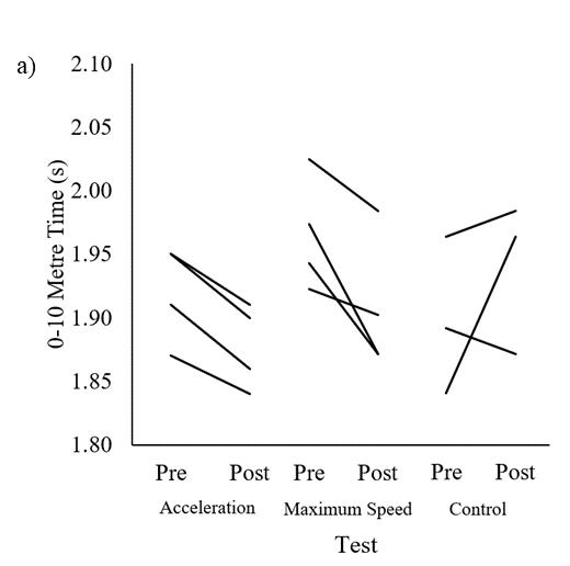
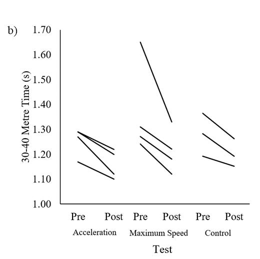
Note: Diff (%): differences between pre- and post-test in percentage.
Intra-rater reliability results of the sonographer are presented in Table 4 (Freeman, 2022). The absolute changes in BFLH fascicle length and eccentric knee flexor strength from pre-test to post-test are visualised in Figure 3 and representative images are included in Supplementary Figure 1. All eight of the participants that took part in structured acceleration or maximum speed sprint training increased fascicle length and decreased pennation angle, as detailed in Table 4. Fascicle lengths were tracked weekly to provide more detailed information. This is reported in Figure 4 which details changes in BFLH fascicle length for acceleration and maximum speed participants. Means and standard deviation for the soreness scores for the acceleration and maximum speed training interventions are displayed in Supplementary Figure 2.
Table 4: Descriptive statistics and test-retest reliability data for the architectural characteristics of the BFLH at rest.
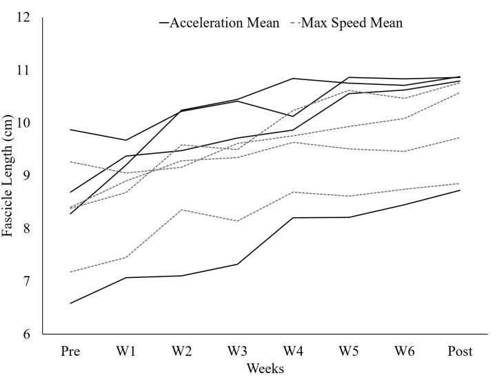
4. Discussion
Note: ICC = Intraclass Correlation Coefficient, TE = Typical Error, MDC95 = Minimal Detectable Change at 95% Confidence Intervals
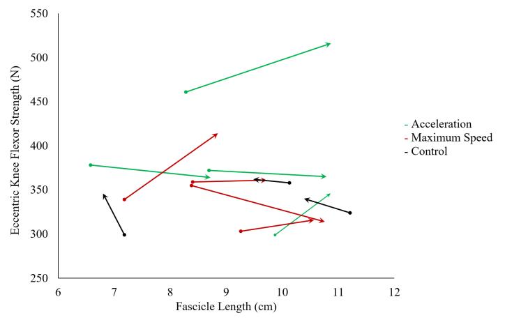
Figure 3. Pre and post-test
This is the first study to investigate the effects of an acceleration specific and maximum speed specific sprint training programs on eccentric knee flexor strength, BFLH fascicle length, sprint performance. Previous investigations have utilised a combination of both acceleration and maximum speed in the intervention design (Freeman et al., 2019),or havealso included resisted sprint training (Mendiguchia et al., 2020). All participants in this study complete each of the 12 allocated sessions across the 6-week training period. Furthermore, the means from 4 participants only represent trends, and any conclusions presented with caution. However, findings from both interventions suggest that sprint training improves eccentric knee flexor strength (Freeman et al., 2021; Mendiguchia et al., 2020) and increase BFLH fascicle length (Mendiguchia et al., 2020). The inclusion of an acceleration specific and maximum speed stimulus is entirely novel in this study design. Prior investigations highlighted the differences between the two aspects of sprint mechanics in relation to the hamstrings (Higashihara et al. 2017) The current study employed a more specific free-sprint (no additional resistance, assistance, or gradient change) acceleration stimulus, which is similar training methods used in field sport athletes. The allocated time to speed trainingperweekalignswiththetimeallocationforsprinttraining reported by high-performance managers in Australian Rules football (Freeman et al., 2021).
4.1. Eccentric knee flexor strength
Eccentric knee flexor strength remained unchanged, as described by the 1% increases for both interventions. This finding is contrary to previously reported findings assessing the influence of sprint training on eccentric knee flexor strength, where a combination of sprint training methods increased eccentric knee
flexor strength in adolescent athletes (Freeman et al., 2019). The baseline measurements for both training groups were above previously reported thresholds (Opar et al., 2014). Therefore, an explanation may be that the stimulus provided by free sprinting alone may not be enough to elicit adaptations in eccentric knee flexor strength, particularly in athletes with high initial values of eccentric knee flexor strength.
4.2. BFLH architecture
Large increases in BFLH fascicle length were observed for both acceleration and maximum speed interventions (23% and 20%, respectively) in this study. Conversely, the control participants in this study decreased by 0.62 cm (7%). These findings are supportive of Mendiguchia et al. (2020), where athletes completing a sprint training program involving resisted sprints, accelerations and maximum speed repetitions increased fascicle length by 1.66 cm (16%). The increases in fascicle length for both training groups were complemented by decreases in pennation angle. The inverse is true for the control group who decreased fascicle length and increased pennation angle. This indicates that the sprint exposure for both groups was likely adequate to improve fascicle length and may prove as viable training strategy to address this modifiable risk factor. Furthermore, the information displayed in Figure 4 should be considered from a trainability perspective. Twelve sessions of either acceleration or maximum speed sprinting was sufficient to improve BFLH fascicle length, a known modifiable risk factor for HSI (Timmins, et al., 2015b), by more than the MDC95
Subsequently, a key finding from this study is the short period of hamstring architectural adaptation. Approximately three weeks of training was sufficient to see an increase in fascicle length outside the MDC95 (Figure 4). This is an important point for strength and conditioning practitioners, as six sessions of either acceleration or maximum speed training is compliant with typical speed training time allocations in elite field sport (Freeman et al., 2021). Moreover, given the low time constraints, low levels of soreness, and minimal equipment requirements, sprint training is applicable across a range of different elite and non-elite field sports.
4.3. Sprint performance
As expected, both acceleration training and maximum speed training improved sprint performance outcomes (Figure 2). This finding was expected and supports previous investigations in this area (Lockie et al., 2012; Rumpf et al., 2015). The relatively large improvement in maximum speed may be explained by participant 3.However, with this individual’s data removed, maximum speed qualities improved by 7% for the acceleration training group. Additionally, the maximum speed training intervention improved acceleration qualities by3% and maximum speed qualities by 9%. Whilst the relationship between improved acceleration and improved maximum velocity has previously been established (Morin et al., 2012; Rabita et al., 2015; Robbins & Young, 2012; Slawinski et al., 2017; Vescovi & McGuigan, 2008), sprint training studies have typically only assessed the influence of acceleration training on acceleration performance or maximum speed training on maximum speed sprinting performance (Rumpf
et al., 2015). The control intervention in this study also tested slightly faster for 0 – 10-metre time and 0 – 40-metre time. The slight increase in eccentric strength observed in the control group may be attributed to the acceleration increase, as previously documented (Ishøi et al., 2017). However, at present there is no explanation for the increase in eccentric strength, as control subjects were instructed to maintain normal training loads, and not to add or increase the load of any specific hamstring or sprinting exercises. This is an unexpected finding; therefore, larger scale studies should be performed to further confirm these findings.
5. Practical applications
This study provides a pilot protocol to assess changes in sprint performance, eccentric knee flexor strength, and BFLH architecture in field sport athletes. The training interventions were completed with minimal soreness (Supplementary Figure 2) and were of an appropriate duration to see changes in sprint performance and muscle architecture in this instance. This should be repeated on a larger scale to confirm initial findings. In addition, this study directly outlines the effect of training a specific speed quality (e.g., maximum speed) on a separate speed quality (e.g., acceleration maximum speed).
In addition, this study is also the first to document the effects of a maximum speed training using gradual build ups, opposed to maximum speed training with a hard acceleration. As neither of these findings havebeenpreviously documented inpeer-reviewed literature, it presents as a novel finding from this study. This may be best explained however, by a retrospective investigation into the 40-yard dash completed at the National Football League Combine, that determined that a higher maximum velocity was important for higher acceleration (Clarke et al., 2019). Theoretically, Vmax may serve as a barrier to acceleration performance, therefore a high Vmax will facilitate a better acceleration phase (Clarke et al., 2019). This theory supports the findings of this study, where the average improvement in the maximum speed split (30 – 40 m) was 0.11 s, coinciding with an improvement of 0.06 s in the acceleration component (0 – 10 m).
Broadly, this study aimed to determine the effects of acceleration andmaximum speed sprint training on eccentricknee flexor strength, BFLH fascicle length and sprint performance. The results indicate that both interventions; acceleration and maximum speed, likely increase BFLH fascicle length and reduce pennation angle. This is in support of the one other study to investigate a combined sprint training protocol on BFLH adaptations (Mendiguchia et al., 2020) . There was no clear change in eccentric knee flexor strength observed, however strength has previously been reported to increase following spring training (Freeman et al., 2019; Ishøi et al., 2017; Mendiguchia et al., 2020; Timmins et al., 2021). As expected, both sprint training methods improved sprint performance, however maximum speed sprint training appeared to improve acceleration and maximum speed sprint times to a greater degree.
This study has a number of limitations. Firstly, the reduced sample power as result of the COVID-19 pandemic and resultant restrictions make drawing broader conclusions more difficult. However, the preliminary findings of increased fascicle length and sprint performance cannot be easily dismissed. More so, this
provides further evidence that sprint training still needs to be pursued at a larger, higher-powered level. Secondly, the athletes in this study were amateur footballers with a regular training history. Therefore, this protocol should be repeated with the same methodology, with a larger sample size, in both elite populations and athletes with very little training history. The practical applications of these findings add further weight to the notion that sprint training is a possible intervention to reduce the risk of HSI in field sport athletes. This study exposed athletes to approximately 150 m to 300 m of sprinting per week for six weeks, volumes that align withbest practice recommendations for sprint training (Haugen et al., 2019)
Furthermore, a potential key benefit is the influence sprinting produces on BFLH architecture. This study reported an average increase in fascicle length of 1.95 cm and 1.67 cm for acceleration and maximum speed interventions respectively. This increase is only slightly smaller than the findings of Presland et al (2018). The additional benefit of sprinting is the concomitant injury and performance benefits associated with sprinting, which is vital to success in field sports (den Hollander et al., 2016; Faude et al., 2012;Rossetal.,2015).Thereisaclearneedforbothacceleration and maximum speed sprinting in field sports. As both interventions delivered increased fascicle length, both may be appropriate at different times during a cycle of training. However, given the carryover benefits of maximum speed training on improvements in acceleration, perhaps earlier prioritisation of maximum speed training may provide the best ‘bang for buck’ stimulus to reduce the risk of HSI, and improve sprint performance in field sport athletes.
Conflict of Interest
The authors declare no conflict of interests.
Acknowledgment
The authors would like to thank Dr. David Opar for his assistance with study design and analysis of the data collected. The authors would also like to thank Ballarat Basketball for their assistance with the testing venue and Dr. Kirsten Porter for her assistance with data collection.
References
AFL. (2021). 2020 AFL Injury Report. Retrieved from www.afl.com.au/news/632528/afl-releases-2020-injuryreport
Askling, C. M., Malliaropoulos, N., & Karlsson, J. (2012). Highspeed running type or stretching-type of hamstring injuries makes a difference to treatment and prognosis. BritishJournal of Sports Medicine, 46(2), 86–87
Bahr, R., Thorborg, K., & Ekstrand, J. (2015). Evidence-based theory hamstring injury prevention is not adopted by the majority of Champions League or Norwegian Premier League football teams: The Nordic Hamstring survey. British Journal of Sports Medicine, 49(22), 1466–1471.
Blazevich, A., J., Gill, N., D, & Zhou, S. (2006). Intra- and intermuscular variation in human quadriceps femoris
architecture assessed in vivo. Journal of Anatomy, 209, 289–310.https://doi.org/10.1111/j.1469-7580.2006.00619.x
Bourne, M. N., Timmins, R. G., Opar, D. A., Pizzari, T., Ruddy, J.D., Sims, C., Williams, M. D., & Shield, A. (2017). An evidence-based framework for strengthening exercises to prevent hamstring injury. Sports Medicine, 48(2), 251–267.
https://doi.org/10.1007/s40279-017-0796-x
Chumanov, E. S., Heiderscheit, B. C., & Thelen, D. G. (2007). The effect of speed and influence of individual muscles on hamstring mechanics during the swing phase of sprinting. Journal of Biomechanics, 40, 3555–3562.
https://doi.org/10.1016/j.jbiomech.2007.05.026
Clarke, K. P., Rieger, R. H., Bruno, R. F., & Stearne, D. J. (2019). The National Football League Combine 40-yd dash: How important is maximum velocity? Journal of Strength and Conditioning Research, 33(6), 1542–1550.
https://doi.org/10.1519/JSC.0000000000002081
den Hollander, S., Brown, J., Lambert, M., Treu, P., & Hendricks, S.(2016). Skills associated with line breaks in elite Rugby Union. Journal of Sports Science and Medicine, 15(3), 501–508.
Dorn, T., W., Schache, A., G., & Pandy, M., G. (2012). Muscular strategy shift in human running: Dependence of running speed on hip and ankle muscle performance. Journal of Experimental Biology, 215, 1944–1956.
https://doi.org/10.1242/jeb.064527
Dow, C. L., Timmins, R. G., Ruddy, J. D., Williams, M. D., Maniar, N., Hickey, J. T., Bourne, M. N., & Opar, D. A. (2021). Prediction of hamstring injuries in Australian Football using biceps femoris architectural risk factors derived from soccer. The American Journal of Sports Medicine, 49, 1–9. https://doi.org/10.1177/03635465211041686
Edouard, P., Mendiguchia, J., Guex, K., Lahti, J., Samozino, P., & Morin, J.-B. (2019). Sprinting: A potential vaccine for hamstring injury? Sports Performance and Science Reports, 48, 1–2. https://doi.org/10.16603/ijspt20170718
Ekstrand, J., Spreco, A., Bengtsson, H., & Bahr, R. (2021). Injury rates decreased in men’s professional football: Year prospective cohort study of almost 12,000 injuries sustained during 1 8 million hours of play. British Journal of Sports Medicine, 55, 1–9. https://doi.org/10.1136/bjsports-2020103159
Faude, O., Koch, T., & Meyer, T. (2012). Straight sprinting is the most frequent actionin goal situations in professional football. Journal of Sports Sciences, 30, 625–631.
https://doi.org/10.1080/02640414.2012.665940
Fiorentino, N. M., Rehorn, M.R, Chumanov, E. S., Thelen, D. G., & Blemker, S. S. (2014). Computational models predict larger muscle tissue strains at faster sprinting speeds. Medicine and Science in Sports and Exercise, 46, 776–786.
https://doi.org/10.1249/MSS.0000000000000172
Franchi, M. V., Fitze, D. P., Raiteri, B. J., Hahn, D., & Sporri, J. (2020). Ultrasound-derived biceps femoris long head fascicle length: Extrapolation pitfalls. Medicine & Science in Sports& Exercise, 52(1), 233–243.
https://doi.org/10.1249/mss.0000000000002123
Freeman, B. W. (2022). The role of sprint training in hamstring injury prevention in field sports. [Doctoral dissertation, Federation University Australia] Federation ResearchOnline.
http://researchonline.federation.edu.au/vital/access/HandleRe solver/1959.17/185191
Freeman, B. W., Talpey, S. W., James, L. P., & Young, W. B. (2021). Sprinting and hamstring strain injury: Beliefs and practices of professional physical performance coaches in Australian football. Physical Therapy in Sport, 48, 12–19. https://doi.org/10.1016/j.ptsp.2020.12.007
Freeman, B. W., Young, W. B., Talpey, S. W., Smyth, A. M., Pane, C. L., & Carlon, T. A. (2019). The effects of sprint training and the Nordic hamstring exercise on eccentric hamstring strength and sprint performance in adolescent athletes. Journal of Sports Medicine and Physical Fitness, 59(7), 1119–1125.
https://doi.org/10.23736/S00224707.18.08703-0
Haugen, T. A., Seiler, S., Sandbakk, Ø., & Tønnessen, E. (2019). The training and development of elite sprint performance: An integration of scientific and best practice literature. Sports Medicine, 5, 44–50.
https://doi.org/10.1186/s40798-0190221-0
Healy, R., Kenny, I. C., & Harrison, A. J. (2019). Profiling elite male 100-m sprint performance: The role of maximum velocity and relative acceleration. JournalofSportandHealth Science, 11(1), 75–84
https://doi.org/10.1016/j.jshs.2019.10.002
Hickey, J. T., Shield, A. J., Williams, M. D., & Opar, D. A. (2014). The financial cost of hamstring strain injuries in the Australian Football League. British Journal of Sports Medicine, 48(8), 729–730. http://doi.org/10.1136/bjsports2013-092884
Higashihara, A., Nagano, Y., Ono, T., & Fukubayashi, T. (2017). Differences in hamstring activation characteristics between the acceleration and maximum-speed phases of sprinting. Journal of Sports Sciences, 36(12), 1313–1318 https://doi.org/10.1080/02640414.2017.1375548
Impellizzeri, F. M., McCall, A., & van Smeden, M. (2021). Why methods matter in a meta-analysis: A reappraisal showed inconclusive injury preventive effect of Nordic hamstring exercise. Journal of Clinical Epidemiology, 140, 111–124
https://doi.org/10.1016/j.jclinepi.2021.09.007
Ishøi, L., Hölmich, P., Aagaard, P., Thorborg, K., Bandholm, T., & Serner, A. (2017). Effects of the Nordic Hamstring exercise on sprint capacity in male football players: A randomized controlled trial. Journal of Sports Sciences, 36(14), 1663–1672. https://doi.org/10.1080/02640414.2017.1409609
Kellis, E., Galanis, N., Natsis, K., & Kapetanos, G. (2009). Validity of architectural properties of the hamstring muscles: Correlation of ultrasound findings with cadaveric dissection. Journal of Biomechanics, 42, 2549–2554. https://doi.org/10.1016/j.jbiomech.2009.07.011
Krommes, K.,Petersen, J.,Nielsen, M. B.,Aargaard, P.,Hölmich, P., & Thorborg, K. (2017). Sprint and jump performance in elite male soccer players following a 10-week Nordic Hamstring exercise Protocol: A randomised pilot study. BMC Research Notes, 10, 1–6. https://doi.org/10.1186/s13104-0172986-x
Lockie,R.G.,Murphy,A.J.,Schultz,A.B.,Knight,T.J.,&Janse De Jonge, X. A. K. (2012). The effects of different speed training protocols on sprint acceleration kinematics and muscle strength and power in field sport athletes. Journal of Strength and Conditioning Research, 26(6), 1539–1550
Mendiguchia, J., Conceição, F., Edouard, P., Fonseca, M., Pereira, R., Lopes, H., Morin, J. B., & Jiménez-Reyes, P. (2020). Sprint versus isolated eccentric training: Comparative effects on hamstring architecture and performance in soccer players. PLoS One, 15, 1–19. https://doi.org/10.1371/journal.pone.0228283
Morgan, D. L. (1990). New insights into the behaviour of muscle during active lengthening. Biophysical Journal, 57(2), 209–221.
Morin, J.-B., Bourdin, M., Edouard, P., Peyrot, N., Samozino, P., & Lacour, J.-R. (2012). Mechanical determinants of 100-m sprint running performance. European Journal of Applied Physiology, 112(11), 3921–3930.
Morin, J.-B., Gimenez, P., Edouard, P., Arnal, P., Jiménez-Reyes, P., Samozino, P., Brughelli, M., & Mendiguchia, J. (2015). Sprint acceleration mechanics: The major role of hamstrings in horizontal force production. Frontiers in Physiology, 6, 404–418. https://doi.org/10.3389/fphys.2015.00404
Opar, D. A., Piatkowski, T., Williams, M. D., & Shield, A. J. (2013). A novel device using the Nordic hamstring exercise to assess eccentric knee flexor strength: A reliability and retrospective injury study. Journal of Orthopaedic & Sports Physical Therapy, 43(9), 636–640.
Opar, D. A., Williams, M. D., Timmins, R. G., Hickey, J., Duhig, S.J., & Shield, A. J. (2014). Eccentric hamstring strength and hamstring injury risk in Australian footballers. Medicine and Science in Sports and Exercise, 47(4), 857–865
https://doi.org/10.1249/MSS.0000000000000465
Presland, J., D., Timmins, R., G., Bourne, M., N., Williams, M., D., & Opar, D. A. (2018). The effect of Nordic hamstring exercise training volume on biceps femoris long head architectural adaptation. Scandinavian Journal of Medicine and Science in Sports, 28, 1775–1783.
https://doi.org/10.1111/sms.13085
Presland, J. D., Timmins, R. G., Bourne, M. N., Williams, M. D., & Opar, D. A. (2018). The effect of the Nordic hamstring exercise training volume on biceps femoris long head architectural adaptation. Scandinavian Journal of Medicine & Science in Sports, 28, 1775–1783.
https://doi.org/10.1111/sms.13085
Prince, C., Morin, J.-B., Mendiguchia, J., Lahti, J., Guex, K., Edouard, P., & Samozino, P. (2021). Sprint specificity of isolated hamstring-strengthening exercises in terms of muscle activity and force production. Frontiers in Sport and Active Living, 2, 1–10. https://doi.org/10.3389/fspor.2020.609636
Rabita, G., Dorel, S., Slawinski, J., Sàez-de-Villarreal, E., Couturier, A., Samozino, P., & Morin J-B. (2015). Sprint mechanics in world‐class athletes: A new insight into the limits of human locomotion. Scandinavian Journal of Medicine & Science in Sports, 25(5), 583–594.
https://doi.org/10.1111/sms.12389
Robbins, D. W., & Young, W. B. (2012). Positional relationships between various sprint and jump abilities in elite American football players. Journal of Strength and Conditioning Research, 26(2), 388–397.
Ross, A., Gill, N., Cronin, J., & Malcata, R. (2015). The relationship between physical characteristics and match performance in Rugby Sevens. European Journal of Sports Science, 15(6), 565–571.
https://doi.org/10.1080/17461391.2015.1029983
Rumpf, M. C., Lockie, R. G.,Cronin, J. B., & Jalilvand, F. (2015). Effect of different sprint training methods on sprint performance over various distances: A brief review. Journal of Strength and Conditioning Research, 30(6), 1767–1785.
Sarto, F., Monti, E., Simunic, B., Pisot, R., Narici, M. V., & Franchi, M. V. (2021). Changes in biceps femoris long head fascicle length after 10-d bed rest assessed with different ultrasound methods. Medicine and Science in Sport and Exercise, 53(7), 1529–1536.
https://doi.org/10.1249/MSS.0000000000002614
Schache, A. G., Dorn, T. W., Blanch, P. D., Brown, N. A. T., & Pandy, M. G. (2012). Mechanics of the human hamstring muscles during sprinting. Medicine and Science in Sports and Exercise, 44(4), 647–658.
https://doi.org/10.1249/MSS.0b013e318236a3d2
Schache, A. G., Dorn, T. W., Wrigley, T. V., Brown, N. A. T., & Pandy, M. G. (2013). Stretch and activation of the human biarticular hamstrings across a range of running speeds. European Journal of Applied Physiology, 113(11), 2813–2828. https://doi.org/10.1007/s00421-013-2713-9
Slawinski, J., Termoz, N., Rabita, G., Guilheim, G., Dorel, S., Morin, J.-B., & Samozino, P. (2017). How 100-m event analyses improve our understanding of world-class men’s and women’s sprint performance. Scandinavian Journal of Medicine & Science in Sports, 27, 45–54.
https://doi.org/10.1111/sms.12627
Specialty, J. (2017, 24 July 2017). Injury time: Injuries cost Premier League clubs £177M during last season, up £20 on previous year. https://www.jltspecialty.com/mediacentre/press-release/2017/june/injury-costs-for-premierleague-clubs
Thelen, D. G., Chumanov, E. S., Best, T. M., Swanson, S. C., & Heiderscheit, B. C. (2005). Simulation of biceps femoris musculotendon mechanics during the swing phase of sprinting. Medicine and Science in Sports and Exercise, 37, 1931–1938.
https://doi.org/10.1249/01.mss.0000176674.42929.de
Timmins, R. G., Filopoulis, D., Nguyen, V., Giannakis, J., Ruddy, J.D., Hickey, J. T., Maniar, N., & Opar, D. A. (2021). Sprinting, strength and architectural adaptations following hamstring training in Australian footballers. Scandinavian Journal of Medicine & Science in Sports, 31(6), 1276–1289 https://doi.org/10.1111/sms.13941
Vescovi, J., & McGuigan, M. R. (2008). Relationships between sprinting, agility and jump ability in female athletes. Journal of Sports Sciences, 26(1), 97–107.
https://doi.org/10.1080/02640410701348644
Watsford, M.L., Murphy, A.J., McLachlan, K.A., et al. (2010). A prospective study of the relationship between lower body stiffness and hamstring injury in professional Australian rules footballers. American Journal of Sports Medicine, 38(10), 2058–2064.
Young,W. B., Duthie, G. M.,James, L. P., Talpey, S. W., Benton, D.T., & Kilfoyle, A. (2018). Gradual vs. maximal acceleration: Their influence on the prescription of maximal speed sprinting in team sport athletes. Sports, 6, 66–74
https://doi.org/10.3390/sports6030066
Young, W., Russell, A., Burge, P., Clarke, A., Cormack, S., & Stewart, G. (2008). The use of sprint tests for assessment of speed qualities of elite Australian Rules footballers. InternationalJournalofSportsPhysiology & Performance, 3, 199–206.
Supplemental materials
Supplementary Figure 1: Representative ultrasound images of the BFLH from Participant 1 (a) and Participant 6 (b).
Timmins, R. G., Bourne, M. N., Shield, A. J., Williams, M. D., Lorenzen, C., & Opar, D. A. (2015). Short biceps femoris fascicles and eccentric knee flexor weakness increases the risk of hamstring injury in elite football (soccer): A prospective cohort study, British Journal of Sports Medicine, 50(24), 1524–1535.
http://bjsm.bmj.com/content/50/24/1524.abstract
Timmins, R. G., Shield, A. J., Williams, M. D., Lorenzen, C., & Opar, D. A. (2015). Biceps Femoris long head architecture: A reliability and retrospective injury study. Medicine and Science in Sports and Exercise, 47(5), 903–915.
https://doi.org/10.1249/MSS.0000000000000507
van den Tillaar, R., Solheim, J. A. B., & Bencke, J. (2017). Comparison ofhamstringmuscle activationduringhigh-speed running and various hamstring strengthening exercises. InternationalJournalofSportsPhysicalTherapy, 12(5), 718–727.
http://www.ncbi.nlm.nih.gov/pmc/articles/PMC5685404/
Supplementary Figure 2: Soreness reported 24h post sprint exposure for the acceleration intervention (black lines) and maximum speed intervention (grey dashed lines).

The Journal of Sport and Exercise Science, Vol. 7, Issue 3, 31-36 (2023)
www.jses.net
JSES
ISSN: 2703-240X
Assessment of the efficiency of intermittent and continuous walking strategies in hypoxia
Kimberly Ashdown1* , Alex Wright2 , Stephen Myers1,2
1Occupational Performance Research Group, Institute of Applied Sciences, University of Chichester, UK
2Birmingham Medical Research Expeditionary Society, Birmingham Medical School, UK
Received: 10.02.2022
Accepted: 19.09.2023
Online: 31.12.2023
Keywords:
Normobaric hypoxia
Exercise
Work efficiency
In normoxia, work efficiency (total oxygen cost of work) is lower in intermittent than continuous exercise, however, this has yet to be confirmed in hypoxia. We measured the efficiency of two matched-work, hypoxic uphill walks: (1) continuous, slow-steady walk, and (2) intermittent, high-speed walk and rest. Fourteen volunteers naive to high-altitude (8 females, 6 males; age = 21 ± 2 years; height = 170 ± 11 cm; body mass = 66.7 ± 12.5 kg; peak walking speed [PWS] = 7.3 ± 0.6 km·h-1) completed two experimental sessions, in a normobarbic chamber at an equivalent altitude of 3,500 m (FiO2 0.135; FiCO2 0.04). The continuous strategy required participants to complete a 30-min walk at 50% of their pre-determined PWS (i.e., transition point between walking and running) with distance recorded. During the intermittent strategy participants completed the 30-min distance at their PWS, interspersed with self-determined rests. Breath-by-breath oxygen consumption, heart rate, and arterial oxygen saturation were measured throughout. The intermittent walk resulted in a 13% shorter 30-min distance completion time (1584 ± 301 s, p = 0.019) compared to the continuous walk, but at a 37% higher total VO2 (continuous VO2 = 73.2 ± 35.2 L vs. intermittent VO2 = 46.1 ± 26.2 L, p = 0.001) and 39% greater energy expenditure (continuous energy expenditure = 1,473 ± 724 kJ vs. Intermittent energy expenditure = 962 ± 548 kJ, p < 0.001). The lower efficiency of the intermittent strategy compared to the continuous, agrees with those data reported for normoxia. It provides useful information for high altitude trekkers and potentially for those looking to identify exercise conditions to maximise weight loss.
1. Introduction
Adventure tourism is becoming increasingly popular (Kurtzman & Caruso, 2018) and transport links to remote destinations mean virtually anyone, rather than just experienced mountaineers, can visitanywhereintheworld,given sufficientfunds(Apollo,2017). Hypoxia limits exercise tolerance (Wehrlin & Hallén, 2006), this coupled with low cardiorespiratory fitness, is likely to increase rating of perceived exertion (RPE) (Rossetti et al., 2017). Mellor et al. (2014) found that participants who exhibited a higher RPE
during a 10-day trek to 5,129 m had a higher incidence of acute mountain sickness (AMS). These factors could have serious implications for those unfamiliar with high altitude environments, or the exercise naïve.
Aerobic exercise in hypoxic conditions is less efficient than the equivalent in normoxia due to the reduction in availability of oxygen caused by thereducedbarometricpressure.This reduction in available oxygen lowers an individual’s maximal rate of oxygen uptake resulting in an increase in relative exercise intensity and reliance on anaerobic energy systems (Campbell et
*Corresponding Author: Kimberly Ashdown, Institute of Applied Sciences, University of Chichester, UK, K.Ashdown@chi.ac.uk
al., 2015; Fulco et al., 1998; Roi et al., 1999; Sutton et al., 1988; Wehrlin & Hallén, 2006). When needing to complete a given amount of work (e.g., summit a mountain within a set time frame) evidence suggests an interval approach may be more beneficial than exercising continuously (Fornasiero et al., 2020). Typically, shorter exercise periods and more frequent breaks are adopted (Fornasiero et al., 2020), however this approach does not support the old adage that “slow and steady wins the race” (Roach et al., 2000).
Edwards et al. (1973) investigated the difference between continuous and intermittent exercise matched for work during a cycling exercise. They found that intermittent exercise equating to 50% maximum power output, separated with unloaded pedaling recovery bouts, resulted in an 8% higher oxygen intake (VO2) compared to continuous cycling (2.16 ± 0.18 vs. 1.98 ± 0.14 L·min-1). Heart rate (HR), ventilation (VE), respiratory exchange ratio (RER) and blood lactate were also higher for the intermittent exercise.
The oxygen cost associated with different walking pacing strategies at high altitude has received limited investigation. Fornasiero et al. (2020) compared self-selected pace walk bouts with predetermined walks and rests of 6 min walk with 2 min rest or 3 min walk with 1 min rest. They found that the 6 min walk protocol resulted in higher HR and RPE and lower HR recovery. However, self-selection of walk-rest ratio at predetermined individualized paces has not yet been considered in relation to potential pacing strategies for high-altitude trekking. Quantifying the associated oxygen costs of continuous or intermittent walking when high-altitude trekking would help to inform decisions regarding pace setting when guiding large groups. Therefore, the aim of this study was to measure the efficiency (total oxygen cost) of two matched-work high-altitude uphill walks, (1) continuous, slow-steady walk, and (2) intermittent, high-speed walk and rest.
2. Methods
2.1. Participants
Fourteen volunteers (8 females, 6 males; age = 21 ± 2 years; height = 170 ± 11 cm; body mass = 66.7 ± 12.5 kg; peak walking speed [PWS] = 7.3 ± 0.6 km·h-1) provided written informed consent prior to completing a health screening questionnaire and participating in the study, which was approved by the University of Chichester Ethics Committee.
2.2. Task
Participants visited the laboratory on three occasions, first completing a peak walking speed test and then completing two exercise pacing sessions (continuous and intermittent) separated by a minimum of five days. All testing was carried out in a normobaric hypoxic chamber (TISS Model 201003-1, TIS Services UK, Medstead, UK) at conditions equivalent to an altitude of 3,500 m (FiO2 0.135; FiCO2 0.04; N2 balance; ambient temperature 10ºC; relative humidity 20%). All tests were completed on a motorized treadmill (HP Cosmos Pulsar, h/p/cosmos sport and medical gmbh, Nussdorf-Traunstein, Germany)setata10%gradient,withparticipantswearingasafety harness. Participants wore the same clothing ensemble
(walking/training shoes, lightweight trousers, t-shirt) for all visits, with the addition of a 7 kg rucksack for the exercise pacing sessions.
2.3. Peak Walking Speed test
The test commenced with participants walking at 0.5 km·h-1 and the speed was increased by 0.5 km·h-1 every 30 s until the point where they transitioned into a run, at which point the test terminated. Participants were given verbal instructions not to run until they definitely could no longer maintain walking. Peripheral arterial oxygen saturation (SpO2) and HR (Datex Ohmeda 3800, GE Healthcare, Hatfield, UK) were monitored continuously and peak speed recorded.
2.4. Exercise pacing sessions
The order of the exercise pacing session was not counter-balanced since the distance completed during continuous walk (where participants were permitted to adjust the treadmill speed) was required to match the distance to complete during the intermittent walk where the pace was not fixed.
For the continuous walk, participants were required to complete a 30 min continuous treadmill walk. The walk commenced at 50% of individual peak walking speed, if participants felt unable to maintain this pace, they were permitted to reduce the treadmill speed in increments of 5% until they felt able to walk continuously. At 30 min the total distance walked was recorded. Pilot work had suggested that the speed reduction option would be required, however, all study participants were able to complete 30 min walking at 50% peak walking speed.
For the intermittent session, the treadmill was set at participants’ individual peak walking speed, and they were required to walk the same distance covered during their continuous walk. Participants walked until they felt unable to maintain the prescribed pace, at which point they stepped astride the treadmill belt to rest. Participants then rested and restarted walking when they felt able; no verbal instruction or encouragement to do this was provided. Total time to complete the distance was recorded and the walk-to-rest ratio was calculated.
For both sessions, HR, SpO2, RPE (Borg, 1982), and breathlessness (Burdon et al., 1982) were recorded every minute. Expired gas was analyzed breath-by-breath using a portable respiratory gas analysis system, which was carried in the rucksack (Cosmed K4b2, Cosmed, Rome, Italy) forming part of the carried load. Total oxygen consumption (L) was calculated by integration using the trapezium rule (Williams et al., 2008).
2.5. Statistical approach
Data are reported as mean ± standard deviation, and were analyzed using GraphPad Prism, (Version 8 for Windows, GraphPad Software, San Diego, California USA). Paired t-tests were used to compare time to complete the walk in each of the pacing strategies. Breath-by-breath variables (VO2, VE, RER, breathing frequency [Rf]), HR, and energy expenditure for walk vs. rest during the intermittent session were compared by paired t-test. The HR and SpO2 were analyzed using a repeated measures
ANOVA with fixed effects for pacing strategy (2: continuous and intermittent) and time (10: where time points were analyzed as a percentage of total walk completion using 10% increments). Additionally, HR and SpO2 throughout the walks were converted to both means and peak values and analyzed using paired t-tests. Mean fat and carbohydrate oxidation (Frayn, 1983) were compared between the twowalks using paired t-test. The RPE and breathlessness ratings were analyzed using a Wilcoxon test. To control for the false discovery rate and correct for multiple comparisons, the two-stage step-up method was employed (Benjamini & Hochberg, 1995). Work completed was calculated from individual body mass and distanced covered.
3. Results
The mean total distance walked in the continuous session was 3.6 ± 0.3 km equating to a mean work completed of 120 ± 31 kJ. For the intermittent session, the time taken to complete the 30-min distance reduced by 217 s (13%) to 1584 ± 301 s (p = 0.019). The analysis of the walk-to-rest ratio showed that participants walked for longer than they rested (walk-to-rest ratio = 1023 ± 49 s : 561 ± 259 s, p < 0.001), but this decreased after the initial walk (first walk time: 118 ± 42 s), and then fell with rest time increasing.
The total oxygen cost of exercise was 37% greater for intermittent than for continuous (Figure 1; intermittent = 73.2 ± 35.2 L vs. continuous = 46.1 ± 26.2 L, p = 0.001; 95% CI [13.26, 41.02]). Exercise efficiency was greater for the continuous walk (continuous = 12.5% vs. intermittent = 8.2%).
0.1 ± 0.1 g·min-1 , p = 0.003; 95% CI [0.2, 0.6]); fat oxidation was similar for both walks (intermittent: 1.1 ± 0.8 g·min-1 vs. continuous: 0.8 ± 0.6 g·min-1 , p = 0.541; 95% CI [-0.2, 0.1]).
Table 1:Comparativeresults for continuous vs. intermittentwalks
ContinuousIntermittent
Figure 1: Oxygen cost of complete continuous and intermittent walk. Bars display the mean group response, and lines show each individual data point. *p = 0.001.
Breathing frequency was higher for the intermittent walk than continuous walk (meantotal time including rests) (p <0.001;95% CI [12.81,23.19])(Table1),as was VE (p <0.001;95% CI [27.92, 48.79]) (Table 1). Energy expenditure was 39% greater for the intermittent walk (p < 0.001, 95% CI [76.56, 217.80]; Table 1). Carbohydrate oxidation was higher for the intermittent walk than thecontinuouswalk(intermittent:0.5±0.4g·min-1 vs.continuous:
Note: Data are presented in mean ± standard deviation.
For the intermittent walk, there were no differences between the walk and rest periods for breathing frequency (Rf) (walk = 49 ± 10 breaths·min-1 vs. rest = 49 ± 12 breaths·min-1 , p = 0.581; VE (walk = 71 ± 20 L·min-1 vs. rest = 69 ± 21 L·min-1 , p = 0.072; oxygen cost (walk = 35.71 ± 12.82 L vs. rest = 34.60 ± 14.77 L, p = 0.167); RER (walk = 0.8 ± 0.3 vs. rest 0.8 ± 0.3, p = 0.077; HR (walk = 158 ± 14 BPM vs. rest 156 ± 20 BPM, p = 0.555); or energy expenditure (walk = 749 ± 268 kJ vs. rest 724 ± 310 kJ, p = 0.167).
3.1. Physiological and perceptual responses
Mean HR and SpO2 responses are presented in Figure 2. Heart rate was elevated throughout exercise during the intermittentwalk compared to the continuous walk (main effect for both pacing strategies (F(1, 26) = 57.95, p < 0.001, ƞ2 = 0.03) and time, (F(2.1, 54.36) = 12.25, p < 0.001, ƞ2 = 0.08), though no pacing strategy × time interaction effect was found (F(9, 234) = 1.35, p = 0.214, ƞ2 = 0.01). Mean HR was 33 ± 11 BPM higher using the intermittent pacing strategy (intermittent: 160 ± 11 vs. continuous: 127 ± 15 BPM, p < 0.001). Similarly, peak HR was 24 ± 12 BPM higher during the intermittent walk compared to the continuous walk (intermittent: 189 ± 12 vs continuous: 165 ± 28 BPM, p = 0.001; Figure 2 upper inset). No main effect for pacing strategy (F(1, 24) = 1.364 p = 0.254, ƞ2 = 0.03) or pacing strategy × time interaction (F(9, 216) = 0.520, p = 0.859, ƞ2 = 0.01) was observed for SpO2 during exercise. Mean SpO2 (continuous: 80.3 vs. intermittent: 80.1%, p = 0.829) and lowest recorded SpO2 (continuous: 78.3% vs intermittent: 78.7%, p = 0.799) were similar between pacing strategies (Figure 2 lower inset). The intermittent walk was completed at a higher rating of both perceived exertion (between hard and very hard 16 ± 2, vs. very light 9 ± 2, p < 0.001) and sensation of breathlessness (severe/heavy 5.0 ± 1.1 vs. very slight 1.4 ± 0.8, p < 0.001).
Figure 2: Group mean ± standard deviation, 95% confidence intervals for heart rate (A) and SpO2 (B) responses throughout exercise (n = 13). The exercise time is represented as a proportion of theexercise load completed(%). *p <0.010. Upperinset shows a box and whisker plots for the mean and peak heart rate and the lower inset shows a box and whisker plot for the mean and lowest recorded SpO2 throughout both pacing strategies. Box edges illustrate the upper and lower quartiles, the midline denotes the median value and the cross denotes the mean. The upper and lower observed values are shown by the whiskers.
4. Discussion
The aim of this study was to compare the efficiency of two walking strategies, a continuous and slow steady walk, and an intermittent rush and rest walk, in terms of time to complete a matched distance (work), total oxygen cost and energy expenditure. The intermittent strategy led to the walk distance being completed approximately three minutes faster than when employing a continuous pacing strategy, however at a ~37% greater oxygen cost and approximately 39% higher energy expenditure.
The benefit of the approximately 14% reduction in time to complete the walk distance observed for the intermittent strategy was arguably offset by the significantly greater oxygen cost and higher energy expenditure required, thus highlighting its inefficiency. While it has been reported that in normoxia the oxygen cost of intermittent exercise is greater than that of continuous exercise (Edwards et al., 1973), the magnitude of this inefficiency in hypoxia was worthy of investigation to establish whether the potential negative physiological impact would be offset by a meaningful improvement in work (walking distance) completion time.
On initial consideration of the results, the approximately 3.5 min faster walk completion time achieved during the intermittent approach could be interpreted as a beneficial gain. Although speculative from an approximately 30 min test this improvement would gain a person about 7 min per hour so for a 5-hour trekking day around 35 min if you were able to maintain this pace. For a summit day this kind of improvement may be meaningful, but for a trek it could be argued less so. The higher energy expenditure, and it being supplied predominately via carbohydrate oxidation, for the intermittent walking strategy is of practical interest, equating as it does to approximately an extra 240 kcal per hour or 1,200 kcal for a 5-hour trekking day. This is a substantially higher expenditure and important at high altitude given the reported difficulty of maintaining body weight at altitude (Kayser & Verges, 2013), typically high-altitude exposure alters substrate utilisation to favour carbohydrate rather than fat oxidation (Braun et al., 2000; Brooks et al., 1991). In the context of the present study, a pacing strategy which further increases carbohydrate oxidation may have negative implications for weight maintenance and therefore require additional dietary measures to counter the effects (Brooks et al., 1991; Roberts et al., 1996) and ultimately increasing pack weight due to needing to carry more food.
Again, accepting that the 30-min exercise window is not fully representative of a day’s trekking or climbing, it is unclear how long individuals wouldhave been abletomaintain the intermittent pacing strategy beyond the 30-min in this study. The walk times decreased, and rest times increased after the initial walk which is suggestive of a finite end to this strategy, or at least a substantial rest would be required before it could be recommenced. There was a tendency for the last intermittent walk time to be longer, but this is likely to be a consequence of the spurt effect (Catalano, 1973) as participants were aware of the distance they had remaining. Future studies should consider increasing the overall walk distance to mimic a typical day’s trekking or climbing to improve ecological validity.
The lack difference for Rf, VO2, and VE between the walk and rest periods of the intermittent walk indicate that while the participant was resting, they were repaying the oxygen debt accrued during the walk. Our results are similar to those found by Edwards et al. (1973) who suggested that the elevated values throughout the rest periods are indicative of repletion of oxygen and phosphagen stores, aerobic metabolism of lactate, and a disturbance to basal metabolism possibly brought about by an increase in core temperature. Heart rates also remained high during the rest phases of the intermittent walk, remaining throughout approximately 22% higher than during the continuous walk, further suggesting that recovery was minimal during the intermittent walk.
The SpO2 was similar between the two walks. Desaturation at rest is indicative of the development of AMS (Roach et al., 1998) and in general is an important predictor of AMS (Burtscher et al., 2019). During exercise SpO2 lowers, particularly if the exertion is high. The similarity between the two pacing strategies in the presentstudycouldhavebeenan effectofthenormobarichypoxia. Previous work has found that SpO2 values are lower in hypobaric hypoxia, either in a chamber or in the field compared to normobaric hypoxia (Levine et al., 1988; Netzer et al., 2017; Self et al., 2011).
Subjective measures were reflective of the higher oxygen cost and HR in the intermittent pacing strategy. Sensations of breathlessness may directly influence ratings of perceived exertion. The interoceptive response is likely to increase the sensations of breathlessness when exercising at a higher intensity compared to lower intensity because of the expectation of having to work harder (Marlow et al., 2019). Although in the present study the physiological responses confirm that the participants were breathing harder when working intermittently, the psychological impact could have disproportionately heightened the sensation. In a field setting, higher RPE or sensation of breathlessness could have a negative impact on affect (overall emotion)anddampentheoverallexperience (Rossettietal.,2017) Since individuals typically visit high altitude destinations as a leisure activity, this negative affect is not desirable.
The main limitation of this study was the acute nature of the exposure to a single simulated altitude. It would be of interest to see whether the magnitude of difference in oxygen cost was greater at a higher simulated altitude, or if an intermittent strategy could be adopted for longer to replicate a day’s high-altitude trekking allowing the advantage of completing the walk distance more quickly to be maintained. The conditions were not counterbalanced for the reason highlighted in the methods, however given that no participants needed to reduce their treadmill speed, 50% PWS at 3,500 m appears to be achievable, so future studies should look to use this intensity for a counterbalanced protocol. The hypoxic exposure was acute in this setting so the results may differ if the participants were acclimatised;thiscouldbeassessed ifasimilarstudywasrepeated in the field at high altitude with a more chronic exposure. If this study was repeated it would be of interest to continue to collect breath-by-breath measures following the completion of both walks to assess the time taken to return to resting levels for all variables. This information would provide a greater understanding of the detriment to the individual and therefore possible negative impact in a field setting.
Though not one of the aims of this study, the identification of a potentially time efficient, energy expenditure inefficient exercise strategy (approximately 26 min 1473 kJ vs. 30 min 962 kJ) is of interest to researchers working in the area of weight reduction. Research in this area should look to explore the acceptability of intermittent exercise in hypoxia to overweight/obese individuals, including what mode of exercise and prescription (controlled versus self-paced) is preferred.While prescribing a high altitude trekking trip to those looking to lose weight (adipose mass) is clearly neither practical or cost effective, the increasing availability ofhypoxic chambers at universities and sports centres may facilitate a hypoxic exercise intervention as part of a weight loss plan.
We have shown that an intermittent peak walking speed pacing strategy decreases completion time of a fixed distance walking task by approximately 3.5 min (13.7%), but that this is offset by approximately 27 L (37%) increase in oxygen cost, a 39% increase in energy expenditure and elevated cardiovascular strain. Our results suggest that the adoption of a more economical, continuous pacing strategy when high altitude trekking, while slightly increasing time to complete the distance, is less metabolically stressful. Due to the difference in time to complete the distance being only a modest increase for the peak walk speed pace, the continuous slower pacing strategy is recommended as preferable. However, in an emergency situation, one could consider the intermittent pacing strategy if necessary.
Conflict of Interest
The authors declare no conflict of interests.
Acknowledgment
This work was funded by the University of Chichester. The authors would like to thank Ben Lee for his contribution to the results section, and all of the participants for their efforts.
References
Apollo, M. (2017). The true accessibility of mountaineering: The case of the High Himalaya. Journal of Outdoor Recreation and Tourism, 17, 29–43.
https://doi.org/10.1016/J.JORT.2016.12.001
Benjamini, Y., & Hochberg, Y. (1995). Controlling the false discovery rate: A practical and powerful approach to multiple testing. Journal of the Royal Statistical Society: Series B (Methodological), 57(1), 289–300.
https://doi.org/10.1111/j.2517-6161.1995.tb02031.x
Borg, G. A. (1982). Psychophysical bases of perceived exertion. Medicine andScience inSportsand Exercise, 14(5), 377–381.
http://www.ncbi.nlm.nih.gov/pubmed/7154893
Braun, B., Mawson, J. T., Muza, S. R., Dominick, S. B., Brooks, G.A., Horning, M. A., Rock, P. B., Moore, L. G., Mazzeo, R. S., Ezeji-Okoye, S. C., & Butterfield, G. E. (2000). Women at altitude: Carbohydrate utilization during exercise at 4,300 m. Journal of Applied Physiology, 88(1), 246–256. https://doi.org/10.1152/jappl.2000.88.1.246
Brooks, G. A., Butterfield, G. E., Wolfe, R. R., Groves, B. M., Mazzeo, R. S., Sutton, J. R., Wolfel, E. E., & Reeves, J. T. (1991). Increased dependence on blood glucose after acclimatization to 4,300 m. Journal of Applied Physiology, 70(2), 919–927. https://doi.org/10.1152/jappl.1991.70.2.919
Burdon, J. G. W., Juniper, E. F., Killian, K. J., Hargreave, F. E., & Campbell, E. J. (1982). The perception of breathlessness in asthma. American Review of Respiratory Disease, 126(5), 825–828
Burtscher, M., Philadelphy, M., Gatterer, H., Burtscher, J., Faulhaber, M., Nachbauer, W., & Likar, R. (2019). Physiological responses in humans acutely exposed to high altitude (3480 m): Minute ventilation and oxygenation are predictive for the development of acute mountain sickness. High Altitude Medicine and Biology, 20(2), 192–197. https://doi.org/10.1089/ham.2018.0143
Campbell, A. D., McIntosh, S. E., Nyberg, A., Powell, A. P., Schoene, R. B., & Hackett, P. (2015). Risk stratification for athletes and adventurers in high-altitude environments: Recommendations for preparticipation evaluation. Wilderness and Environmental Medicine, 26(4), 30–39. https://doi.org/10.1016/j.wem.2015.09.016
Catalano, J. F.(1973).Effect of perceived proximity to endoftask upon end-spurt. Perceptual and Motor Skills, 36(2), 363–372 https://doi.org/10.2466/pms.1973.36.2.363
Edwards, R. H. T., Ekelund, L. G., Harris, R. C., Hesser, C. M., Hultman, E., Melcher, A., & Wigertz, O. (1973). Cardiorespiratory and metabolic costs of continuous and intermittent exercise in man. The Journal of Physiology, 234(2), 481–497. https://doi.org/10.1113/jphysiol.1973.sp010356
Fornasiero, A., Savoldelli, A., Stella, F., Callovini, A., Bortolan, L., Zignoli, A., Low, D. A., Mourot, L., Schena, F., & Pellegrini, B. (2020). Shortening work-rest durations reduces physiological and perceptual load during uphill walking in simulated cold high-altitude conditions. High Altitude Medicine & Biology, 21(3), 249–257. https://doi.org/10.1089/ham.2019.0136
Frayn, K. N. (1983). Calculation of substrate oxidation rates in vivo from gaseous exchange. Journal of Applied Physiology Respiratory Environmental and Exercise Physiology, 55(2), 628–634. https://doi.org/10.1152/jappl.1983.55.2.628
Fulco, C. S., Rock, P. B., & Cymerman, A. (1998). Maximal and submaximal exercise performance at altitude. Aviation Space and Environmental Medicine, 69(8), 793–801.
Kayser, B., & Verges, S. (2013). Hypoxia, energy balance and obesity: From pathophysiological mechanisms to new treatment strategies. Obesity Reviews, 14(7), 579–592. https://doi.org/10.1111/obr.12034
Kurtzman, R. A., & Caruso, J. L. (2018). High-altitude illness death investigation. Academic Forensic Pathology, 8(1), 83–97.https://doi.org/10.23907/2018.006
Levine, B. D., Kubo, K., Kobayashi, T., Fukushima, M., Shibamoto, T., & Ueda, G. (1988). Role of barometric pressure in pulmonary fluid balance and oxygen transport. Journal of Applied Physiology, 64(1), 419–428. https://doi.org/10.1152/jappl.1988.64.1.419
Marlow, L. L., Faull, O. K., Finnegan, S. L., & Pattinson, K. T. S. (2019). Breathlessness and the brain. Current Opinion in Supportive and Palliative Care, 13(3), 200–210. https://doi.org/10.1097/spc.0000000000000441
Mellor, A. J., Woods, D. R., O’Hara, J., Howley, M., Watchorn, J., & Boos, C. (2014). Rating of perceived exertion and acute mountain sickness during a high-altitude trek. Aviation Space and Environmental Medicine, 85(12), 1214–1216. https://doi.org/10.3357/ASEM.4083.2014
Netzer,N.C.,Rausch,L.,Eliasson,A.H.,Gatterer,H.,Friess,M., Burtscher, M., & Pramsohler, S. (2017). SpO2 and heart rate during a real hike at altitude are significantly different than at its simulation in normobarichypoxia. FrontiersinPhysiology, 8, 1–9.
https://doi.org/10.3389/fphys.2017.00081
Roach, R. C., Greene, E. R., Schoene, R. B., & Hackett, P. H. (1998). Arterial oxygen saturation for prediction of acute mountain sickness. Aviation, Space, and Environmental Medicine, 69(12), 1182–1185.
http://www.ncbi.nlm.nih.gov/pubmed/9856544
Roach, R. C., Maes, D., Sandoval, D., Robergs, R. A., Icenogle, M.,Hinghofer-Szalkay,H.,Lium,D.,&Loeppky,J.A.(2000). Exercise exacerbates acute mountain sickness at simulated high altitude. Journal of Applied Physiology, 88(2), 581–585.
https://doi.org/10.1152/jappl.2000.88.2.581
Roberts, A. C., Butterfield, G. E., Cymerman, A., Reeves, J. T., Wolfel, E. E., & Brooks, G. A. (1996). Acclimatization to 4,300-m altitude decreases reliance on fat as a substrate. Journal of Applied Physiology, 81(4), 1762–1771.
https://doi.org/10.1152/jappl.1996.81.4.1762
Roi, G. S., Giacometti, M., & Von Duvillard, S. P. (1999). Marathons in altitude. Medicine and Science in Sports and Exercise, 31(5), 723–728. https://doi.org/10.1097/00005768199905000-00016
Rossetti, G. M. K., Macdonald, J. H., Smith, M., Jackson, A. R., Callender, N., Newcombe, H. K., Storey, H. M., Willis, S., Van Den Beukel, J., Woodward, J., Pollard, J., Wood, B., Newton, V., Virian, J., Haswell, O., & Oliver, S. J. (2017).
MEDEX2015: Greater sea-level fitness is associated with lower sense of effort during Himalayan trekking without worse acute mountain sickness. High Altitude Medicine and Biology, 18(2), 152–162. https://doi.org/10.1089/ham.2016.0088
Self, D. A., Mandella, J. G., Prinzo, O. V., Forster, E. M., & Shaffstall, R. M. (2011). Physiological equivalence of normobaric and hypobaric exposures of humans to 25,000 feet (7620 m). AviationSpaceandEnvironmentalMedicine, 82(2), 97–103.
https://doi.org/10.3357/ASEM.2908.2011
Sutton, J. R., Reeves, J. T., Wagner, P. D., Groves, B. M., Cymerman, A., Malconian, M. K., Rock, P. B., Young, P. M., Walter, S. D., & Houston, C. S. (1988). Operation Everest II: Oxygen transport during exercise at extreme simulated altitude. Journal of Applied Physiology, 64(4), 1309–1321.
https://doi.org/10.1152/jappl.1988.64.4.1309
Wehrlin, J. P., & Hallén, J. (2006). Linear decrease in VO2max andperformancewithincreasingaltitudeinenduranceathletes. European Journal of Applied Physiology, 96(4), 404–412.
https://doi.org/10.1007/s00421-005-0081-9
Williams, C. A., James, D. V., & Wilson, C. (2008). Mathematics and science for exercise and sport: The basics. Routledge

The Journal of Sport and Exercise Science, Vol. 7, Issue 3, 37-44 (2023)
www.jses.net
JSES
ISSN: 2703-240X
Youth field hockey coaches’perspectives and use of sports recovery strategies
Cameron M. Collins1 , Frans H. van der Merwe2, Aleisha J. Rainey1, Kesava Kovanur Sampath1, Suzanne C. Belcher1*
1Centre for Health and Social Practice, Waikato Institute of Technology, Te Pūkenga Hamilton, New Zealand
2Centre for Sport Science and Human Performance, Waikato Institute of Technology, Te Pūkenga Hamilton, New Zealand
Received: 12 08.2023
Accepted: 11.11.2023
Online: 31.12.2023
Keywords:
Field hockey Coach Perception Recovery strategy
1. Introduction
Field hockey, a physically demanding sport gaining popularity among New Zealand's youth, necessitates a balanced approach to training load and recovery to minimise injury risk and performance decline. Youth sports coaches are vital in implementing injury prevention programs prioritising sports recovery and the health and wellbeing of young field hockey players. This study aimed to investigate New Zealand field hockey coaches’ practices, beliefs and perceived barriers, and benefits of sports recovery protocol implementation. Twenty-three New Zealand youth field hockey coaches (female n = 15, male n = 7, non-binary n = 1) completed the 21-question Qualtrics questionnaire distributed via 25 New Zealand field hockey associations. Data were analysed using Microsoft Excel and presented as proportions (%) and means ± standard deviation. Coaches illustrate a positive view towards sports recovery and appear to understand why sports recovery is performed in youth field hockey. Stretching was the most frequently used (100%) and perceived to be the most beneficial (61.5%) form of sports recovery; however, the prescription of sports recovery amongst participants was low (57%). Limited knowledge, time, and resources have been highlighted as critical barriers to implementing sports recovery. Therefore, providing more coach education and resources may be beneficial, allowing youth field hockey coaches to manage time and space to prescribe sports recovery post-games and training more effectively.
In the contemporary landscape of youth sports in New Zealand, field hockey, an internationally and Olympic-recognised sport, plays a pivotal role for many youths. However, sport in New Zealand, in common with the global context, has witnessed a noticeable surge in competitive play and training demands imposed upon young players, propelled by their personal aspirations and expectations of their support systems to achieve sporting success (Gould et al., 2012; Walters et al., 2022). This engagement reflects a broader societal shift toward structured and highly competitive sporting experiences for young players
(Brenner et al., 2016). Notably, this transformation of the sport has led to a significant amplification in the demands associated with weekly training, playing, and tournament commitments, creating a dynamic and demanding environment known as ‘organised chaos’ (Phibbs et al., 2018; Van der Merwe et al., 2019). This evolved landscape presents a multifaceted challenge for youth coaches responsible for guiding young players through the paths of skill development, performance optimisation, and injury prevention.
One of the central challenges faced by youth field hockey coaches within this ‘organised chaos’ is striking the delicate balance between optimising player performance and preventing
*Corresponding Author: Suzanne Belcher, Waikato Institute of Technology, Te Pūkenga Hamilton, New Zealand, Suzie.Belcher@wintec.ac.nz
overexertion (Phibbs et al., 2018). This challenge encompasses both the physical and mental demands placed on young players as they strive to meet the ever-increasing expectations of a highly competitive environment (Brenner et al., 2019; Phibbs et al., 2018). Coaches grapple with the fine line between functional over-reaching, characterised by controlled training stress that leads to performance improvement, and non-functional overreaching, which increases the risk of injury and burnout (Clive et al., 2018). Recognising when and how to push players to their limits while safeguarding their well-being is paramount. It is within this intricate framework that the importance of sports recovery strategies becomes evident (Ihsan et al., 2016). Effective integration of recovery techniques has been found to enhance players resilience which in turn prevented injury risk as well as optimising some performance outcomes, enabling them to thrive intheyouthfieldhockeyenvironment(Kellmannetal.,2018;Van der Does et al., 2017).
Globally, coaches and athletes routinely implement a wide range of sports recovery methods. Active-land-based (e.g. walking, jogging, low-intensity cycling), active-water-based (e.g. swimming, pool walking), stretching (e.g. static, dynamic, yoga), cold water immersion, contrast water immersion (alternating hot and cold water), and tissue release modalities (e.g. foam rolling, massage, and massage guns) have commonly been reported within research as those strategies frequently used by coaches and players (Crowther et al., 2017; Murray et al., 2017; Shell et al., 2020).InastudybyShellandcolleagues(2020),commonreasons for coaches (n = 10) prescribing active-land-based, active-water based, stretching and massage were to ‘decrease muscle soreness’, ‘increase blood flow’, ‘reduce muscle soreness’, and ‘increase subsequent performance’. Common reasons for prescribing cold water immersion and contrast water therapy were to ‘reduce muscle soreness’ and ‘enhance blood flow and circulation (Shell et al., 2020). However, conflicting evidence exists regarding which strategy is most effective and how to prescribe recovery.
Despite limited research outlining the trends and perceptions of sports recovery strategies used by youth coaches in New Zealand, youth sports coaches are known to play a vital role in facilitating recovery (Rees et al., 2021). However, appropriate coach education is key to successfully delivering appropriate sportsrecoverystrategies(Gianottietal.,2010).Theimpactyouth coaches have on player adherence to sports recovery remains unclear (McKay et al., 2014); however, it is thought adherence to sports recovery and injury prevention programmes is greater if coaches are actively implementing them into their training, games, and tournaments (Lindblom et al., 2014). In addition, it has been reported that younger players who are often inexperienced or knowledgeable regarding effective post-game recovery strategies look to coaches toguidethem (Rees et al., 2021;Shell etal.,2020). Coaches’ priorities and behaviours could be seen as paramount in creating safe sporting environments for youth players; however, the levelof injuryprevention knowledgeinyouthcoaches appears limited as youth coaches are often volunteers and cannot be expected to be experts in sports recovery (Rees et al., 2021). Therefore, youth coaches must be educated accordingly to provide safe and effective strategies to help improve recovery (McKay et al., 2014).
Many sports recovery strategies exist in a broader attempt to optimise player performance, reduce player fatigue, aid in injury prevention, and assist in overall player well-being. A key
influence on youth players' uptake of sports recovery is the selfefficacy, knowledge, attitudes, and behaviours of youth coaches implementing the strategies. Therefore, the primary aim of this study was to determine what recovery strategies are currently being used by New Zealand youth field hockey coaches during a regular field hockey season. The secondary aim was to gain insight into why participants are using or not using recovery strategies by examining beliefs, barriers, and benefits of sports recovery. This research will provide valuable insight into the perspectives and practices of recovery strategies and may be used to inform educational strategy in the future.
2. Methods
2.1. Design
An online questionnaire was developed and distributed using Qualtrics software (Qualtrics© 2022, v.05/22, Provo, UT) to measure participants' usage, knowledge, perceptions, and barriers of sports recovery of New Zealand youth field hockey coaches. The questionnaire incorporated mixed response types, including open and closed-ended questions, tick boxes, and a Likert scale. The questionnaire was developed by adapting questions from previously published research questionnaires relating to sports recovery (Crowther et al., 2017; Murray et al., 2017; Shell et al., 2020), and youth field hockey (Rees et al., 2021). The Wintec Human Ethics Committee granted ethics approval before the survey’s release (Approval reference: WTLR16090522), and participants provided informed consent before commencing the questionnaire.
2.2. Participant recruitment
Eligible participants were invited to participate in the study via an invitation sent by 25 New Zealand field hockey associations. To be included in the study, participants had to satisfy two critical inclusion criteria: a field hockey coach, coaching an under-20 school, club, representative, or national team inclusive of players 16 – 19 years of age, and experience coaching over at least one season of field hockey in New Zealand within the last five years. Participants were recruited via the New Zealand Field Hockey Association’s social media platforms and email databases using the researcher’s advertising poster, where participants could directly access the questionnaire.
2.3. Procedure
The questionnaire contained 21 items. The questionnaire comprised three sections: section one encompassed eight questions for collecting demographic data. Section two included four questions investigating whether participants promote sports recovery, what sports recoverystrategies were used, the perceived benefit of the strategy, and any barriers to facilitating sports recovery. Responses to the first three questions were collected using tick boxes with selection options; participants were encouraged toprovideopen-ended responseswhen describingany barriers, they encountered in facilitating sports recovery. Section three constituted nine questions exploring the participant’s perceptions of why they facilitate sports recovery. Section three
utilised a 5-point Likert scale to determine responses (where 1 = strongly disagree, 5 = strongly agree). The participant’s survey was terminated in section three if they did not facilitate sports recovery. Three members of the research team and five volunteers known to the researchers completed the survey before publication to check the survey's clarity, comprehension, timing, and ease of access. The survey was then published through Qualtrics (Qualtrics© 2022, v.05/22, Provo, UT) and distributed via New Zealand Field Hockey Associations between June and September 2022.Noidentifiableparticipantinformationwascollected within thesurveyquestionstomaintaintheanonymityoftheparticipants.
2.4. Statistical approach
Raw data were screened and withdrawn if the data set was less than 90% complete. In accordance with previous research, including missing data sets of greater than 10% is likely to result in statistical analysis bias and is considered substantial in subsequent discussions (Bennett, 2001; Dong et al., 2013). Additionally, data sets were screened for incomplete responses. Demographic responses in section one that did not use whole numbers were rounded up or down to the nearest whole number. Section two, question three, resulted in some respondents giving multiple responses when only one response was required. Only the first response was analysed, as the research team considered the first response to be the participants' priority response.
Analysis was conducted using Microsoft® Excel® 2016 MSO (Version 2210, WA), and box plots were used to analyse normality visually; if the sample size were small, the data would be primarily represented as a proportion (%). Participants' agreement to the 5-point Likert scale (1 = strongly disagree, 5 = strongly agree) statements were presented as mean ± standard deviation (SD).
3. Results
Twenty-three New Zealand field hockey coaches (female n = 15, male n = 7, non-binary n = 1) met the inclusion criteria and provided consent to participate in the online questionnaire; no participants were excluded from this study due to an incomplete questionnaire. A relatively even spread of representation was achieved including coaches from 14 New Zealand field hockey associations nationwide, ensuring a diverse and balanced perspective on sports recovery strategies. The distribution of participants age was 19 – 24 years (n = 6), 25 – 34 years (n = 7), 35 – 44 years (n = 3), 45 – 54 years (n = 4), and 55 – 64 years (n = 3). The average coaching experience was 9.3 years ± 10.3years, with 8.7% of coaches coaching at the national level, 56.5% at a regional level, 34.8% at a club level, and 91.3% at a school level. The range of hours coached over a week was 3 to 13 hours for 3 to 10 months, and coaches attended up to five tournaments within a year. Coaching six hours a week for six months and attending two tournaments a year were the most common coaching training and competition demographics. Though the data approximated normal distribution, the sample size remained small (n = 23 in sections one and two; n = 11 in section three), and therefore the data was primarily presented as proportions (%).
Fifty-seven percent of the participants self-reported prescribing sports recovery strategies during the field hockey
season. Coaches commonly prescribed 3.6 ± 1.0 forms of sports recovery, and the most prescribed sports recovery strategy amongst all levels of coaches was stretching (100%). Active landbased (92.3%) and tissue release modalities (69.2%) were the following most prescribed recovery strategies among coaches (Figure 1).
All coaches (100%) with more than six years of coaching experience prescribed stretching (static/dynamic stretching or activities such as yoga) and active land-based recovery throughout a field hockey season (Figure 2). Interestingly, of the 43% of coaches who did not prescribe sports recovery, 70% of these coaches had somewhere between one and five years of experience (Figure 2).
Stretching, followed by active land-based recovery, was perceived as the most beneficial form of sports recovery. Across all levels of coaching, of the coaches that prescribed stretching (n
= 13), 61.5% of coaches perceived this to be the most beneficial form of recovery strategy. Contrary to this, for coaches who prescribed active-land-based interventions (n = 12), only 25% of coaches perceived this to be the most beneficial. None of the coaches perceived active water-based, contrast water therapy or tissue release modalities as the most beneficial form of sports recovery (Figure 3).
Numerous barriers to sports recovery were highlighted in the study. Of the coaches that prescribed sports recovery (n = 13) during the field hockey season, time (44%), player motivation and mindset (17%), and resources (17%) were the most common barriers to sports recovery prescription. Conversely, of the coaches who did not prescribe sports recovery (n = 10) during the field hockey season, lack of knowledge (40%) and time (33%) were commonly reported as barriers to sports recovery prescription (Figure 4).
Eleven coachescompleted sectionthreeofthesurvey.Perceptions of why coaches prescribe sports recovery can be seen in Figure 5. Overall, coaches appeared to have some understanding as to why sports recovery is performed. The most highly rated reason for coaches prescribing sports recovery was ‘reduces injury rates.’ The statement “Have been advised to by coach/mentor or seen another player/coach do it” was the lowest ranked reason for coaches’ prescribing sports recovery. Physical benefits (mean ± SD = 4.3 ± 0.2) were the most common reason why coaches prescribed sports recovery, followed by physiological benefits (mean ± SD = 4.0 ± 0.1) and psychological benefits (mean ± SD = 3.9 ± 0.6) (Figure 5).
4. Discussion
This study aimed to better understand coaches’ current practices of sports recovery in youth field hockey and examine coaches’ beliefs, barriers, and perceived benefits of sports recovery. We found that coaches across a range of coaching experiences prescribe numerous sports recovery strategies. In addition, varying opinions exist on the perceived benefit of sports recovery as well as the reasons why sports recovery is prescribed. Fiftyseven percent of youth field hockey coaches surveyed prescribed sports recovery throughout the season, indicating that many coaches do not deem sports recovery integral to sports performance (McAtte et al., 2014). In alignment with previous research, several commonly prescribed techniques included stretching, active-land-based activities, and tissue release modalities (Crowther et al., 2017; Murray et al., 2017; Shell et al., 2020). However, it is important to note that participants’ interpretation of stretching both in our study as well as previous research was largely open to interpretation. Conversely, previous studies more commonly used hydrotherapy strategies compared to this research (Murray et al.,2018). This difference could be due to coaches in this study highlighting access to resources, time, and knowledge as significant barriers to prescribing sports recovery.
Stretching was the most common sports recovery strategy used across all coaching levels, keeping with previous literature suggesting that stretching is the most common form of sports recovery. Stretching was also perceived to be the most beneficial form of sports recovery across coaches, rating it significantly more beneficial than active-land-based recovery, which was perceived tobethesecondmostbeneficialformofsportsrecovery. Typically, coaches get limited contact time with players throughout the week, requiring players to recover quickly posttraining or games to maintain peak performance, and stretching may be considered an easily accessible, quick recovery strategy (Rees et al., 2021). In addition, the popularity of stretching across coaches could be due to several factors, including but not limited to the ability to perform stretching as a team, self-administered, mainstream popularity amongst other teams, accessibility, no equipment, and space required, and ease of use (Crowther et al., 2017;Reesetal.,2021).Likewise,decadesofresearch throughout mainstream literature have recommended stretching in post-sport recovery (Afonso et al., 2021; Apostolopoulos et al., 2018; McAtte et al., 2014; Sands et al., 2013). Although stretching was found to be the most frequent and perceived beneficial form of sports recovery, further research is now required to understand how coaches prescribe stretching and whether stretching prescriptions are appropriate.
Active land-based recovery was also primarily used among coaches, with 100% of coaches with more than five years of experience prescribing forms of active land-based recovery. However, many coaches did not perceive it as the most beneficial form of sports recovery. Performance-based research by Rasooli and colleagues (2012) found that active land-based recovery between intermittent performance resulted in worse performance than passiveorcombinedrecovery.Furthermore,Van Hoorenand colleagues (2018) suggested that active land-based recovery does not improve next-day performance or sports performance on the same day if subsequent performances are greater than four hours apart. This aligns with multiple papers that suggest active sports
recovery benefits successive sports performance; however, this wasonlywhen thetimebetween sportsperformancewasshort(10 – 20 minutes; Franchini et al., 2009; Heyman et al., 2009). Based on New Zealandfield hockey tournament scheduling at secondary school’s tournament week and U18 national tournament week, players are highly unlikely to play two games within four hours (Hockey New Zealand, 2022a; Hockey New Zealand, 2022b). Therefore, coaches’ possible lack of perceived benefit of active land-based recovery aligns with current research suggesting that active land-based recovery does not improve player performance over other sports recovery strategies if the time between sports performance is greater than four hours (Van Hooren et al., 2018). However, given its prescription popularity amongst coaches, subsequent investigations must be completed on field hockey players to determine the influence active land-based recovery has on youth field hockey players. This will ensure that coaches receive appropriate education to prescribe active land-based recovery correctly.
Coaches are likely to encounter barriers when prescribing sports recovery, with many obstacles often out of the coach’s control (Rees et al., 2021). New Zealand field hockey coaches are generally community-based, limiting coaches' time with players (Hockey New Zealand, 2021). It can be suggested that youth players’ schoolwork, employment commitments, and social life may play a part in why coaches struggle to prescribe sports recovery (Venter, 2014). Because many teams train for 1.5 – 2 hours twice a week, coaches may be reluctant to allocate training time to sports recovery,instead relyingonplayers to complete this in their own time (Rees et al., 2021). However, research has suggested that coaches should incorporate recovery into training and tournament planning, which has been shown to improve adherence and subsequent recovery between sports sessions (Kellmann, 2010; Venter, 2014). Therefore, sports recovery programmes would benefit from being short yet ensuring the programmes are beneficial for players to account for the limited time coaches have with their players.
Coaches who completed this survey generally illustrated a positive attitude towards sports recovery with a good understanding of the physical, psychological, and physiological effects sports recovery has on players. However, despite the average participant having 9.3 years of coaching experience, a lack of practical knowledge and skills inhibited many coaches’ ability to implement sports recovery. Furthermore, survey participants with one to five years of experience were less likely to implement sports recovery, which could be attributed to a lack of knowledge (Norcross et al., 2016; Rees et al., 2021). Many youth field hockey coaches in New Zealand are amateurs predominantly hired on a volunteer basis (Hockey New Zealand, 2021). Sports recovery has been suggested to increase undue pressure on coaches who cannot be expected to be knowledgeable in sports recovery (Rees et al., 2021). Amateur coaches are likely to have vocations outside of coaching field hockey, and therefore, it would appear excessive and unfair to make our youth coaches carry the burden of sports recovery without appropriate knowledge (Rees et al., 2021).
Despite a perceived lack of knowledge inhibiting many coaches’ ability to implement sports recovery, coaches who completed the survey preferred to obtain their own knowledge on sports recovery rather than base their practices on other coaches’ recovery practices. The varying responses to the implementation
of sports recovery have highlighted the need for theoretical education on sports recovery and its practical application within the field hockey environment (Murray et al., 2017). Therefore, workshops providing evidence-based educational programmes to coaches could help improve sports recovery prescription and use (Fullagar et al., 2019). This has been proven successful for New Zealand rugby, which implemented a compulsory coaching workshop called ‘RugbySmart’, which focuses on primary and secondary injury prevention, including aspects relating to sports recovery (Quarrie et al., 2020). In a survey by New Zealand Rugby in 2017, 84% of respondents either agreed or somewhat agreed with the appropriate and relevant content (New Zealand Rugby, 2017).
The research has some limitations. One limitation of this study pertains to the method of data collection for sports recovery strategies, specifically related to stretching. Participants were asked to tick a box that mentioned "stretching (static/dynamic or activities such as yoga)." This approach could potentially lead to over-reporting or under-reporting of responses, as the generic nature of the options may not capture the full range and nuances of stretching practices utilised by participants. Additionally, multiple questions were misinterpreted, which decreased the clarity of responses. Multiple responses were provided when only one answer was required. In these cases, researchers only accepted the first result, which may have biased the results. Social desirability bias may have influenced the participant’s responses. In alignment with research by Perinelli and colleagues (2016), coaches may have answered questions about what they perceived as correct or socially acceptable. Future research studies using observation methodologies could help minimise social desirability bias. Despite participants from 14 New Zealand field hockey associations being represented within the data, many regions were not, and therefore, generalising the findings to the wider population of New Zealand youth field hockey coaches is difficult. Finally, this study used a small sample size; therefore, future research would benefit from a larger sample of youth field hockey coach responses.
Conclusion
Thisresearch consistedofan originalstudyinvestigatingcoaches’ current practices of sports recovery in youth field hockey in New Zealand and examining coaches’ beliefs, barriers, and perceived benefits of sports recovery. Generally, coaches illustrate a positive view towards sports recovery and show some understanding of the benefits of why sports recovery is performed in youth field hockey. Like past research, stretching was the most common sports recovery strategy prescribed and perceived as the most beneficial form of sports recovery across all coaching levels. However, youth coaches with greater coaching experience were more likely to prescribe sports recovery. Finally, knowledge and time constraints are key barriers to implementing sports recovery in youth field hockey. Future research focussing on the key findings and limitations addressed in this study must further understand the landscape of youth coaches’ practices, attitudes, and knowledge towards sports recovery in youth field hockey. This may help guide the development of suitable sports recovery educational programmes and resources for youth field hockey coaches in New Zealand.
Practical Implications
Based on the key findings of this study, the following recommendations should be considered by key stakeholders involved in New Zealand field hockey regarding youth coaches and sports recovery:
• Increase youth coaches’ knowledge of sports recovery through resources and online/face-to-face workshops, emphasising delivering to less experienced coaches.
• Develop a hockey-specific post-training and game recovery routine/strategy for regional associations to deliver to youth coaches.
• Adapt turf booking schedules for training, games, and tournaments to allocate time for coaches to implement effective sports recovery programmes.
• Allocate designated space at turf locations with appropriate resources (if available) to ensure youth coaches can implement sports recovery programmes successfully.
• Educate players and parents on the importance of sports recovery and the possible implications of not performing sports recovery.
Conflict of Interest
The authors declare no conflict of interests.
Acknowledgment
The author would like to thank all the participants of this study.
References
Afonso, J., Clemente, F. M., Nakamura, F. Y., Morouço, P., Sarmento, H., Inman, R. A., & Ramirez-Campillo, R. (2021). The effectiveness of post-exercise stretching in short-term and delayed recovery of strength, range of motion and delayed onset muscle soreness: A systematic review and meta-analysis of randomized controlled trials. Frontiers in Physiology, 12, 1–25
https://doi.org/10.3389/fphys.2021.677581
Apostolopoulos, N. C., Lahart, I. M., Plyley, M. J., Taunton, J., Nevill, A. M., Koutedakis, Y., Wyon, M., & Metsios, G. S. (2018). The effects of different passive static stretching intensities on recovery from unaccustomed eccentric exercise – arandomized controlled trial. AppliedPhysiology,Nutrition, and Metabolism, 43(8), 806–815.
https://doi.org/10.1139/apnm-2017-0841
Bennett, D. A. (2001). How can I deal with missing data in my study? Australian and New Zealand Journal of Public Health, 25(5), 464–469.
Brenner, J. S., & Council on Sports Medicine and Fitness. (2016). Sports specialization and intensive training in young athletes. Pediatrics, 138(3), 1–9. https://doi.org/10.1542/peds.20162148
Brenner, J. S., LaBotz, M., Sugimoto, D., & Stracciolini, A. (2019). The psychosocial implications of sport specialization in pediatric athletes. Journal of Athletic Training, 54(10), 1021–1029. https://doi.org/10.4085/1062-6050-394-18
Clive, C., Pope, D., & Smith, T. (2018). Overtraining and the complexities of coaches’ decision-making: Managing elite
athletes on the training cusp. Reflective Practice, 19(2), 145–166.
https://doi.org/10.1080/14623943.2017.1361923
Crowther, F., Sealey, R., Crowe, M., Edwards, A., & Halson, S. (2017). Team sport athletes’ perceptions and use of recovery strategies: A mixed-methods survey study. BMC Sports Science, Medicine & Rehabilitation, 9, 1–10 https://doi.org/10.1186/s13102-017-0071-3
Dong, Y., & Peng, C. Y. (2013). Principled missing data methods for researchers. SpringerPlus, 2(1), 1–17.
https://doi.org/10.1186/2193-1801-2-222
Franchini, E., de Moraes Bertuzzi, R. C., Takito, M. Y., & Kiss, M. A. (2009). Effects of recovery type after a judo match on bloodlactate and performancein specific andnonspecificjudo tasks. European Journal of Applied Physiology, 107(4), 377–383.
https://doi.org/10.1007/s00421-009-1134-2
Fullagar, H. H., McCall, A., Impellizzeri, F. M., Favero, T., & Coutts, A. J. (2019). The translation of sport science research to thefield: Acurrentopinionandoverview ontheperceptions of practitioners, researchers and coaches. Sports Medicine, 49(12), 1817–1824. https://doi.org/10.1007/s40279-01901139-0
Gianotti, S., Hume, P. A., & Tunstall, H. (2010). Efficacy of injury prevention related coach education within netball and soccer. Journal of Science and Medicine in Sport, 13(1), 32–35. https://doi.org/10.1016/j.jsams.2008.07.010
Gould, D., Flett, R., & Lauer, L. (2012). Psychosocial aspects of youth sports: The role of parents and coaches. Psychology of Sport and Exercise, 13(1), 80–87. https://doi.org/10.1016/j.psychsport.2011.07.005
Heyman, E., De Geus, B., Mertens, I., & Meeusen, R. (2009). Effects of four recovery methods on repeated maximal rockclimbing performance. Medicine and Science in Sports and Exercise, 41(6), 1303–1310. https://doi.org/10.1249/MSS.0b013e318195107d
Hockey New Zealand. (2021). 2021 Hockey NZ Annual Report. https://hockeynz.co.nz/wp-content/uploads/2022/03/HockeyNZ-Annual-Report-2021-FINALMAR29.pdf
Hockey NewZealand.(2022a). 2022FederationCup& Marie Fry Trophy. https://hockeynz.altiusrt.com/competitions/457
Hockey New Zealand. (2022b). 2022 Vantage National U18 Women’s Championship. https://hockeynz.altiusrt.com/competitions/455
Ihsan, M., Watson, G., & Abbiss, C. R. (2016). What are the physiological mechanisms for post-exercise cold water immersion in the recovery from prolonged endurance and intermittent exercise? Sports Medicine, 46(8), 1095–1109. https://doi.org/10.1007/s40279-016-0483-3
Kellmann, M. (2010). Preventing overtraining in athletes in highintensity sports and stress/recovery monitoring. Scandinavian Journal of Medicine & Science in Sports, 20(2), 95–102. https://doi.org/10.1111/j.1600-0838.2010.01192.x
Kellmann, M., Bertollo, M., Bosquet, L., Brink, M., Coutts, A. J., Duffield, R., Erlacher, D., Halson, S. L., Hecksteden, A., Heidari, J., Kallus, K. W., Meeusen, R., Mujika, I., Robazza, C., Skorski, S., Venter, R., & Beckmann, J. (2018). International Journal of Sports Physiology Performance, 13(2), 240–245. https://doi.org/10.1123/ijspp.2017-0759
Lindblom, H., Carlfjord, S., & Hägglund, M. (2018). Adoption and use of an injury prevention exercise program in female football: A qualitative study among coaches. Scandinavian
Journal of Medicine & Science in Sports, 28(3), 1295–1303.
https://doi.org/10.1111/sms.13012
McAtte, R. E., & Charland, J. (2014). Facilitated stretching (4th ed.). Human Kinetics.
McKay, C. D., Steffen, K., Romiti, M., Finch, C. F., & Emery, C. A. (2014). The effect of coach and player injury knowledge, attitudes and beliefsonadherence to the FIFA 11+programme in female youth soccer. British Journal of Sports Medicine, 48(17), 1281–1286. https://doi.org/10.1136/bjsports-2014093543
Murray, A. M., Turner, A. P.,Sproule, J., & Cardinale, M. (2017). Practices & attitudes towards recovery in elite Asian & UK adolescent athletes. Physical Therapy in Sport: Official Journal of the Association of Chartered Physiotherapists in Sports Medicine, 25, 25–33. https://doi.org/10.1016/j.ptsp.2016.12.005
Murray, A., Fullagar, H., Turner, A. P., & Sproule, J. (2018). Recovery practices in Division 1 collegiate athletes in North America. Physical Therapy in Sport: Official Journal of the AssociationofCharteredPhysiotherapistsinSportsMedicine, 32, 67–73. https://doi.org/10.1016/j.ptsp.2018.05.004
New Zealand Rugby. (2017). Coach survey [unpublished NZ Rugby report]. New Zealand Rugby.
Norcross, M. F., Johnson, S. T., Bovbjerg, V. E., Koester, M. C., & Hoffman, M. A. (2016). Factors influencing high school coaches’ adoption of injury prevention programs. Journal of Science and Medicine in Sport, 19(4), 299–304.
https://doi.org/10.1016/j.jsams.2015.03.009
Phibbs, P. J., Jones, B., Roe, G., Read, D., Darrall-Jones, J., Weakley, J., Rock, A., & Till, K. (2018). The organised chaos of English adolescent rugby union: Influence of weekly match frequency on the variability of match and training loads. European Journal of Sport Science, 18(3), 341–348. https://doi.org/10.1080/17461391.2017.1418026
Post, E. G., Trigsted, S. M., Schaefer, D. A., Cadmus-Bertram, L. A., Watson, A. M., McGuine,T. A., Brooks, M. A., & Bell, D. R. (2020). Knowledge, attitudes, and beliefs of youth sports coaches regarding sport volume recommendations and sport specialisation. JournalofStrengthandConditioningResearch, 34(10), 2911–2919.
https://doi.org/10.1519/JSC.0000000000002529
Quarrie, K., Gianotti, S., Murphy, I., Harold, P., Salmon, D., & Harawira, J. (2020). RugbySmart: Challenges and lessons from the implementation of a nationwide sports injury prevention partnership programme. Sports Medicine, 50(2), 227–230. https://doi.org/10.1007/s40279-019-01177-8
Rasooli, S. A., Jahromi, M. K., Asadmanesh, A., & Salesi, M. (2012). Influence of massage, active and passive recovery on swimming performance and blood lactate. The Journal of Sports Medicine and Physical Fitness, 52(2), 122–127.
https://pubmed.ncbi.nlm.nih.gov/22525646/
Rees, H., Matthews, J., McCarthy Persson, U., Delahunt, E., Boreham, C., & Blake, C. (2021). Coaches’ attitudes to injury and injuryprevention: Aqualitative study of Irish fieldhockey coaches. BMJ Open Sport and Exercise Medicine, 7(3), 1–9. https://doi.org/10.1136/bmjsem-2021-001074
Sands, W. A., McNeal, J. R., Murray, S. R., Ramsey, M. W., Sato, K., Mizuguchi, S., & Stone, M. H. (2013). Stretching and its effects on recovery: A review. Strength and Conditioning
Journal, 35(5), 30–36. https://doi.org/10.1519/SSC.0000000000000004
Shell, S. J., Slattery, K., Clark, B., Broatch, J. R., Halson, S., Kellmann, M., & Coutts, A. J. (2020). Perceptions and use of recovery strategies: Do swimmers and coaches believe they are effective? Journal of Sports Science, 38(18), 2092–2099. https://doi.org/10.1080/02640414.2020.1770925
Van Hooren, B., & Peake, J. M. (2018). Do we need a cool-down after exercise? A narrative review of the psychophysiological effects and the effects on performance, injuries and the longterm adaptive response. Sports Medicine, 48(7), 1575–1595.
Van der Does, H. T., Brink, M. S., Otter, R. T., Visscher, C., & Lemmink, K. A. (2017). Injury risk is increased by changes in
perceived recovery of team sport players. Clinical Journal of Sport Medicine, 27(1), 46–51.
Van der Merwe, F., & Haggie, M. (2019). GPS analysis of a team competing in a national under 18 field hockey tournament Journal of Australian Strength & Conditioning, 27(05), 6–11.
Venter, R. E. (2014). Perceptions of team athletes on the importance of recovery modalities. EuropeanJournalofSport Science, 14(1), S69–S76.
https://doi.org/10.1080/17461391.2011.643924
Walters, S. R., Minjares, V., Bradbury, T., Lucas, P., Lenton, A., Spencer,K.,&Spencer,S.(2022).Promotingaculturechange in junior and youth sport in New Zealand. Frontiers in Sports and Active Living, 4, 1–24
https://doi.org/10.3389/fspor.2022.811603
