3 minute read
How Would YOU Treat This Patient?
A healthy 13 year old young lady presented for orthodontic evaluation. She had been seen by two other orthodontists, each of whom suggested a different treatment plan. The parent was very aware of the problems of crowding, openbite and the congenitally missing mandibular left second premolar. Initial records were obtained so that a treatment plan could be formulated that would address the patient’s needs, the parent’s concerns and correct the malocclusion.
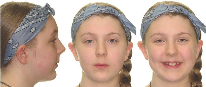
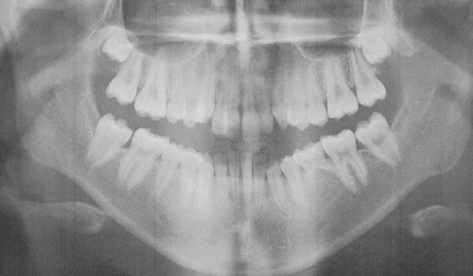
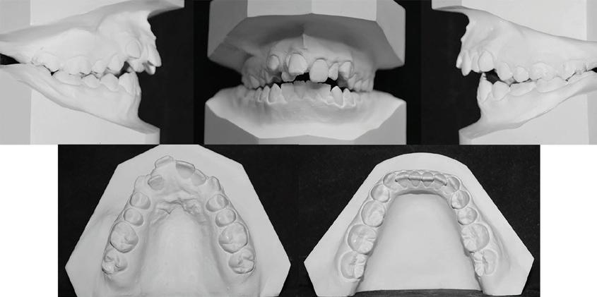
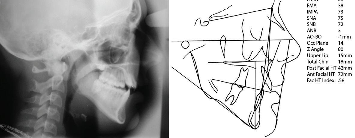
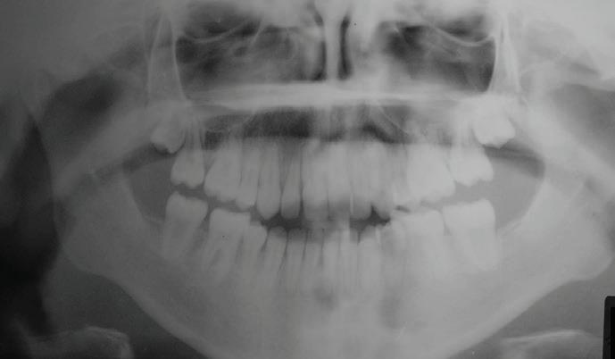
The initial facial photographs (Figure 1) exhibit an obtuse nasolabial angle and significant crowding upon smiling. The patient has very mild facial asymmetry with chin deviation to the left. Lower facial height is excessive. The pretreatment casts (Figure 2) confirm a Class II dental relationship, maxillary and mandibular crowding, small maxillary lateral incisors and an anterior openbite. The panoramic radiograph (Figure 3) highlights the missing mandibular left second premolar and exhibits blocked out maxillary canines as well as unerupted but developing maxillary third molars. Mandibular third molars appear to be absent. The cephalogram and its tracing (Figure 4a, 4b) confirm excessive vertical dimension. Even though an anterior openbite and a significant overjet exists, the patient has an ANB of only 3º. Both maxilla and mandible are slightly retruded to cranial base. Significantly, incisor mandibular plane angle is only 73º. For this reason, the patient has acceptable facial esthetics because the higher the mandibular plane angle, the more the mandibular incisors need to be upright in order for the patient to have good facial esthetics. The other significant finding was that the chin thickness is greater than normal. Total chin is 18 mm whereas upper lip is 15 mm. The upright mandibular incisors and the chin thickness camouflage the significant vertical problem as well as the Class II dental relationship.
HOW WOULD YOU TREAT THIS PATIENT?
The priority for the parents was to correct the dental malocclusion and close the anterior openbite. The parents had been given surgical options by the other practitioners whom they visited. Surgery was to consist of a mandibular osteotomy and a maxillary impaction to address the skeletal vertical discrepancy. The crowding was to be resolved by expansion and an implant by one practitioner and with extractions by another practitioner, but both suggested surgery. After careful and thoughtful consideration of the records and the wishes of the parent, the treatment plan that was devised was to extract the maxillary right and left first premolars, the mandibular right second premolar and the mandibular left second deciduous molar and to treat without the surgical option. It was explained to the parent that this treatment plan would require excellent patient cooperation, many appliance adjustments and many archwire changes. The parent chose this plan for the child.
The teeth were extracted and the patient was bonded/ banded with .022 conventional orthodontic appliances. The maxillary canines were retracted with headgear and elastic chain. The mandibular space was closed with power chain and mandibular crowding was resolved concurrently. The maxillary incisors were retracted with a closing loop archwire after maxillary canine retraction. A requirement during treatment was to hold mandibular incisors and the maxillary molars in their pretreatment positions.
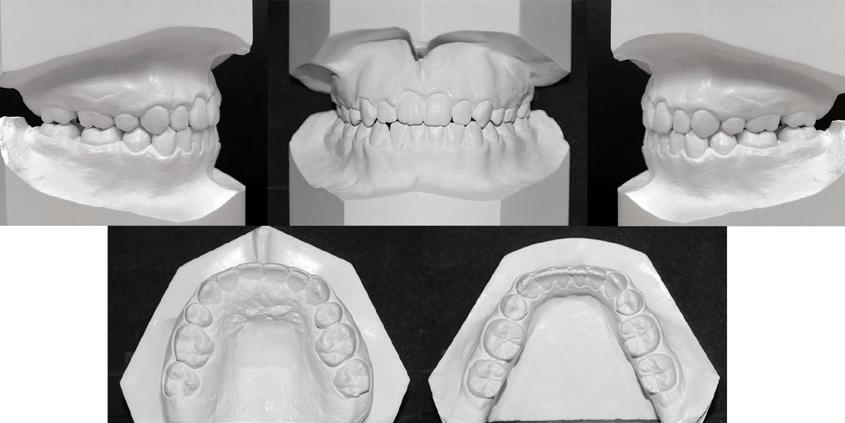
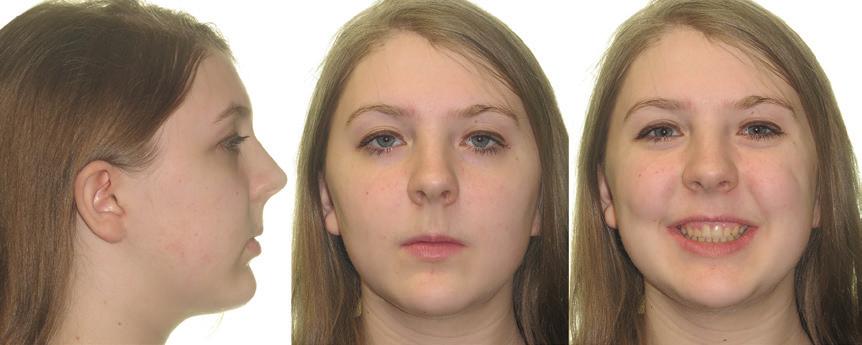
The final facial photographs (Figure 5) confirm maintenance of the facial profile and of the vertical dimension. Nasolabial angle seems to be not quite as obtuse. The smile exhibits about 2 mm of gingiva, more than the pretreatment smile, but it must be noted that the posttreatment smile is much more of a smile! The posttreatment casts (Figure 6) confirm correction of the Angle’s Class II occlusion, elimination of crowding and extraction space closure. Arch form and arch width were maintained. Mandibular canines were not expanded.
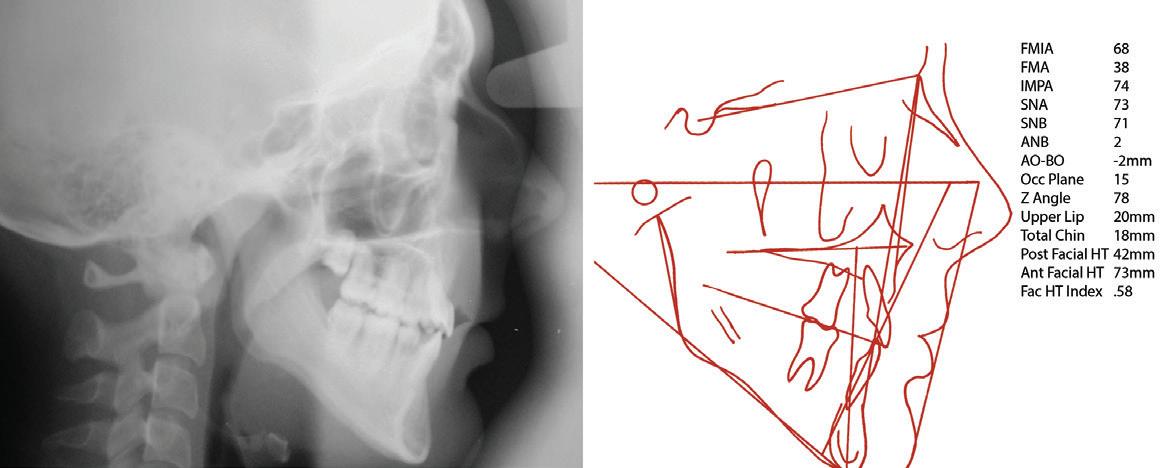
Mandibular second molars were left distally tipped so they can upright into a functional occlusion. The posttreatment panoramic radiograph (Figure 7) exhibits uprighting of teeth into the extraction spaces and correct long axial position of the teeth. The posttreatment cephalogram and its tracing (Figures 8a & 8b) confirm maintenance of mandibular incisor position and of vertical dimension. FMA did not open.
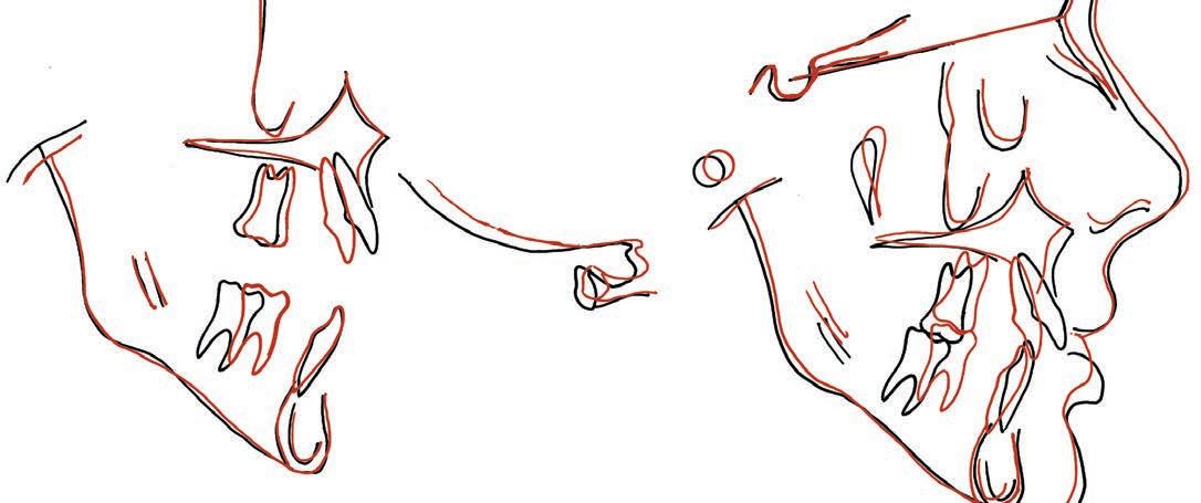
The ANB remained constant. Upper lip thickened due to retraction of the maxillary anterior teeth. Merrifield’s Z angle changed only 2º. The goal of maintaining the face was achieved. The malocclusion was corrected. The pretreatment/posttreatment superimpositions (Figure 9) confirm maintenance of mandibular incisor position, protraction of mandibular posterior teeth, control of the vertical dimension with no maxillary molar extrusion and retraction of the maxillary anterior teeth. Treatment time was 26 months. The patient was retained with maxillary and mandibular Hawley retainers.
You may have treated this patient differently. Although the diagnosis among all practitioners who looked at the patient was Angle’s Class II malocclusion with crowding and openbite, the practitioner’s opinions differed on how to best treat the young lady.
Updates from our State Associations
This year has started with many successful Component (State) meetings. Members and exhibitors are enjoying their time together. We strongly encourage you to attend your component meetings.
Alabama, Florida, Georgia, Mississippi, North Carolina, South Carolina and Tennessee have hosted their meetings.
Please note the upcoming Component meetings
Kentucky
Louisiana
Virginia
West Virginia
August 25, 2023
March 31 -
April 1, 2023
March 31 -
April 1, 2023
July 21 - 22, 2023
The Brown Hotel, Louisville, KY
The Westin Canal Place, New Orleans, LA
The Greenbrier, White Sulpher Spring, WV
The Greenbrier, White Sulpher Spring, WV


