14 minute read
Know your Equipment: Measuring Cardiac Output
by TEAM
Verghese T. Cherian, MD, FFARCSI
Professor of Anesthesiology Penn State Health College of Medicine Hershey, PA 17033 mailto:vcherian@pennstatehealth.psu.edu
Advertisement
Cardiac output (CO) is the volume of blood ejected by each ventricle per minute and is mathematically, the product of stroke volume (SV) and heart rate (HR). The factors that can modify the CO are the heart rate, the cardiac rhythm, the preload, the contractility, and the afterload of the ventricles and their proclivity to ischemia. CO provides a measure of systemic oxygen delivery and global tissue perfusion.
Adolph Fick (1829–1901), in 1870, was the first to describe the method to estimate the cardiac output. [1] He suggested that if the amount of a marker taken up by an organ is known, then the blood flow to the organ can be calculated by measuring the concentration of the marker in the arterial supply and the venous drainage of that organ. Applying this principle, if the oxygen consumption (VO2) of a person is measured by a spirometer using a closed rebreathing circuit with a CO2 absorber, and the oxygen content of the arterial (Ca) and the mixed venous blood (Cv) is measured, then the CO can be calculated. VO2 = (CO x Ca) – (CO x Cv); CO = VO2 / (Ca – Cv) E.g. Cardiac Output = (250 ml O2/minute) / (200 ml O2/L - 150 ml O2/L) = 5 L/minute Measuring the oxygen consumption in a clinical situation is not practical. However, CO can be measured using other techniques, namely the indicator dilution method, arterial pulse waveform analysis, ultrasonography, and the bioimpedance method.
Indicator dilution technique: An indicator is injected into a vein and is then sampled over time, in either the pulmonary artery or a systemic artery. The concentration of the indicator will display a rapid rise followed by a logarithmic decay. The volume of blood the indicator gets diluted in determines the rate of decay. The Stewart Hamilton equation states that if a known amount of a substance is injected upstream, the change in its concentration downstream is related to the flow. (Figure 1)
Figure 1:An indicator is injected upstream and measured by a detector downstream. The integral of its concentration over time can be used to measure the cardiac output using the Stewart-Hamilton equation
Known volume of indicator Concentration of indicator over time
Detector Stewart-Hamilton equation
�������� =
I – Amount of indicator in moles ∫���� ���������������� – Integral of indicator concentration over time (Area under the curve)
The flow (volume over time) in this situation is the cardiac output. The indicators used could be thermodilution of a cold or warm thermal bolus, or lithium.
The pulmonary artery catheter (PAC) was initially developed to measure the pressure in the various chambers of the heart and was later modified by Jeremy Swan and William Ganz to measure the central filling pressures and the CO.
The PAC is a multi-channel catheter, about 110 cm long, that is inserted through the internal jugular or the subclavian vein into the right atrium. It is then gently
continued on page 7
continued from page 6
‘floated’, aided by an inflatable balloon at the tip, through the right ventricle into the pulmonary artery. (Figure2)
Figure 2: The Pulmonary artery catheter (PAC) is a multi-lumen catheter, which is gently ‘floated’ through the right ventricle into the pulmonary artery.
Proximal port
Distal port Balloon inflation port
Thermistor PAC
The passage of the PAC can be guided by transducing the channel that opens at the tip of the PAC and observing the pressure waves at the different locations. The aim is to sit the tip of the catheter in the middle branch of the left or right pulmonary artery to measure the pressure in the pulmonary artery. In that position, if the terminal balloon is inflated, the flow through that arteriole is blocked and the tip is ‘wedged’ and displays the blood pressure distal to it. This pulmonary artery wedge pressure correlates to the left ventricular end-diastolic pressure or the preload. One of the channels of the PAC opens into the right atrium and reflects the central venous pressure. A rapid response thermistor, to measure the temperature of the blood, is located at the tip of the catheter.
The PAC is the gold standard of CO measurement against which other methods are compared. A 10 ml bolus of cold (~0 °C) saline or isotonic dextrose solution is injected into the right atrium, and the temperature of the blood in the pulmonary artery is measured continuously by the thermistor. After the initial decrease in temperature, there is a slow return to the baseline temperature. The rate of return depends on the flow of blood that dilutes the bolus of cold saline. The area under the ‘temperature’ curve represents the change in temperature over time which is inversely proportional to the CO.
A continuous CO monitoring PAC uses the same thermodilution principle, but instead of a bolus of cold injectate, an electric filament located along the proximal part of the catheter emits pulses of thermal energy and the change in the blood temperature is recorded by the thermistor at the tip of the PAC. Another advance in PAC construction is the incorporation of a tiny light source near the catheter tip which can measure the oxygen saturation of the surrounding blood using reflectance oximetry. When the catheter is in the appropriate position, it measures mixed venous oxygen saturation (SvO2).
Arterial Pressure Waveform Analysis: ‘Windkessel’ effect: When the left ventricle (LV) contracts it ejects a stroke volume (≈70ml) under (systolic) pressure (≈120mmHg). The elastic wall of the aorta distends to accommodate the stroke volume and subsequently recoils to push this volume forward at (diastolic) pressure (≈80mmHg). Otto Frank, the German physiologist who first described this phenomenon, wherein a cyclical ventricular contraction generates a constant flow through the blood vessels, called it the ‘Windkessel’ effect. The fire engines of those times used an air chamber (Windkessel chamber) that converted the sinusoidal pressure generated by the rotatory pump into a constant water stream.
Mathematical analysis: The general principle of the arterial pressure waveform analysis is that the Systolic Pressure-time Integral (SPI) or the area under the systolic part of the arterial curve is directly proportional to the stroke volume and inversely proportional to the total arterial compliance. The estimate of aortic compliance is based on the three-element Windkessel model, which includes the compliance of the great arteries (C), the resistance of the vascular system, principally in the arterioles (R), and the aortic impedance (Z) due to the elastic, reflective pressure wave. (Figure 3)
Figure 3: The Systolic Pressure-time Integral (SPI) or the area under the systolic part of the arterial curve (A), is directly proportional to the stroke volume and inversely to the arterial compliance. The estimate of aortic compliance is modeled on the three-element Windkessel (B). [Z- impedance, C- capacitor, R- resistor]
Systolic Pressure-time Integral (SPI)
A Z
C R
3-Element Windkessel
B
continued on page 8
continued from page 7
Therefore, to measure the CO from the arterial pressure waveform, the arterial compliance should be known. This can either be estimated based on the patient’s biometric values such as gender, age, height and weight (uncalibrated technique), or by calculating it by first measuring the CO using the Fick’s principle (calibrated technique). [3]
• Flo-Trac / Vigileo: The Flo-Trac sensor is attached to a standard arterial line. The monitor analyzes the arterial waveform and calculates the CO by incorporating the value of aortic compliance based on the population demographics (e.g. age, weight, height and gender). A newer model (Clear-Sight®) generates the arterial waveform by using a finger cuff and vascular unloading technique.
• Pulse index Continuous Cardiac Output (PiCCO):
This system uses a transpulmonary thermodilution method to calibrate its algorithm. It requires a standard central line and a thermistor-tipped arterial cannula which is placed within the femoral or the brachial artery. Cold saline is injected into the central line and the temperature change is measured at the arterial cannula. The CO is measured using the
Stewart Hamilton equation. This information is then used to calibrate the program that analyzes the pulse pressure waveform.
• Lithium Dilution Cardiac Output (LiDCO): This system uses a lithium dilution method to calibrate its algorithm and requires only a standard arterial cannulation. Lithium chloride is injected into a central or even a peripheral vein and the change in concentration is measured by an electrode sampling at the arterial cannula site.
Ultrasonography: Although ultrasound are sound waves greater than 20,000 hertz (Hz), the frequency used for medical imaging is 2-20 MHz. The speed of ultrasound through tissue is 1540 m/s. Echocardiography uses ultrasound to delineate the structure and function of the heart and the ultrasound probe can be placed either on the chest wall, known as the trans-thoracic echocardiography (TTE), or inserted into the esophagus, the trans-esophageal echocardiography (TEE).
Doppler principle: When an ultrasound wave strikes a moving object some of it gets reflected with a change in its frequency. The reflected waves are at a higher frequency if the object is moving toward the probe and lower if it is moving away. The shift in frequency is proportional to the velocity of the moving object. The frequency of the reflected wave can be detected by the ultrasound probe and using the Doppler frequency shift equation, the velocity of the reflector can be calculated.
Frequency (received) = (2 x Frequency (transmitted) x Velocity of reflector x Cosθ)/ (Speed of sound in tissue)
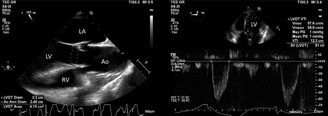
The basic premise of using ultrasonography to calculate the CO is to measure the cross-sectional area of the left ventricular outflow tract (LVOT) and the velocity of blood expelled through it using the Doppler principle. (Figure 4)
Figure 4: Trans-esophageal Echocardiographic (TEE) views showing (A) the diameter at the left ventricular outlet tract, and (B) measuring the Velocity Time integral (VTI) using the pulsed-wave Doppler and calculating the storke volume (SV).

A B
The diameter of the LVOT, about 1cm from the aortic valve, is measured by specific orientation of the ultrasound probe, namely the mid-esophageal long axis (TEE) or the parasternal long axis (TTE) view. Assuming the LVOT to be circular, the area is calculated using the formula for area of a circle (A=πr2). Using a deep trans-gastric (TEE) or the apical five-chamber (TTE) view, a pulsed-wave Doppler is used to measure the velocity of the red cells at the LVOT. Care should be taken to measure it at the same location the LVOT diameter was measured. The area under the curve of this spectral Doppler profile is known as the velocity time integral (VTI). This depicts the distance a red cell travels per beat or the stroke distance, which when multiplied by the estimated cross-sectional area of the aorta, gives the stroke volume. As evident in Figure 4, most echocardiogram machines are programed to calculate the LVOT area and the stroke volume, and the accuracy of the measurement depends on the expertise of the proceduralists to attain the appropriate view.
continued from page 8
Bioimpedance & Bioreactance: The human body is able to conduct electrical current because of the presence of charged ions within the body fluids. According to Ohm’s law (I= V/R), the current passing through a substance is inversely proportional to the resistance it offers to the potential difference. This resistance is termed ‘impedance’ when it is an alternating current. Impedance can be defined as a combination of Ohmic resistance and reactance, or resistance to an alternating current, and is denoted as Z. The ‘bio-impedance’ varies between different tissues, e.g. blood (150 ohm/cm), plasma (63 ohm/cm), lungs (1275 ohm/cm) and fat (2500 ohm/cm). [4]
The basic principle of the bioimpedance cardiography is that the thorax is viewed as two concentric cylinders, a low impedance, inner one containing blood and a higher impedance, outer one constituting the lung and other body tissues. Since the volume of blood increases with each systole, the bioimpedance decreases during the ejection phase and returns to baseline (Zo) during diastole. Electrodes are applied at the base of the neck (thoracic inlet) and the costal margins (thoracic outlet), and a high-frequency, low-amplitude current (1.4-1.8 mA at 30-75 kHz) is transmitted between these electrodes and the change in their amplitude is measured. (Figure 5)
Figure 5: The bioimpedance model views the chest as two concentric cylinders, a low impedance, inner one containing blood, and a higher impedance, outer one constituting the lung and other thoracic tissues. The electrodes are applied at the base of the neck (thoracic inlet) and the costal margins (thoracic outlet), and a high-frequency (30-75 kHz), low-amplitude current (1.4-1.8 mA) is transmitted between them.
This information can be used to calculate the rate of the change of Z (∆Z/∆t), which correlates to the flow of blood into the aorta, during systole.
The stroke volume is proportional to the product of ∆Z/∆t, the ventricular ejection time (VET), and the volume of electrically participating tissue (VEPT), which is essentially the thoracic volume. The VET is calculated from the R-R interval on the ECG and the VEPT is predicted from the height, weight, and gender of the subject. SV= ∆Z/∆t x VET x VEPT
Although this technique of measuring CO was originally used in astronauts in the 1960’s, several modifications have been incorporated over the years. Two significant modifications are substitution of the cylindrical model of the chest to a truncated cone and use of the formula that accounts for deviation from ideal body weight in calculating the thoracic volume. The bioimpedance technique is unreliable during arrhythmias (VET is measured from the R-R interval), acute changes in tissue water (pulmonary oedema or pleural effusion would alter the bioimpedance), changes in temperature and humidity (impact on the electric conductivity between the electrodes and the skin), and mechanical factors such as noise from electrocautery, mechanical ventilation, and surgical manipulation.
Bioreactance is an improved technology which analyzes the relative phase shifts in the voltage of the alternating currents. According to bioreactance, the human thorax is considered an electric circuit with resistors (R) and capacitors (C), which together generate the two components of thoracic impedance (Zo), namely amplitude and phase (phi, o). The pulsatile ejection of blood from the heart modifies the value of R and of C, leading to instantaneous changes in the amplitude and the phase of Zo. Since phase shifts can occur only because of pulsatile flow, and the overwhelming component of thoracic pulsatility stems from the aorta, the bioreactance signal is strongly correlated with aortic flow. This is impacted less by movement artefact, patient body variance, and thoracic fluid, which would be relatively static.
The bioreactance device is made up of a highfrequency (75-kHz) sine wave generator and four dual-electrodes to establish electrical contact with the body. Each electrode sticker contains one electrode that transmits the high-frequency sine wave into the body, while the other electrode is used by the voltage input amplifier. The signal processing unit detects the relative phase shift (∆o of the input signal. The peak rate of change in phase (∆o/dt) is proportional to the peak aortic flow.
SV = K x VET x ∆o / dt
K is a constant of proportionality which is determined by bioreactance
continued from page 9
Monitoring the CO guides administration of fluids and vasopressors, especially in critically ill patients or during major abdominal surgery with large fluid shifts. It can be measured using invasive, minimally invasive and non-invasive techniques. This was an attempt to explain the physical principles used in each of the techniques and to give a better understanding of their strengths and limitations. References Betteridge N, Armstrong F. Cardiac output monitoring. Anaesthesia and intensive care medicine 2021; 23: 101-110
Westerhof N, Lankhaar J-W, Westerho BE. The arterial Windkessel. Med Biol Eng Comput 2009; 47:131–141. DOI 10.1007/s11517-008-0359-2
Esper SA, Pinsky MR. Arterial waveform analysis. Best Practice & Research Clinical Anaesthesiology 2014; 28: 363-380. https://doi.org/10.1016/j.bpa.2014.08.002
Jakovljevic DG, Trenell MI, MacGowan GA. Bioimpedance and bioreactance methods for monitoring cardiac output. Best Practice & Research Clinical Anaesthesiology 2014; 28: 381-394
May is Mental Health Awareness Month!
Make sure you take time to care for yourself. Get outdoors and get some sunlight! Here are some neat places to visit in our great Commonwealth!
Austin Dam Memorial Park https://austindam.mailchimpsites.com/ Go hiking, camping, fishing, or picnicking and overlook the grassy meadow that memorializes the 2nd worst dam break in Pennsylvania history.
Ricketts Glen State National Park https://www.dcnr.pa.gov/StateParks/FindAPark/RickettsGlenStatePark/Pages/default.aspx With 11 trails and 22 waterfalls, Ricketts Glen is sure to double your pleasure and double your fun with all the scenic views it has to offer.
Blue Marsh Lake https://visitpaamericana.com/partner/blue-marsh-lakerecreation-area/ Biking, walking, hiking, horseback riding, boating, fishing —take your pick of outdoor recreation. Blue Marsh Lake is a great spot for lovers of land and water alike. Valley Forge State Park https://www.nps.gov/vafo/index.htm Take a trip back in time and visit this 3,500-acre park that was an encampment site of the Continental Army during the Revolutionary War. Enjoy the museum, monuments, and meadows while listening to the many bird species that abide there.
Bushkill Falls https://www.visitbushkillfalls.com/ Visit the Niagara of Pennsylvania. This Pocono Mountains hideaway boasts a series of eight waterfalls and a plethora of activities for a full day of exploration and enjoyment.
Presque Isle State Run Park https://www.dcnr.pa.gov/StateParks/FindAPark/PresqueIsleStatePark/pages/default.aspx This 3,112-acre state park in Erie offers numerous trails, colorful sea glass, and 11 miles of beach so that you can swim or sunbathe, bike or hike, and enjoy nature to your heart’s desire.


