49 minute read
Antiviral Finishing on Textiles - An Overview
Goutam BAR1*, Debjit BISWAS1, Shrutirupa PATI1, Kavita CHAUDHARY2, Mahadev BAR3
1Department of Textile Design, National Institute of Fashion Technology, Bhubaneswar, India 2Banasthali Institute of Design, Banasthali University, Banasthali, Rajasthan, India 3Laboratoire Génie de Production, LGP, Université de Toulouse, INP-ENIT, Tarbes, France *goutam.bar@nift.ac.in
Advertisement
Review UDC 677.027:578.4 DOI: 10.31881/TLR.2020.17
Received 4 August 2020; Accepted 21 October 2020; Published Online 31 October 2020; Published 2 March 2021
ABSTRACT
Antiviral textiles are one of the most promising areas of protective textiles. Antiviral textiles are important in the field of health and hygiene. They become an essential part of our daily-life when a pandemic situation arises. The present paper critically analyses and summarizes various researches of the production of antiviral textiles. Different classes of the virus, how the virus transmits and replicates, various antiviral agents for textiles and their working mechanism, and the application procedure of various synthesized and bio-based antiviral compounds on textiles have been discussed in this paper. Finally, the present paper compares the existing antiviral finishing on textiles in terms of its effectiveness, durability and skin-friendliness and, following that, discusses the possibilities of using antiviral textiles in various sectors.
KEYWORDS
Virus, Textiles, Antiviral agent, Antiviral finish, Antiviral textiles
INTRODUCTION
History reveals that a virus is the reason behind most of the global pandemics that occurred in the near past, for instance the H3N2 virus behind the influenza pandemic in 1968, the SARS virus behind the 2003 pandemic, the H1N1 virus behind the swine flu pandemic in 2009, the Ebola virus behind the 2014 pandemic and the most recent novel coronavirus behind the 2020 pandemic [1,2]. The World Health Organization (WHO) reported that the diseases caused by viral infections are the second leading cause of human death [3]. Around 250,000 to 500,000 people die every year only due to the influenza virus infection [4]. To prevent or minimize these fatalities, several vaccines are developed by the researchers but these are not sufficient enough to protect human beings from all kinds of viruses. Thus, it is necessary to look for other possible safety measures which can protect us from these deadly viruses, prior to an appropriate vaccine being developed. Viruses fall in the category of microbes and they are omnipresent [5,6]. NASA researchers have found microbes even at the height of 32 km above the sea level and at the depth of 11 km under the sea level. It is estimated that all microbes present in the earth are approximately 25-fold the mass of all animals [7]. Microbes have an outer polysaccharide cell wall and a semi-permeable membrane, which protects their inner substrates and preserves their functionality [8,9]. Whenever a microbe finds itself in favourable conditions in terms of temperature and humidity or a host cell, it starts reproducing itself [10]. However, all microbes
are not the same, they differ from each other in terms of their structure, sizes, habitat etc. In the context of their cellular nature a microbe can be unicellular, multicellular or acellular. Viruses are acellular. In the domain of life, living cells are classified in three categories called Archaea, Bacteria and Eukarya. Most of the microbes fall in the category of Archaea or in the category of Bacteria, but a virus falls in none of these three categories, they are considered to be a grey area between the living and the non-living [11]. Hence, a substrate that provides a perfect protection against a bacterial or other microbial attack, may or may not be useful against a virus. Whenever we think about human protection, textiles come to our mind first. Textiles protect human beings from extreme weather conditions. With suitable structural design and chemical finishes, textiles can protect a human body from sharp objects, impact thrust, fire accidents, electric shocks etc. [12]. However, textiles, in their parent form (i.e. without any suitable chemical treatment) cannot protect a person from a microbial attack. In fact, textiles are susceptible to microbial growth due to their high moisture retention and surface area [13,14]. The presence of starch, protein and fats in natural fibres makes them more susceptible to a microbial attack. The degeneration of fibre, foul odour, colour change, unwanted stains etc. are the signs of a microbial attack on textiles [7,15,16]. These microbes (microbial attack signs) mainly belong to the bacteria family. Textiles are treated with various synthetic as well as bio-based antibacterial agents in order to protect them and their user from a bacterial attack [8,17,18]. However, a textile fabric treated with antibacterial agents may not act as an antiviral fabric, as the virus and the bacteria are completely different from each other [8,11]. As mentioned earlier, foul odour, colour change, unwanted stains etc. are the signs of bacterial infection on textiles but no such signs are observed when textiles are infected with a virus. Viruses do not replicate or grow on textiles because they require a host cell to be able to do that but viruses are deadlier for the human race than bacteria [19,20]. The world is currently going through a pandemic situation resulting from the novel coronavirus. There are many deadlier viruses present in the world, which are inactive now but can affect the human race at some other time [20]. Hence, it is very much necessary to have a clear concept of antiviral textiles as they act as the first layer of protection against the unknown viruses, before the development of a vaccine or a medicine. The present paper critically analyses and summarizes various researches on producing antiviral textiles. The paper furthermore discusses different classes of viruses, how they transmit and replicate, various antiviral agents for textiles and their working mechanism, and the application procedure of various synthesized and bio-based antiviral compounds on textiles.
Viruses and their classification
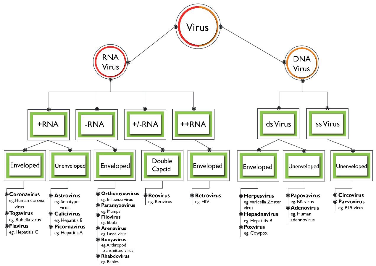
As mentioned earlier, viruses are acellular, considered to be a grey area between the living and the nonliving. Viruses are small protein capsids which store some genetic information. All viruses consist of nucleic acid and a protein coat [21]. The nucleic acid may be either a Deoxyribonucleic Acid (DNA) or a Ribonucleic acid (RNA) but a virus never has both of them. The protein coat encases the nucleic acid while some viruses are also enclosed by a layer of fat and protein molecules called envelopes. The size of the virus varies depending on its structure. In order to reproduce themselves, viruses are dependent on their host cell. Initially, they replicate the genetic information in the cell and produce viral progenies to infect the other cells [22]. Viruses are broadly classified based on their structure, symmetry, chemical composition, structure of genome and on their mode of replication. Based on their genome structure and chemical composition, viruses that infect human beings are divided into twenty-one families [21]. The classification of viruses causing infections in humans is presented in Figure 1.
Figure 1. Classification of viruses Figure 1. Classification of viruses
Transmission of viruses
A virus can be transmitted in various ways. Virus transmission mainly depends on their ability to overcome a barrier. A virus can transmit from cell to cell, animal to animal, person to person or even from one species to another. It can be transmitted either by a direct contact or by an indirect contact. In the case of a direct contact, the virus is spread from the infected host to an uninfected person by means of a physical touch. While in the case of an indirect contact, the virus proliferates through different mediums such as textiles, contaminated surfaces etc., sometimes the disease-bearing organisms known as vectors transmit viruses to an uninfected person in the form of blood-sucking insects such as mosquitos [23]. Figure 2 explains the most probable and significant ways in which a virus can infect human beings. Here, infected animals, contaminated areas, and infected persons are considered as major sources of a virus. The droplets released by an infected person or an animal in the form of a cough, sneeze, blood stains etc. make a place or a person contaminated. Droplets having a virus particle of the size less than 1 μm contribute to the spread of airborne diseases [22]. According to Wells [24], the aqueous part of a droplet evaporates quickly in the air and the residue called the droplet nuclei spreads around the corners and ultimately contaminates the place and the people who are sharing the same air supply. Some viruses have large particles, which makes them heavy, and as a result they don’t contribute to the spread of airborne diseases. The transmission of these heavy viruses is greatly influenced by their lifespan outside a host cell. For instance, a SARS-CoV-2 can survive about 24 hours on a cardboard and 3 to 4 days on plastics and stainless steel [25], while an astrovirus, Hepatitis A and Polio virus can survive up to 2 months [26]. The long lifespan outside a host cell makes these viruses deadlier for a human being. However, the researchers have observed that most of the health-related issues in human beings are caused by micro-sized virus particles with the aerodynamic diameter less than 1 μm [22].
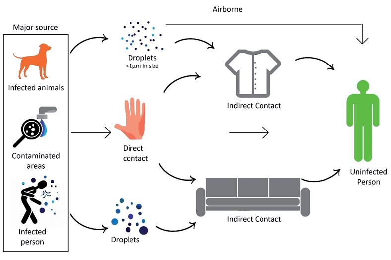
Figure 2. Modes of virus transmission
Replication of viruses
Figure 2. Modes of virus transmission
Viral replicati on is a biological process that produces new viruses during the infecti on of the host cell. Replication of viruses Hence, prior to replicati on, a virus has to enter its host cell fi rst. Without a host-cell a virus cannot repliViral replication is a biological process that produces new viruses during the infection of the host cell. cate itself as it doesn’t have any organelles such as nuclei, mitochondria, ribosomes and the cytoplasmic
Hence, prior to replication, a virus has to enter its host cell first. Without a host-cell a virus cannot components which are necessary for the synthesis of its own structure [23]. During viral replicati on, initi ally, a virus parti cle or a virion att aches itself to the host-cell membrane and injects its DNA or RNA into replicate itself as it doesn’t have any organelles such as nuclei, mitochondria, ribosomes and the the host cell to initi ate the infecti on. In general, a virus enters a human host cell through the membrane cytoplasmic components which are necessary for the synthesis of its own structure [23]. During viral fusion method. Once a virion enters its host cell, the viral genome gets access to its host cell organelles and replication, initially, a virus particle or a virion attaches itself to the host-cell membrane and injects enzymes. This ulti mately initi ates the process of the replicati on of the viral nucleic acid (either DNA or RNA) its DNA or RNA into the host cell to initiate the infection. In general, a virus enters a human host cell and the structural protein with its code. This newly synthesized nucleic acid and the structural proteins are then joined together as new virus parti cles. Finally, these new viruses leave the host cell either by cell lysis through the membrane fusion method. Once a virion enters its host cell, the viral genome gets or by the budding process and become ready to infect other host cells [27]. access to its host cell organelles and enzymes. This ultimately initiates the process of the replication of the viral nucleic acid (either DNA or RNA) and the structural protein with its code. This newly synthesized nucleic acid and the structural proteins are then joined together as new virus particles.
Finally, these new viruses leave the host cell either by cell lysis or by the budding process and become ready to infect other host cells [27].
Role of textiles in protection against viruses
As menti oned earlier, viruses are considered to be a grey area between the living and the non-living, and they need a host cell to replicate themselves. Thus, a virus will not be able to replicate itself on a texti le surface. However, texti les can act as an acti ve medium for the viral transmission [28,29]. Frequent disinfecti on or disposal of the contaminated texti les could be an eff ecti ve way of restricti ng viral transmission through texti les [30]. The second possible way would be to impart anti viral properti es to texti les. A normal Role of textiles in protection against viruses texti le fabric does not have any anti viral properti es but the incorporati on of suitable components into As mentioned earlier, viruses are considered to be a grey area between the living and the non-living, texti les can make them anti viral. The incorporati on of an anti viral agent into texti les can be done at diff erent stages and in diff erent ways. Various ways of developing anti viral texti les are discussed in the next and they need a host cell to replicate themselves. Thus, a virus will not be able to replicate itself on a secti on of this paper. Anti viral agents make the treated texti les anti viral by employing either of the following textile surface. However, textiles can act as an active medium for the viral transmission [28, 29]. two mechanisms or by combining them. In the fi rst mechanism, the applied chemical makes the surface Frequent disinfection or disposal of the contaminated textiles could be an effective way of restricting energy of a texti le surface relati vely low. Doing so will stop the viral transmission via texti les by restricti ng viruses to the texti le surface. This low surface energy of texti les can also destroy the outer lipid barrier of
the same by breaking it into fragments. As a result of these interactions, the disintegration of the
virus ensues, manifesting itself in the leakage of the viral genome and a loss of infectivity, leaving the viral particle inactive on the treated textile surface [7, 32, 33]. A schematic diagram explaining the
3.
a virus which will make the viral genome inacti ve by making it unable to penetrate into a host cell [31]. In the second approach, when a virus comes in contact with a texti le surface treated with an anti viral agent, these acti ve agents bind with the outer layer of the virus and inhibit its vital mechanisms. These anti viral agents oxidize and dissolve the lipid or the glycoprotein layer and enter inside the virus structure. Finally, these anti viral agents adhere to the genome (i.e. with the virus DNA or RNA) and deacti vate the same by breaking it into fragments. As a result of these interacti ons, the disintegrati on of the virus ensues, manifesti ng itself in the leakage of the viral genome and a loss of infecti vity, leaving the viral parti cle inacti ve on the treated texti le surface [7,32,33]. A schemati c diagram explaining the anti viral mechanism through virus destructi on (i.e. through the second approach) is shown in Figure 3.

Figure 3. Antiviral mechanism through virus destruction on textile fabric surface
Figure 3. Antiviral mechanism through virus destruction on textile fabric surface
Antiviral treatments for textiles
Inherently, texti le materials are not anti viral. Texti le materials are suscepti ble to microbial growth due to their high moisture retenti on and surface area [14]. The additi on of suitable anti viral agents onto texti les can make them anti viral. The incorporati on of an anti viral agent into the texti les can be done at diff erent stages and in diff erent ways. Anti viral fi nishing on the texti le material can be done onto the texti le fi bre, yarn, fabric, and fi nal products depending upon the anti viral agents and their applicati on technique. For ease of applicati on and conti nuous producti on, anti viral applicati on onto the fabric is preferable. For fabric like cott on, wool, silk, and other manmade fi bre, anti viral treatment is done by surface treatments, like the exhaust method, the pad-dry-cure method, microencapsulati on, and the coati ng technique, alone or in combinati on, depending on the anti viral compositi on and the fabric quality. Anti viral agents can also be incorporated in manmade fi bres by mixing suitable anti viral agents into the polymer matrix before the fi bre extrusion [34,35]. The exhaust method is the most common and popular method of dyeing and fi nishing texti le materials. This method is also well suited for the anti viral fi nishing of natural fi bres and polyamide fabrics. The process is very similar to the dipping technique and applicati on is carried out below the boiling temperature. For polyester fabric, the high-temperature exhaust method is preferable. The process is very similar to disperse dyeing and the applicati on is carried out at 120-130 °C. The pad-dry-cure method is another way of applying
an antiviral composition onto the textile material. The fabric is padded with the antiviral composition in an aqueous medium along with a suitable binder, followed by drying and curing for proper fixation. This method is used alone or in combination with other techniques [36,37,38]. Micro-encapsulation is a process in which an active ingredient as a core substance is generally stored within a polymeric shell. For antiviral finishing on the textile material, a microcapsule solution is prepared by mixing and stirring the antiviral composition and the polymeric coating material in an emulsion reactor at 1000–10000 rotations per minute for 6–48 hours. The size of a microencapsulated shell generally varies within the micrometre and the millimetre range. This antiviral microcapsule solution is applied onto the textile substrate along with other binding compositions in an agitator, followed by drying and curing. This technique is gaining popularity in the area of textile finishing, especially for the fragrance finish, antimicrobial finish etc. for sportswear and medical textiles [39,40,41]. In the coating technique, the covering material is deposited onto the textile substrate to enhance the surface properties. Likewise, in textiles, the fabric can be coated with an anti-pathogenic component along with a suitable binding agent. The preferred coating techniques are reverse roll, rod, spray, and gravure coating techniques. The coating technique requires a substrate with minimum porosity. Most of the non-woven substrates are porous; therefore, alternate versions of the coating technique can be used, such as spray and dip-coating techniques. In certain applications, there might be a need for the controlled release of antiviral agents. In that case, the coating may include microencapsulation. The coating can be of a single layer as well as multi-layered. In multi-layered coating, initially, the substrate is coated with an active ingredient solution by using the spray-coating method and on top of that, the second layer is coated with the binding solution by using the rod-coating method [42].
Triclosan based antiviral finish
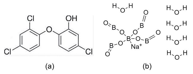
Triclosan is well known for its antimicrobial activity, especially against bacteria [36, 43]. However, a textile fabric can exhibit an antiviral property when it is treated with triclosan along with sodium pentaborate pentahydrate. The chemical structures of triclosan and sodium pentaborate pentahydrate molecules are shown in Figure 4. Iyigundogdu et al. [36] have prepared a triclosan-based antiviral solution which contains 0.03% triclosan, 3% sodium pentaborate pentahydrate and 7% glucapon 215 CS UP. A cotton fabric, treated with the abovementioned antiviral solution for 30 minutes by the exhaustion method at pH 5 exhibits promising results against the adenovirus and poliovirus. Glucapon 215 CS UP acts as an emulsifying agent and the triclosan inhibits the growth of viruses by restricting lipid biosynthesis. The function of sodium pentaborate pentahydrate in the above composition is not well understood but its presence enhances the antiviral activity of the triclosan. According to the Spearman–Karber test method, a cotton fabric treated with the abovementioned solution can reduce the viral titre by 60%. Iyigundogdu et al. [36] have predicted that the textiles treated with the combination of triclosan and sodium pentaborate pentahydrate can be effective against the enveloped and non-enveloped DNA and non-enveloped RNA viruses, such as hepatitis B, HIV, HCV, Ebola, MERS and SARS.
BAR G, BISWAS D, PATI S, CHAUDHARY K, BAR M. Antiviral Finishing on Textiles … TLR 0 (0) 2020 00-00.
Treatment with honeysuckle extract
Since ancient times, honeysuckle has been used as a traditional Chinese medicine for the treatments
of wind-heat, respiratory tract infection, fever and inflammatory conditions [46]. The honeysuckle
viral mRNA transcription and the subsequent protein translation. The chemical structure of
several viruses, including HIV, adenovirus, hepatitis B and HSVs. [2, 47]. Textile material can be
CN101324026B, 2011) has observed that treating a cotton fabric with a drug-component solution
to 50%) extracts by using the microencapsulation technique imparted antiviral properties to the
onto a cotton fabric along with a nano zinc oxide solution (5% to 20%), and an aqueous polyurethane solution (10% to 15%), followed by curing at 150 °C [41]. The preparation of microcapsules and their
zinc oxide to the cotton fabric. The finished fabric is friendly to the skin and it shows an antiviral
activity even after 20 washes with a virus inhibitory rate of more than 80% against the influenza and
herpes simplex virus [41].
Figure 4. Chemical structure (a) Triclosan (b) Sodium pentaborate pentahydrate [44, 45]
Figure 4. Chemical structure (a) Triclosan (b) Sodium pentaborate pentahydrate [44, 45]
Treatment with honeysuckle extract
Since ancient ti mes, honeysuckle has been used as a traditi onal Chinese medicine for the treatments of windheat, respiratory tract infecti on, fever and infl ammatory conditi ons [46]. The honeysuckle extract is rich in chlorogenic acid, which acts as an anti viral agent. Chlorogenic acid suppresses the viral mRNA transcripti on and the subsequent protein translati on. The chemical structure of chlorogenic acid is shown in Figure 5. It is reported that chlorogenic acid can be eff ecti ve against several viruses, including HIV, adenovirus, hepati ti s B and HSVs. [2,47]. Texti le material can be treated with the honeysuckle extract by the microencapsulati on technique. In microencapsulati on, an acti ve chemical agent is generally stored within a polymeric shell. The size of a microencapsulated shell generally varies within the micrometre and the millimetre range [40,41]. Jinmei (Patent No. CN101324026B, 2011) has observed that treati ng a cott on fabric with a drugcomponent soluti on containing honeysuckle (30% to 60%), Radix Glycyehizae (20% to 50%) and Weeping forsythia (20% to 50%) extracts by using the microencapsulati on technique imparted anti viral properti es to the fabric surface. The microcapsules were prepared using the above drug soluti on along with polylacti c acid (mass rati o 1:2) in an emulsion reactor. Further, the microcapsules (30% to 60%) were applied onto a cott on fabric along with a nano zinc oxide soluti on (5% to 20%), and an aqueous polyurethane soluti on (10% to 15%), followed by curing at 150 °C [41]. The preparati on of microcapsules and their applicati on onto the cott on fabric is illustrated in Figure 6. The nano zinc oxide has excellent pathogen killing properti es, whereas the aqueous polyurethane binds the microcapsules and nano zinc oxide to the cott on fabric. The fi nished fabric is friendly to the skin and it shows an anti viral acti vity even aft er 20 washes with a virus inhibitory rate of more than 80% against the infl uenza and herpes simplex virus [41].

Figure 5. Chemical structure of chlorogenic acid [48] Figure 5. Chemical structure of chlorogenic acid [48]
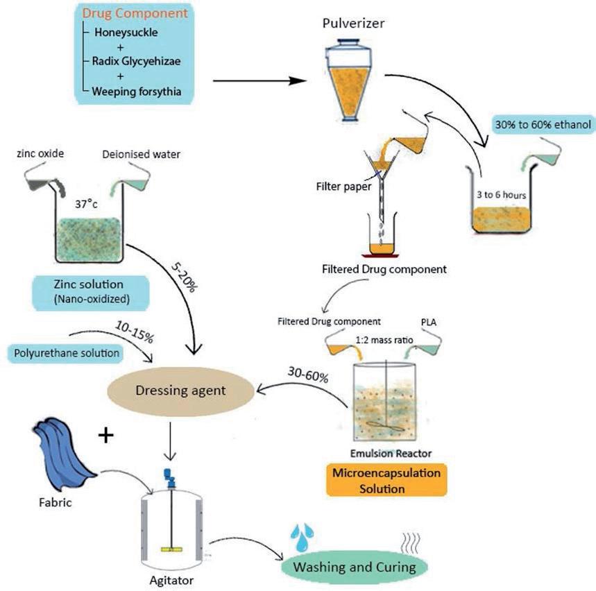
Figure 6. Preparation of antiviral microcapsules and their application onto cotton fabric by using honeysuckle extract Figure 6. Preparation of antiviral microcapsules and their application onto cotton fabric by using honeysuckle extract
Povidone iodine-based coated textiles Povidone iodine-based coated textiles
Ripa et al. [43] have reported that iodophors i.e. povidone iodine and ti nctures of iodine have excellent Ripa et al. [43] have reported that iodophors i.e. povidone iodine and tinctures of iodine have anti microbial eff ecti veness against a broad spectrum of viruses. Almost all microorganisms causing human disease are suscepti ble to free iodine released by the povidone iodine complex. Povidone iodine can be excellent antimicrobial effectiveness against a broad spectrum of viruses. Almost all microorganisms applied onto the texti le materials to make it anti viral by using the coati ng technique. Coati ng is a technique causing human disease are susceptible to free iodine released by the povidone iodine complex. through which functi onal materials are layered onto a substrate to enhance the surface properti es. Uniform Povidone iodine can be applied onto the textile materials to make it antiviral by using the coating coati ng over a texti le surface can be applied by using reverse roll, rod and gravure coati ng techniques. technique. Coating is a technique through which functional materials are layered onto a substrate to Coati ng can be of a single layer as well as multi -layered. The multi -layer coated fabric shows bett er anti viral properti es. Snyder (Patent No.US5968538A, 1999) prepared a coati ng soluti on which contains an acti ve enhance the surface properties. Uniform coating over a textile surface can be applied by using ingredient (71.5%) and a premix soluti on (28.5%). The acti ve ingredient comprised of 6.7% of Nonoxynol reverse roll, rod and gravure coating techniques. Coating can be of a single layer as well as multi9 (N9) and 93.3% of polyvinyl-pyrrolidone-iodine complex (PVP-I). The premix soluti on which acted as a layered. The multi-layer coated fabric shows better antiviral properties. Snyder (Patent binder was comprised of 54.6% of polyethylene glycol, 5.4% of hydroxypropyl methylcellulose, 7.2% of No.US5968538A, 1999) prepared a coating solution which contains an active ingredient (71.5%) and polyoxyethylene sorbitan, 11.5% of hydrous magnesium silicate and 21.3% of ethanol. The combinati on of PVP-I and N9 imparted anti viral and hydrophilic properti es to the coated substrates. Upon absorpti on of a premix solution (28.5%). The active ingredient comprised of 6.7% of Nonoxynol 9 (N9) and 93.3% of moisture by the coati ng material, PVP-I releases free iodine which acts against the viruses [42]. According polyvinyl-pyrrolidone-iodine complex (PVP-I). The premix solution which acted as a binder was to Bigliardi et al. [32] free iodine has excellent penetrati on capacity against biofi lms, which accelerates the comprised of 54.6% of polyethylene glycol, 5.4% of hydroxypropyl methylcellulose, 7.2% of
penetration of iodine through the virus cell wall. The virucidal activity of iodine involves the inhibition of vital mechanisms by oxidizing the fatty/amino acid and deactivating the proteins as well as DNA or RNA [49]. It is observed that antiviral effectiveness of the 10%-povidone-iodine exhibits promising results against certain viruses as compared to polyhexanide, chlorhexidine and the 70%-ethanol [42].
Antiviral textiles treated with copper compound
Textile materials treated with copper-based compounds show excellent antibacterial properties which inspire the researchers to study its influence against the viruses. Organic and inorganic colloidal, nanosized copper-compound particles can be applied onto textile substrates in various ways, such as the hightemperature exhaust process, sol-gel process, foulard, and spray method [37]. According to Borneman [37], a polyester fabric was treated with copper pigments in mild acidic condition by using the high-temperature exhaust process. Further, the treated fabric was padded with a polymer binder and was cured subsequently to fix the copper pigment over the fabric surface. The antiviral test result indicated that the finished fabric was efficiently hygienic when tested against the bacteriophage MS2. It absorbed 91% of the virus from the affected source and at the same time 90% of the virus concentration was reduced in the cloth [37]. The copper released electrically positive charged particles which broke the outer membrane of the virus. They also destroyed the genetic compound, thereby making it impossible for the virus to replicate [50,51]. According to current research, copper particles interact with the oxygen molecule to form a reactive oxygen species (ROS). The ROS reacts to inactivate the virus, it results in fragmentation of the genome of the virus on the copper surface, ensuring that the inactivation is irreversible [52]. Fujimori et al. (Patent No. EP2786760A1, 2014) have observed that when a fibre having a carboxyl functionalgroup is impregnated in the dispersed divalent copper compound solution, a salt-stabilizer is required to control the uniform attachment of the divalent copper ion onto the fibre surface. This restricts the amount of the copper compound that can be attached to the fibre, thus leading to insufficient antiviral performance. Antiviral activity of these fibres can be improved further by using a monovalent copper compound like CuCl, CUI, CuBr etc. or an iodide compound of Cu, Ag, Sb, Ir, Ge, Sn, Tl, Pt, Pd, Bi, Au, Fe, Co, Ni, Zn, In or Hg, instead of a divalent copper compound. Test result indicates that copper (I) chloride (CuCl) gives best antiviral effectiveness compared to the other compounds mentioned above. The dispersed CuCl solution can be applied over all kinds of textile surfaces with the help of a binder, which immobilizes the CuCl on the fabric surface. Antiviral synthetic fibre can be produced by adding CuCl to the molten polymer before the extrusion of fibre [53]. According to Gabbay (Patent No. US7169402B2, 2007), an antiviral synthetic fibre can be produced by adding ionic copper powder having particle size below 10 microns to the polymer slurry before the extrusion with a solid content range from 0.25% to 10%. It was observed that the active copper particles encapsulated in the fibre with certain portions protrude and expose from the surface of the polymeric fibre and make the fibre antiviral. Test result proclaims that viral inactivation rate of 0.25% of CuCl is 99.9999% against influenza and Feline Calicivirus with the exposure time of only 1 minute [54]. Diaz (Patent No. WO2015035529A2, 2015) has claimed that an antiviral fabric can be produced by using copper filaments. A plied yarn was produced by using copper filament along with a textile yarn, as shown in Figure 7. Further, this yarn was converted to a textile fabric. In contact with the atmospheric oxygen, the copper filament oxidizes into cuprous oxide and cupric oxide. These copper oxides form a layer on the copper surface and release positively charged copper ions which further inactivate the virus [55].
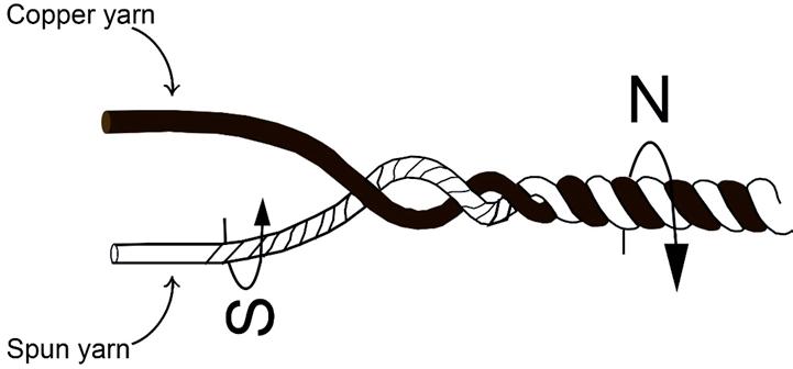
Figure 7. Plied yarn with copper as a component Figure 7. Plied yarn with copper as a component
Metal phthalocyanine based antiviral textiles Metal phthalocyanine based antiviral textiles
Phthalocyanines have been used for photodynamic therapy in medical science since long ago. PhthalocyaPhthalocyanines have been used for photodynamic therapy in medical science since long ago. nines can photosensiti ze by absorbing light in the presence of oxygen leading to the generati on of an excited sensiti zer. The excited sensiti zer modifi es the biomolecules, such as amino acids, protein, lipid and nucleic Phthalocyanines can photosensitize by absorbing light in the presence of oxygen leading to the acid by oxidati on reacti on which leads to the inacti vati on of the microbes [56,57]. Metal phthalocyanine generation of an excited sensitizer. The excited sensitizer modifies the biomolecules, such as amino BAR G, BISWAS D, PATI S, CHAUDHARY K, BAR M. Antiviral Finishing on Textiles … TLR 0 (0) 2020 00-00. compounds can be applied onto the texti les by using the ionic dyeing method. Matsushita et al. (Patent No. acids, protein, lipid and nucleic acid by oxidation reaction which leads to the inactivation of the EP2243485A1, 2010), cati onized a rayon fabric by dipping it into a soluti on containing 50g/L of cati on UK, microbes [56, 57]. Metal phthalocyanine compounds can be applied onto the textiles by using thephthalocyanine derivative and enhances the antiviral effect of the metal phthalocyanine. Cobalt (II) a cati onic agent and 15g/L of sodium hydroxide for 45 min, at 85 °C having material to liquor rati o of 1:10. Further, the cati onized fabric was treated with 1% cobalt (II) phthalocyanine monosulfonic acid and cobalt ionic dyeing method. Matsushita et al. (Patent No. EP2243485A1, 2010), cationized a rayon fabric by phthalocyanine monosulfonic and cobalt (II) phthalocyanine disulfonic acid can be replaced by iron (II) phthalocyanine disulfonic acid in a strong alkaline medium adjusted by sodium hydroxide soluti on (pH 12) dipping it into a solution containing 50g/L of cation UK, a cationic agent and 15g/L of sodium (III) phthalocyanine tetracarboxylic acid to obtain similar antiviral properties. Figure 8 shows the at 80 °C by using the exhaust method. The cati onizati on treatment improves the carrier eff ect of the metal hydroxide for 45 min, at 85 ℃ having material to liquor ratio of 1:10. Further, the cationized fabric chemical structure of cobalt (II) phthalocyanine monosulfonic acid, cobalt (II) phthalocyanine phthalocyanine derivati ve and enhances the anti viral eff ect of the metal phthalocyanine. Cobalt (II) phthawas treated with 1% cobalt (II) phthalocyanine monosulfonic acid and cobalt (II) phthalocyanine disulfonic acid and iron (III) phthalocyanine tetracarboxylic acid. Metal phthalocyanines have locyanine monosulfonic and cobalt (II) phthalocyanine disulfonic acid can be replaced by iron (III) phthalocyanine tetracarboxylic acid to obtain similar anti viral properti es. Figure 8 shows the chemical structure disulfonic acid in a strong alkaline medium adjusted by sodium hydroxide solution (pH 12) at 80 °C byenzyme-like catalytic activity and it has been found that protein denaturation is induced by the of cobalt (II) phthalocyanine monosulfonic acid, cobalt (II) phthalocyanine disulfonic acid and iron (III) phthausing the exhaust method. The cationization treatment improves the carrier effect of the metal adsorptive properties and redox catalytic functions of the metal phthalocyanines. Virus titre test locyanine tetracarboxylic acid. Metal phthalocyanines have enzyme-like catalyti c acti vity and it has been result of the rayon fabric treated by metal phthalocyanine indicates virus reduction rate of 99% or found that protein denaturati on is induced by the adsorpti ve properti es and redox catalyti c functi ons of the above when tested against the avian influenza virus. However, only cationized rayon fabric shows a metal phthalocyanines. Virus ti tre test result of the rayon fabric treated by metal phthalocyanine indicates virus reducti on rate of 99% or above when tested against the avian infl uenza virus. However, only cati ovirus reduction rate of 96.83% which explains the impact of cationization [38]. nized rayon fabric shows a virus reducti on rate of 96.83% which explains the impact of cati onizati on [38].

Figure 8. Chemical structure of (a) cobalt (II) phthalocyanine monosulfonic acid (b) cobalt (II) phthalocyanine disulfonic Figure 8. Chemical structure of (a) cobalt (II) phthalocyanine monosulfonic acid (b) cobalt (II) phthal acid (c) iron (III) phthalocyanine tetracarboxylic acid [38] ocyanine disulfonic acid (c) iron (III) phthalocyanine tetracarboxylic acid [38]
Cationic surfactant-based treatment
Cati onic surfactants synthesized from the condensati on of esterifi ed dibasic amino acids and fatt y acids are widely used as fi nishing agents for texti les and as disinfectants in detergents [58]. However, they can BAR G, BISWAS D, PATI S, CHAUDHARY K, BAR M. Antiviral Finishing on Textiles … TLR 0 (0) 2020 00-00. also be used as an eff ecti ve anti microbial agent because of their biocidal properti es. Quaternary ammonium compounds, a type of a cati onic surfactant, are eff ecti ve against many bacteria as well as enveloped viruses, whereas non-enveloped viruses are highly resistant against it [59]. Cati onic surfactant having a Ethanol improves the permeability of the cationic surfactant into the fibre and helps enhance the polyoxyalkylene group in combinati on with a water miscible solvent exhibits anti viral acti vity against both antiviral effect. A 99.9% drop in virus concentration on the surface of the textile material was enveloped and non-enveloped viruses on texti le materials [60]. Tobe et al. (Patent No. JP6519080B2, 2019) observed when the textile material treated with the abovementioned antiviral composition was prepared an anti viral compositi on containing 0.2% of didecyl methyl poly (oxyethyl) ammonium propionate, tested against Feline calicivirus after leaving it for 1 hour [60]. a cati onic surfactant, 10% of ethanol and a water miscible solvent and have treated a texti le fabric by using the aerosol spray method, followed by air-drying. The weight pick-up of the texti le fabric was restricted to Chitosan based antiviral treatment 50%. Ethanol improves the permeability of the cati onic surfactant into the fi bre and helps enhance the anti viral eff ect. A 99.9% drop in virus concentrati on on the surface of the texti le material was observed when The use of biomaterials in developing protective textiles has increased rapidly due to their ecothe texti le material treated with the abovementi oned anti viral compositi on was tested against Feline califriendly, biodegradable and non-toxic properties. Among biomaterials, chitosan is widely utilized for civirus aft er leaving it for 1 hour [60]. multi-functional finishes on textile materials such as the antimicrobial finish, mosquito repellent finish, crease resistant finish etc. [61, 62]. Chemical structure of chitosan is shown in Figure 9.
Chitosan is generally obtained from crab shells, shrimp shells, lobsters as well as the exoskeleton of the zooplankton like corals, jellyfish etc. [63, 64, 65]. It contains a large amount of positively charged nucleophilic amino groups which damage the virus’ cell membrane. The effectiveness of chitosan’s antiviral activity is enhanced when a textile material is treated with chitosan along with organic acid crosslinking agent, plant extract and sodium phosphate. Xinming (Patent No CN105506984A, 2016) prepared an antiviral solution containing 0.5% of hydroxypropyl chitosan, 0.6% of O-carboxymethyl-
N, N, N-trimethyl ammonium chloride chitosan, 1% of butane tetracarboxylic acid, 1% of citric acid, 2% of sodium phosphate, 0.6% of folium artemisiae argyi extract, 0.4% of dandelion extract, 0.6% of tea extract and 0.8% of aloe vera extract and has applied the same over a textile surface through the pressure-rolling process having the weight pick-up 70-100%. After that, the fabric was dried at about 100-120 °C and baked at 150-190°C for 2.5-5 minutes. On evaluation, it was observed that the treated fabric had high efficacy (more than 99.9%) against a broad spectrum of viruses [66].
Chitosan based antiviral treatment
The use of biomaterials in developing protecti ve texti les has increased rapidly due to their eco-friendly, biodegradable and non-toxic properti es. Among biomaterials, chitosan is widely uti lized for multi -functi onal fi nishes on texti le materials such as the anti microbial fi nish, mosquito repellent fi nish, crease resistant fi nish etc. [61,62]. Chemical structure of chitosan is shown in Figure 9. Chitosan is generally obtained from crab shells, shrimp shells, lobsters as well as the exoskeleton of the zooplankton like corals, jellyfi sh etc. [63,64,65]. It contains a large amount of positi vely charged nucleophilic amino groups which damage the virus’ cell membrane. The eff ecti veness of chitosan’s anti viral acti vity is enhanced when a texti le material is treated with chitosan along with organic acid crosslinking agent, plant extract and sodium phosphate. Xinming (Patent No CN105506984A, 2016) prepared an anti viral soluti on containing 0.5% of hydroxypropyl chitosan, 0.6% of O-carboxymethyl-N, N, N-trimethyl ammonium chloride chitosan, 1% of butane tetracarboxylic acid, 1% of citric acid, 2% of sodium phosphate, 0.6% of folium artemisiae argyi extract, 0.4% of dandelion extract, 0.6% of tea extract and 0.8% of aloe vera extract and has applied the same over a texti le surface through the pressure-rolling process having the weight pick-up 70-100%. Aft er that, the fabric was dried at about 100-120 °C and baked at 150-190°C for 2.5-5 minutes. On evaluati on, it was observed that the treated fabric had high effi cacy (more than 99.9%) against a broad spectrum of viruses [66].

Figure 9. Chemical structure of chitosan [67] Figure 9. Chemical structure of chitosan [67]
Antiviral treatment for nonwoven textiles
Antiviral treatment for nonwoven textiles
Nonwoven fabrics are a wide range of fibrous materials, which are formed through direct fibre web formation. They are widely used for filtration purposes for their high air permeability, abrasion resistance, uniform structure, etc. [68]. A nonwoven fabric can also be used for inhibiting the growth of microbes, especially viruses, when treated with acidic polymers. Poly carboxylic acid polymer is preferable as an acidic polymer to treat nonwoven fabric. The acidic polymer can be applied on nonwoven fabric in combination with organic acid, plasticizers or surfactants in different ratios in order to enhance the antiviral performance of the treated fabric. Biedermann et al. (Patent No. WO2008009651A1, 2008) prepared a loading solution containing 2% (w/w) of carbopol ETD 2020, a poly carboxylic acid polymer and 1% (w/w) of citric acid and have coated a polypropylene nonwoven fabric with the abovementioned loading solution. When a virus came into contact with the coated nonwoven surface, it interacted with the acidic polymer and was subsequently entrapped. The acidic environment (pH 2 to 2.5) of the acidic polymer inactivated and neutralized the virus. A reduction in viral titre around 99.97% was observed when nonwoven fabric coated with the acidic polymer was tested against the avian influenza A NIBRG-14 H5N1 virus with an hour of contact time [69]. Kim (Patent No. KR101317166B1, 2013) treated a nonwoven fabric using the kimchi enzyme to make it antiviral. Kimchi is a type of fermented Korean food. Kimchi enzyme is obtained by culturing the lactic acid bacteria after aging kimchi. An aqueous antiviral solution comprised of kimchi enzyme (10%) and polyvinyl alcohol resin (25%) was prepared and sprayed onto the nonwoven polyester fabric by using the electrospinning method, followed by drying at 100 °C. The finished fabric contained 3% w/w of kimchi enzyme and 7% w/w of polyvinyl alcohol resin. It was observed that the virus log reduction value of the nonwoven fabric treated with kimchi enzyme is more than 4.9 (>99.87%) when tested against the influenza A virus after being incubated for an hour. The ingredients of kimchi, such as green onion and ginger, hinder the virus growth further [70].
Antiviral treatment for nonwoven textiles
The idea of developing antiviral textiles is a novel one. Only a few studies on developing antiviral textiles have been carried out and most of these studies have been patented. The viruses for antiviral testing, test conditions etc. are different from one study to another. Hence, very limited information is available for the comparison of existing antiviral textiles. Here, the effectiveness of antiviral finish and its durability are considered for the comparison of various antiviral finished textile materials. The effectiveness of an antiviral textile material depends on the antiviral compounds used and the virus against which the material is being tested. It is observed that textile materials treated with monovalent copper compound, metal phthalocyanine, acidic polymer and kimchi enzyme showed a virus reduction rate of more than 99% when tested against the influenza virus [38,53,54,69,70]. Textiles treated with a cationic surfactant and textiles treated with chitosan show a similar result when tested against the feline calicivirus [60,66]. Durability of the antiviral finish on the textile material depends on how frequently the product is washed. In the case of single-use health care and hygiene products, durability is least important while in the case of daily used apparel and home textile products, durability influences its effectiveness. It is observed that fabrics treated with the honeysuckle extract, chitosan, kimchi enzyme and metal phthalocyanine are durable for several washes [38,41,66,70]. The premix binder solution makes textile material coated with povidone iodine durable and imparts sanitizing activity to the finished product [42], whereas textile materials treated with a cationic surfactant are semi-durable. Along with effectiveness and durability, skin-friendliness is another important parameter which determines whether the finished textile is suitable for garment manu-
facturing or not. Kimchi enzyme, chitosan and honeysuckle are extracted from bio-sources. The treated textile materials are safe for the skin when these bio-extracts are applied onto the textile fabric within the permissible limit [41,66,70]. Textile materials treated with acidic polymer and a cationic surfactant are also skin-friendly as the abovementioned chemicals exhibit excellent antiviral property at a very low add-on level. Polyester fabric treated with the copper pigment and CuCl gives excellent antiviral activity but these are not recommended for apparel as they induce skin irritation in case of direct and prolonged contact with the skin [53,54,71].
Looking at the outbreak of deadly viruses in the past and considering the ongoing pandemic scenario, the antiviral property should be one of the most desirable requirements for any textile product. However, textile materials are not inherently antiviral, they need to be treated with some suitable chemical agents to impart the antiviral property. This treatment raises the cost of the finished product and, moreover, not all antiviral agents are environment friendly. Thus, one should use antiviral textiles where it is necessary. The healthcare sector should be the prime consumer of antiviral textiles, as the personnel connected to this sector deals with the virus-affected patients directly. Depending upon the number of uses, the textile for healthcare sector are of two types, namely single-use products and multiple-use products. Surgical gowns, surgical caps, facemasks etc. are the example of some single-use textile products while the patients’ bed covers, pillow covers, window curtains etc. are the example of multiple-use products. The singleuse textiles can be treated with non-durable antiviral agents while the durability of the antiviral finishing is one of the major requirements for the multiple-use products. After healthcare, the public transport sector should be the next major consumer of antiviral textiles. Majority of the world’s population depends on public transport on a daily basis which increases the probability of virus transmission followed by surface contamination. Therefore, there is a great scope of use for the antiviral polyester fabric treated with CuCl or polyester fibres embedded with ionic copper powder by dope finishing for seat covers and curtains in buses and trains because of their high antiviral efficacy and excellent dirt-cleaning efficiency. Furthermore, train and bus seat covers can also be coated with povidone iodine and nonoxynol 9 coating solution for their self-sanitizing activity. Like healthcare and public transport sectors, textile materials used in hospitality sectors should have antiviral properties. In hotels, bed linens, table linen and bath linen are frequently used by different customers. To prevent the possibility of any viral transmission the above textile products should be treated with some suitable antiviral agents. During the pandemic situation, antiviral properties are preferable for the garments also. Fabrics treated with the honeysuckle extract, chitosan and kimchi enzyme are durable for up to several washes. The abovementioned antiviral agents are bio-based and do not have any adverse effect on human skin. The fabrics treated with the abovementioned antiviral compounds can be used for outerwear garments, like shirts, T-shirts, trousers, tops, skirts and bottoms. These fabrics can also be used for sportswear and military wear. For instant antiviral effect, any type of textile products can be treated with a cationic surfactant-based antiviral composition by using the dipping or spraying method, followed by air drying.
CONCLUSION
Looking at the current scenario, the need for antiviral textiles is becoming essential. Researches are being carried out to keep up the pace with the market demand and safety of the people. Textile materials are treated with various synthesized chemicals such as triclosan, copper compound, acidic polymer, povi-
done iodine, cationic surfactant, and metal phthalocyanine as well as with some natural extracts such as honeysuckle extract, chitosan and kimchi enzyme, in order to impart antiviral property to them. Most of these textiles treated with chemicals and bio-extracts show excellent antiviral property against a wide spectrum of viruses. Keeping the disastrous viral diseases and pandemics in mind, the above antiviral agents can be applied onto various textile products to fight against the SARS-CoV-2 virus, thereby providing a meaningful solution for saving the mankind. Antiviral textiles continue to be one of the most dynamic fields of research and one that needs to be on the lookout for novel technologies.
Declaration of conflicting interests The authors declare no potential conflicts of interest with respect to the research, authorship, and/or publication of this article.
Funding The author(s) received no financial support for the research, authorship, and/or publication of this article.
REFERENCES
[1] Qiu W, Rutherford S, Mao A, Chu C. The Pandemic and its Impacts. Health, Culture and Society. 2017; 9:1–11. https://doi.org/10.5195/hcs.2017.221 [2] Ding Y, Cao Z, Cao L, Ding G, Wang Z, Xiao W. Antiviral activity of chlorogenic acid against influenza A (H1N1/H3N2) virus and its inhibition of neuraminidase. Scientific Reports. 2017;7(1):1-11. https://doi. org/10.1038/srep45723 [3] Li Y, Pi Q, You H, Li J, Wang P, Yang X, et al. A smart multi-functional coating based on anti-pathogen micelles tethered with copper nano particles via a biosynthesis method using l-vitamin C. RSC Advances. 2018;8(33):18272–18283. https://doi.org/10.1039/C8RA01985A [4] Paget J, Spreeuwenberg P, Charu V, Taylor RJ, Iuliano AD, Bresee J, et al. Global mortality associated with seasonal influenza epidemics: New burden estimates and predictors from the GLaMOR Project.
Journal of Global Health. 2019;9(2):1-12. http://dx.doi.org/10.7189/jogh.09.020421 [5] Yuan G, Cranston R. Recent Advances in Antimicrobial Treatments of Textiles. Textile Research Journal. 2008;78(1):60–72. https://doi.org/10.1177/0040517507082332 [6] Windler L, Height M, Nowack B. Comparative evaluation of antimicrobials for textile applications.
Environment International. 2013;53:62–73. https://doi.org/10.1016/j.envint.2012.12.010 [7] Gupta D, Bhaumik S. Antimicrobial treatments for textiles. India Journal of Fibre &Textile Research. 2007;32:254–63. [8] Radhika D. Review Study on Antimicrobial Finishes on Textiles – Plant Extracts and Their Application.
International Research Journal of Engineering and Technology. 2019;6(11):3581–3588. [9] Morais D, Guedes R, Lopes M. Antimicrobial Approaches for Textiles: From Research to Market.
Materials. 2016;9(6):498. https://doi.org/10.3390/ma9060498 [10] Joshi M, Ali SW, Rajendran S. Antibacterial finishing of polyester/cotton blend fabrics using neem (Azadirachta indica): A natural bioactive agent. Journal of Applied Polymer Science. 2007;106(2):793–800. https://doi.org/10.1002/app.26323 [11] Sapp J. The Prokaryote-Eukaryote Dichotomy: Meanings and Mythology. Microbiology and Molecular
Biology Reviews. 2005;69(2):292–305. https://doi.org/10.1128/mmbr.69.2.292-305.2005
[12] Govarthanam KK, Anand SC, Rajendran S. Development of Advanced Personal Protective Equipment Fabrics for Protection Against Slashes and Pathogenic Bacteria Part 1: Development and Evaluation of Slash-resistant Garments. Journal of Industrial Textiles. 2010;40(2):139–155. https://doi. org/10.1177/1528083710366722 [13] Beşen BS. Production of Disposable Antibacterial Textiles Via Application of Tea Tree Oil Encapsulated into Different Wall Materials. Fibers and Polymers. 2019;20(12):2587–2593. . https://doi.org/10.1007/ s12221-019-9350-9 [14] Shahidi S, Ghoranneviss M, Moazzenchi B, Rashidi A, Mirjalili M. Investigation of Antibacterial Activity on Cotton Fabrics with Cold Plasma in the Presence of a Magnetic Field. Plasma Processes and Polymers. 2007;4(S1):S1098–1103. https://doi.org/10.1002/ppap.200732412 [15] Deshmukh A, Deshmukh S, Zade V, Thakare V. The Microbial Degradation of Cotton and Silk dyed with Natural Dye: A Laboratory Investigation. International Journal of Theoretical & Applied Sciences. 2013;5(2):50–59. [16] Boryo DEA. The effect of microbes on tactile material: a review on the way out so far. International Journal of Engineering Science. 2013;2:9–13. [17] Callewaert C, De Maeseneire E, Kerckhof F-M, Verliefde A, Van de Wiele T, Boon N. Microbial Odor Profile of Polyester and Cotton Clothes after a Fitness Session. Applied and Environmental Microbiology. 2014;80(21):6611–6619. https://doi.org/10.1128/aem.01422-14 [18] Bar G, Bar M. Antibacterial Efficiency of Croton Bonplandianum Plant Extract Treated Cotton Fabric.
Current Trends in Fashion Technology & Textile Engineering. 2020; 7(1):5-9. [19] Freney J, Renaud FNR. Intelligent Textiles and Clothing for Ballistic and NBC Protection. 2012th edition. B.V.: Springer Science+Business Media; 2012. Chapter 3, Textiles and Microbes; p. [53–81]. [20] Wu KJ. There are more Viruses than Stars in the Universe. Why do only some Infect Us? [Internet]. www.nationalgeographic.com. [cited 2020 Jun 25]. Available from: https://www.nationalgeographic. com/science/2020/04/factors-allow-viruses-infect-humans-coronavirus/, 2020. [21] Gelderblom HR. Medical Microbiology. 4th edition. Galveston: Galveston (TX): University of Texas
Medical Branch; 1996. Chapter 42, Structure and Classification of Viruses. [22] Riley RL. Airborne infection. The American Journal of Medicine. 1974;57(3):466–475. https://doi. org/10.1016/0002-9343(74)90140-5 [23] Sattentau Q. Avoiding the void: cell-to-cell spread of human viruses. Nature Reviews Microbiology. 2008;6(11):815–826. https://doi.org/10.1038/nrmicro1972 [24] Wells WF. Airborne Contagion and Air Hygiene: An Ecological Study of Droplet Infection. Cambridge, Mass.: Harvard University Press; 1955. [25] Van Doremalen N, Bushmaker T, Morris DH, Holbrook MG, Gamble A, Williamson BN, et al. Aerosol and Surface Stability of SARS-CoV-2 as Compared with SARS-CoV-1. The New England Journal of Medicine. 2020;382(16):1564–1567. https://doi.org/10.1056/nejmc2004973 [26] Kramer A, Assadian O. Use of Biocidal Surfaces for Reduction of Healthcare Acquired Infections. Switzerland: Springer international publishing; 2014. Chapter 1, Survival of Microorganisms on Inanimate
Surfaces; p. [7–26]. [27] Billiau A. Virus Replication – An Introduction. Paediatric Research. 1980;14(2):165–165. https://doi. org/10.1203/00006450-198002000-00023 [28] Lim SH, Hudson SM. Application of a fiber-reactive chitosan derivative to cotton fabric as an antimicrobial textile finish. Carbohydrate Polymers. 2004;56(2):227–234. https://doi.org/10.1016/j. carbpol.2004.02.005
[29] Yazhini KB, Prabu HG. Antibacterial Activity of Cotton Coated with ZnO and ZnO-CNT Composites.
Applied Biochemistry and Biotechnology. 2015;175(1):85–92. https://doi.org/10.1007/s12010-0141257-8 [30] Ford WB. Disinfection procedures for personnel and vehicles entering and leaving contaminated premises. Revue scientifique et technique (International Office of Epizootics).1995;14(2):393–401. https://doi.org/10.20506/rst.14.2.847 [31] Kampf G, Todt D, Pfaender S, Steinmann E. Corrigendum to “Persistence of coronaviruses on inanimate surfaces and their inactivation with biocidal agents”. Journal of Hospital Infection. 2020;104:246–251. https://doi.org/10.1016/j.jhin.2020.06.001 [32] Bigliardi PL, Alsagoff SAL, El-Kafrawi HY, Pyon J-K, Wa CTC, Villa MA. Povidone iodine in wound healing:
A review of current concepts and practices. International Journal of Surgery. 2017;44:260–268. https:// doi.org/10.1016/j.ijsu.2017.06.073 [33] Hsu BB, Yinn Wong S, Hammond PT, Chen J, Klibanov AM. Mechanism of inactivation of influenza viruses by immobilized hydrophobic polycations. Proceedings of the National Academy of Sciences of the United States of America. 2011;108(1):61–66. https://doi.org/10.1073/pnas.1017012108 [34] Periolatto M, Ferrero F, Vineis C, Varesano A, Gozzelino G. Antibacterial Agents. IntechOpen; 2017.
Chapter 2, Novel Antimicrobial Agents and Processes for Textile Applications; p. [17–37]. [35] Gulrajni ML, Gupta D. Emerging techniques for functional finishing of textile. Indian Journal of Fibre &
Textile Research. 2011;36(4):388-397. [36] Iyigundogdu ZU, Demir O, Asutay AB, Sahin F. Developing Novel Antimicrobial and Antiviral Textile
Products. Applied Biochemistry and Biotechnology. 2017;181(3):1155–1166. https://doi.org/10.1007/ s12010-016-2275-5 [37] Borneman J. Scientists Prove Benefit of Textiles with Antiviral and Antibacterial Effect | Textile World [Internet]. Available from: https://www.textileworld.com/textile-world/textile-news/2014/01/scientistsprove-benefit-of-textiles-with-antiviral-and-antibacterial-effect/, 2014. [38] Matsushita M, Otsuki K, Takakuwa H. Anti-viral agents, anti-viral fibers and anti-viral fiber structures. European Patent No. EP2243485A1, 2010. [39] Nelson G. Microencapsulation in textile finishing. Coloration Technology. 2008;31(1):57-64. [40] Roy Choudhury AK. Principles of Textile Finishing. Cambridge: Woodhead Publishing; 2017. 354 p. [41] Jinmei W. Nanometer traditional chinese medicine microcapsule fabric finishing agent, preparation and fabric finishing method. China Patent No. CN101324026B, 2011. [42] Snyder DE Jr. Anti-bacterial/anti-viral coatings, coating process and parameters thereof. US Patent
No.US5968538A, 1999. [43] Ripa S, Bruno N, Reder RF, Casillis R, Roth RI. Handbook of Topical Antimicrobials: Industrial Applications in Consumer Products and Pharmaceuticals. New York: Marcel Dekker; 2003. Clinical Applications of
Povidone-Iodine as a Topical Antimicrobial; p. [77–98]. [44] Ma D, Wu T, Zhang J, Lin M, Mai W, Tan S, et al. Supramolecular Hydrogels Sustained Release Triclosan with Controlled Antibacterial Activity and Limited Cytotoxicity. Science of Advanced Materials. 2013;5(10):1400–1409. https://doi.org/10.1166/sam.2013.1602 [45] National Center for Biotechnology Information, Compound CID: 129866050 [Internet]. [cited 2020 Jun 25]. Available from: https://pubchem.ncbi.nlm.nih.gov
[46] Liu CW, Chen BC, Chen TM, Chang YY. Effect of Different Extraction Methods on Major Bioactive
Constituents at Different Flowering Stages of Japanese Honeysuckle (Lonicera japonica Thunb.). Journal of Agronomy & Agricultural Science. 2020;3(21):1-9. [47] Zhou Y, Tang RC. Facile and eco-friendly fabrication of AgNPs coated silk for antibacterial and antioxidant textiles using honeysuckle extract. Journal of Photochemistry and photobiology B: Biology. 2018;178:463471. [48] Toyama DO, Ferreira MJP, Romoff P, Fávero OA, Gaeta HH, Toyama MH. Effect of Chlorogenic Acid (5-Caffeoylquinic Acid) Isolated from Baccharis oxyodontaon the Structure and Pharmacological Activities of Secretory Phospholipase A2 from Crotalus durissus terrificus. BioMed Research International. 2014;2014:1–10. https://doi.org/10.1155/2014/726585 [49] Bigliardi P, Langer S, Cruz JJ, Kim SW, Nair H, Srisawasdi G. An Asian Perspective on Povidone Iodine in
Wound Healing. Dermatology. 2017;233(2–3):223–233. https://doi.org/10.1159/000479150 [50] Borkow G. Using Copper to Fight Microorganisms. Current Chemical Biology. 2012;6(2):93–103. https:// doi.org/10.2174/187231312801254723 [51] Borkow G, Gabbay J. Putting copper into action: copper-impregnated products with potent biocidal activities. The FASEB Journal. 2004;18(14):1728–1730. https://doi.org/10.1096/fj.04-2029fje [52] Warnes SL, Little ZR, Keevil CW. Human Coronavirus 229E Remains Infectious on Common Touch Surface
Materials. mBio. 2015;6(6):1-10. https://doi.org/10.1128/mbio.01697-15 [53] Fujimori Y, Nakayama T, Sato T. Antiviral agent. European Patent No. EP2786760A1, 2014. [54] Gabbay J. Antimicrobial and antiviral polymeric materials. US Patent No. US7169402B2, 2007. [55] Diaz MS. Antiviral and antimicrobial material. PCT Patent No. WO2015035529A2, 2015. [56] Ben-Hur E, Chan WS. The Porphyrin Handbook. Amsterdam: Academic Press; 2003, Chapter 117,
Phthalocyanines in Photobiology and their Medical Applications; p. [1-36]. [57] Wiehe A, O’Brien JM, Senge MO. Trends and targets in antiviral phototherapy. Photochemical and
Photobiological Sciences. 2019;18(11):2565–2612. https://doi.org/10.1039/c9pp00211a [58] Bonvila XR, Roca SF, Pons RS. Antiviral use of cationic surfactant. PCT Patent No. WO2008014824A1, 2008. [59] Shirai J, Kanno T, Tsuchiya Y, Mitsubayashi S, Seki R. Effects of Chlorine, Iodine, and Quaternary Ammonium Compound Disinfectants on Several Exotic Disease Viruses. Journal of Veterinary Medical
Science. 2000;62(1):85–92. https://doi.org/10.1292/jvms.62.85 [60] Tobe S, Tobe S, Takehiko M. Antiviral composition for textiles. Japan Patent No. JP6519080B2, 2019. [61] Inamdar MS, Chattopadhyay DP. Chitosan and its versatile applications in textile processing. Man-Made
Textiles in India. 2006;49:211-216. [62] Pillai CKS, Paul W and Sharma CP. Chitosan: Manufacture, Properties, and Usage. New York: Nova
Science Publishers; 2010, Chapter 3, Chitosan: Manufacture, Properties & Uses; p. [133-216]. [63] Zhao Y, Xu Z, Lin T. Antimicrobial Textiles. Cambridge: Woodhead Publishing; 2016, Chapter 12, Barrier textiles for protection against microbes; p. [225-245]. [64] Joshi M, Ali SW, Purwar R, Rajendran S. Eco-friendly antimicrobial finishing of textiles using bioactive agents based on natural products. Indian Journal of Fibre & Textile Research. 2009;34:295-304. [65] Chattopadhyay DP, Inamdar MS. Handbook of Sustainable Polymers: Processing and Applications. Florida: Pan Stanford Publishing; 2015, Chapter 19, Chitosan and Nano Chitosan: Properties and
Application to Textiles; p. [659-741].
[66] Xinming X. Method for performing antibacterial and antivirus treatment on textiles by utilizing natural biomaterials, China Patent No CN105506984A, 2016. [67] Sami El-banna F, Mahfouz ME, Leporatti S, El-Kemary M, A. N. Hanafy N. Chitosan as a Natural Copolymer with Unique Properties for the Development of Hydrogels. Applied Sciences. 2019;9(11):2193. https:// doi.org/10.3390/app9112193 [68] Nallathambi G, Evangelin S, Kasthuri R, Nivetha D. Journal of Textile Engineering & Fashion Technology. 2019;5:81-84 [69] Biedermann K, Deng F, King S, Middleton A, Oths PJ. Anti-viral face mask and filter material. PCT Patent
No. WO2008009651A1, 2008. [70] Kim Y. Antivirus non-woven fabrics, hybrid cabin air filter containing the same and manufacturing method thereof. South Korea Patent No. KR101317166B1, 2013. [71] Li H, Toh PZ, Tan JY, Zin MT, Lee C, Li B, Leolukman M, Bao H, Kang L. Selected Biomarkers Revealed
Potential Skin Toxicity Caused by Certain Copper Compounds. Scientific Reports. 2016;6(1):1-11. https:// doi.org/10.1038/srep37664


