
10 minute read
Progeria: The Fight Against Time

from Issue 12

Hutchinson-Gilford Progeria Syndrome:
The fight against time
Advertisement
Jennifer Gao, Christine Ibrahim, Jennifer Lee, Christina Martorelli, & Penny Yin
hAVIng juST oVer one hunDreD DocumenTeD cASeS worlDwIDe, huTchInSongIlforD progerIA SynDrome (hgpS) IS A fATAl DISeASe whIch cAuSeS premATure AgIng In chIlDren. ThIS ArTIcle AImS To proVIDe A Thorough bAckgrounD on ThIS unDer DIScuSSeD IllneSS by DeScrIbIng ITS pAThogeneSIS, clInIcAl feATureS, DIAgnoSIS, lIfeSTyle effecTS, currenT TreATmenT, AnD poTenTIAl AreAS for furTher reSeArch. SympTomS STem from A SIngle poInT-muTATIon In The lmnA gene ThAT DISrupTS A cell’S normAl nucleAr lAmInA funcTIonS. unforTunATely, There IS no cure for hgpS, howeVer, currenT clInIcAl TrIAlS Show ThAT ThIS DISeASe mAy be conTrolleD SympTomATIcAlly uSIng fArneSylTrAnSferASe InhIbITorS. currenT lITerATure AlSo SuggeSTS ThAT rIbonucleIc AcID InTerference (rnAI) IS A poTenTIAl TreATmenT meThoD ThAT TArgeTS The DISeASe AT A SubcellullAr leVel.
Derived from the Greek word meaning “prematurely old”, progeria denotes a group of disorders that impart signs of
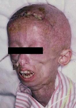
Figure 1 A 15-year-old HGPS patient exhibiting sunken cheeks, narrow nose, and protrusion of eye sockets. premature aging in patients of all ages. Hutchinson-Gilford progeria syndrome (HGPS) is a severe form of progeria that specifically targets infants and children (Progeria Research Foundation, 2006). Originally thought to be an autosomal recessive genetic disorder, HGPS is caused by a single, random nucleic acid mutation in the LMNA gene (Pollex & Hegele, 2004). The LMNA gene codes for lamin A and C proteins, both of which normally serve as structural microfilaments for the nuclear lamina, located within the cell’s nuclear envelope. The nuclear lamina is crucial in maintaining structural stability of the nuclear envelope, as well as regulating the gene transcription process. It embeds nuclear pores and binds to various transcription factors and proteins (Capell & Collins, 2006). Approximately 90% of HGPS cases are caused by a substitution of cytosine for thymine that yields a silent G680G mutation (Glynn & Glover, 2005). Although no change is observed at the amino acid level, the activation of a cryptic splice donor site occurs at the nucleic acid level. Thus, an extra 150 nucleotides are lost during RNA splicing, including an important endoproteolytic cleavage site, the loss of which renders improper maturation of the lamin A protein (Glynn & Glover, 2005). This mutated protein, progerin, then becomes permanently bound to a farnesyl group, a 12-carbon lipid molecule, which is normally cleaved for proper protein function. As a result, progerin becomes erroneously embedded in the nuclear membrane (Capell & Collins, 2006). Drastic events follow, including “nuclear blebbing” - formation of blister-like bubbles on the nuclear envelope, and disruption of the transcription factors essential for DNA replication and protein synthesis (Capell & Collins, 2006). All of these damages accumulate to form an unstable nucleus that may lead to mistranslated proteins, lower cell reproductive ability, and possible cell apoptosis.
Cytoplasm
Nuclear blebbing Nuclear pore clustering
Altered binding and mislocation of nuclear envelope proteins
Nuclear SREBP1
blebbing
Nucleus
Progerin Farnesyl group Nuclear lamina (lamins A,B1,B2,C)
cLinicAL FeATureS
Although infants with HGPS appear healthy at birth, they begin to exhibit signs of abnormal physical growth and common characteristics of aging by twelve months of age (Brown, 1992). The aging process that a HGPS patient undergoes in one year is equivalent to approximately ten years in a normal individual (Pollex & Hegele, 2004). Therefore, by the age of ten, HGPS patients would have various cardiovascular, respiratory, and orthopaedic conditions. On average, children with HGPS die at the age of thirteen due to cerebrovascular and coronary atherosclerosis (Pollex & Hegele, 2004). However, the intellectual development of these children is not affected by HGPS, as the LMNA gene is not expressed in brain cells (Progeria Research Foundation, 2006). In general, HGPS children are characterized by short stature, below average weight, and limited sexual maturation. A ten-year-old HGPS patient will be of same
RB Outer nuclear membrane
Disrupted heterochromatin organization
Misregulated membrane Inner nuclear
gene expression
Chromatin Nuclear pore complex
Heterochromatin Nuclear envelope proteins (nesprin, emerin, LAP2) Transcriptional regulators
Figure 2 Structural and functional consequences due to the improper embedding of lamin A protein in the nuclear lamina.
(GCL, MOK2, RB, SREBP1) height as an average three-year-old child (Sarkar & Shinton 2001). Phenotypes are most notable in the facial area, including a small jaw (micrognathia), proportionally large cranium, protruding eyes, narrow nose, and prominent veins on the scalp (Pollex & Hegele, 2004). Moreover, HGPS children all experience premature loss of hair (alopecia), eyebrows, and eyelashes. Typical dermatological features include dry, wrinkled skin, caused by the hardening of connective tissue and the loss of subcutaneous adipose tissue, as well as the uneven thickening of the skin due to the presence of scar tissue-like lesions (Uitto, 2002; Sarkar & Shinton, 2001).
diAgnoSiS & LiFeSTyLe eFFecTS
Since children afflicted with progeria appear healthy at birth, it is extremely difficult to diagnose this disease in newborns. However, at around six to twelve months, a baby with HGPS will fail to gain weight and display a noticeable discolouration of the skin. The pediatrician may recommend
testing the arterial walls of the child, as patients with HGPS often exhibit early atherosclerosis. The final determinant of the diagnosis is a blood test that confirms the presence of the mutated LMNA gene (Progeria Research Foundation, 2006). Numerous lifestyle adjustments must be made following diagnosis of HGPS. Children with progeria are placed on strict diets to ensure proper nutritional intake (Progeria Research Foundation, 2006). Also, HGPS patients are at an increased risk for cardiovascular disease and are given daily doses of Aspirin, a blood thinner that increases blood circulation and the oxygen-carrying capacity of the body. However, undesirable effects may occur. The constant ingestion of Aspirin, especially at such a young age, may lead to stomach pain, excessive bleeding, or bruising (Gordon, Leslie, Lloyd & Smoot, 2001). Since HGPS patients have weaker and more fragile bones, they are placed in extensive exercise programs to maximize their range of motion, which can become restricted due to tightened skin and arthritis (Progeria Research Foundation, 2006). HGPS patients also undergo hydrotherapy to alleviate pain. Even when all these lifestyle changes are followed, the average lifespan for a child with progeria ranges from eight to twenty-one years.
currenT TreATmenTS
Due to the unique pathogenesis, disease development, and limited cases of HGPS, treatments are currently at the stage of clinical trials and many impart a multitude of unwanted side effects (Pollex & Hegele, 2004). Present research for HGPS treatment focuses on the use of farnesyltransferase inhibitors (FTIs) (Yang et al., 2006). FTIs inhibit a naturally occurring process known as
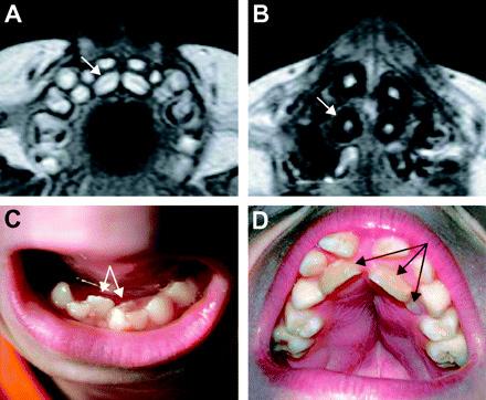
Figure 3 MRI and photographic images of teeth deformations in patients with HGPS.Images A and B are MRIs of an 8-month old girl and a 10.5-year-old boy; images C and D are photographs of a 10.5-year-old boy and a 10-year-old boy. “farnesylation” in the nucleus, which ensures the binding of lamin A protein to the nuclear envelope, catalyzed by farnesyltransferase (Fong et al., 2006). The farnesyl group only serves to aid in this binding of lamin A to the nuclear envelope (Fong et al., 2006). Thus, in normal cells, once the lamin A protein is bound, the farnesyl group is immediately cleaved. In HGPS patients, however, the farnesyl group remains attached to the protein, rendering abnormal nuclear morphology. This malformed structure then results in the manifestation of symptoms characteristic of HGPS patients (Glynn & Glover, 2005). FTI drugs prevent the enzyme farnesyltransferase from catalyzing the reaction between the farnesyl group and the lamin A protein, subsequently preventing lamin A from binding to the nuclear envelope. As a result, the nuclear envelope will possess a normal conformation (Gordon, 2006). Experiments conducted by Yang et al. (2006) indicate that FTIs are effective in treating mice with HGPS. While this treatment is promising, FTIs may impart undesirable side-effects such as diarrhea and liver malfunction (Gordon, 2006). Ongoing research for more effective treatments for the disease is thus required.
FuTure TreATmenT By rnAi
Current literature suggests that ribonucleic acid interference (RNAi) is one of the most promising therapies for HGPS. While eukaryotic cells have single stranded RNA, viruses are comprised of double stranded RNA (dsRNA). When a dsRNA is injected into a eukaryotic cell, the cell recognizes the dsRNA as foreign and attempts to destroy these molecules with a degradation process known as RNA-interference (PBS, 2005). The cell not only destroys the foreign
molecules, but also eradicates any host-made protein that contains the same complementary amino acid sequence. Scientists attempt to exploit this evolutionary process by inserting a synthesized dsRNA with the sequencing of choice. The addition of a synthesized dsRNA with the LMNA sequence would prompt the cell to eliminate all mutated lamin proteins at the post-transcriptional level, thereby reducing progerin expression (Matzke, 2004). The rationale behind attempts to use RNAi as a treatment is based on the suggestion that progeria is not caused by the absence of the correct, functioning LMNA gene
reFerenceS
and its proteins, but rather due to an over-expression of the mutated proteins within certain locations of the cell. It is the over-expression of mutated proteins that causes toxic effects (Fong, Ng, Lammerding, Vickers, & Meta, 2006). Huang et al. (2006) discovered that reduced expression of mutated LMNA proteins not only abated abnormal nuclear morphologies on HGPS cells, but also increased cell longevity. In addition, Fong et al. (2006) showed significant symptomatic improvement in the LMNA gene knock-out mice. Thus, RNAi is a potential treatment that may help to eradicate the pathogenesis of
HGPS.
Antler, C. (2003). The science creative quarterly. Antisense RNA.
Retrieved February 2, 2007 from http://www.scq.ubc.ca/?p=265. Brown, W. T. (1992). Progeria: a human-disease model of accelerated aging. American Society for Clinical Nutrition, 55, 1222-1224. Capell, B. C. & Collins, F. S. (2006). Human laminopathies: nuclei gone genetically awry. Nature Review Genetics, 7, 940-952. Faivre, L. & Cormier-Daire, V. (2005). Progeria. In V. Cormier-Daire,
Orphanet Encylopedia. Retrieved February 4, 2007, from http:// www.orpha.net/data/patho/GB/uk-progeria05.pdf Fong, L. G., Ng, J. K., Lammerding, J., Vickers, T. A., & Meta, M. (2006).
Prelamin A and lamina A appear to bedispensable in the nuclear lamina. American Society for Clinical Investigation, 116, 743-752. Glynn, M.W., & Glover, T.W. (2005). Incomplete Processing of Mutant
Lamin A in Hutchinson-Gilford Progeria Syndrome Leads to
Nuclear Abnormalities, Which are Reversed by Farnesyltransferase
Inhibition. Human Molecular Genetics, 14, 2959-2969. Goldman R. D. et al., (2004). Accumulation of mutant lamin A causes progressive changes in nuclear architecture in Hutchinson–
Gilford progeria syndrome. Proceedings of the National Academy of Sciences, 101(24), 8963-8968. Gordon, L. B. (2006). Update: Farnesyltransferase Inhibitors (FTIs) as Potential Drug Therapy for Children with Progeria: Recent
Research Findings and Frequently Asked Questions. Retrieved
December 4, 2006, from http://www.progeriaresearch.org/assets/ files/pdf/FTIQ&AAugust2006Final.pdf. Gordon, L., Feit, L. R. & Smoot, L. (2001) Important Information for You and Your Doctors About Low-Dose Aspirin Treatment and Progeria.
Retrieved December 20, 2006 from http://www.progeriaresearch. org/assets/files/pdf/AspirinTrtmnt(07-04).pdf. Huang, S., Chen, L., Libina, N., Janes, J., & Martin, G. (2005). Correction of cellular phenotypes of Hutchinson-Gilford Progeria cells by
RNA interference. Human Genetics, 118, 444-450. Jansen, T. & Romiti, R. (2000). Progeria infantum (Hutchinson-Gilford
Syndrome) associated with scleroderma-like lesions and acroostelysis: a case report and brief review of the literature. Pediatric
Dermatology, 17(4), 282-285. Matzke, A. J, & Matzke, M. A (2004). Planting the seeds of a new paradigm. PLOS Biology. 2(5). Retrieved January 4, 2006 from http://biology.plosjournals.org/perlserv/?request=getdocument&doi=10.1371/journal.pbio.0020133. PBS (2005). Sciencenow RNAi interference. Retrieved January 11, 2006 from http://www.pbs.org/wgbh/nova/sciencenow/3210/02-explflash.html. Pollex R. L., & Hegele, R. A. (2004). Hutchinson–Gilford Progeria.
Clinical Genetics, 66(5), 375-381. Progeria (2005). In Medline Plus Medical Encylopedia. U.S. Nationary
Library of Medicine. Retrieved February 4, 2007 from http://www. nlm.nih.gov/medlineplus/ency/article/001657.htm. Progeria Research Foundation, The. (2006). About Progeria. Retrieved
December 20, 2006 from http://www.progeriaresearch.org/ about_progeria.html Progeria Research Foundation, The. (2006). Diagnostic Testing.
Retrieved December 20, 2006 from http://www.progeriaresearch. org/diagnostic_testing.html Sarkar, P. K. & Shinton, R. A. (2001). Hutchinson-Guilford progeria syndrome. Postgraduate Medicine Journal, 77, 312-317. Uitto, J. (2002). Searching for clues to premature aging. Trends in
Molecular Medicine 8(4), 155-157. Yang, S. H., Meta, M., Qiao, X., Frost, D., Bauch, J., Coffinier, C.,
Majumdar, S., Bergo, M.O., Young, S.G., Fong, L.G. (2006). A farnesyltransferase inhibitor improves disease phenotypes in mice with a Hutchinson-Gilford progeria syndrome mutation.
Journal of Clinical Investigation, 116, 2115-2122.
