The Modern
Equine Vet www.modernequinevet.com
Vol 10 Issue 4 2020
Biomechanics Can Improve Rehabilitation Ask the Nutritionist? Balancing Diet for Sport Horses With Allergies Ask the Immunologist? Why are Some Horses More Reactive Than Others? Technician Update: Critical Attention for Post-Op Colic
CHECK OUT: ASK THE IMMUNOLOGIST? YOUR VACCINE QUESTIONS ANSWERED
TABLE OF CONTENTS
COVER STORY
4 Biomechanics Can Improve Rehabilitation Protocols Cover: Shutterstock/gibleho
ASK THE NUTRITIONIST
How Do We Provide a Balanced Diet to a Performance Horse with a Suspected Food Allergy?...................................................................................... 3 NEW COLUMN: ASK THE THE IMMUNOLOGIST
How to Explain the Benefits vs. Risks of Vaccination........................................................... 7 WOUND CARE
Topical CBD May Stimulate Wound Healing in Horses.......................................................10 INFECTIOUS DISEASES
Not Just for People: Equine Coronavirus.................................................................................13 TECHINICIAN UPDATE
Critical Care of a Post-Op Colic....................................................................................................14 FDA Approves Aservo EquiHaler for Severe Equine Asthma............................................12 What Does the COVID Stimulus Package Do for You?.........................................................19 ADVERTISERS Purina Sponsored Content.........................................3 Merck Animal Health........................................5 Merck Animal Health Sponsored Content..............7
American Regent Animal Health/Adequan...........9 Heska............................................................................11 AAEVT............................................................13
The Modern
Equine Vet SALES: Matthew Todd • Lillie Collett EDITOR: Marie Rosenthal ART DIRECTOR: Jennifer Barlow CONTRIBUTING WRITERS: Paul Basillo • Adam Marcus COPY EDITOR: Patty Wall Published by PO Box 935 • Morrisville, PA 19067 Marie Rosenthal and Jennifer Barlow, Publishers PERCYBO media publishing
2
Issue 4/2020 | ModernEquineVet.com
LEGAL DISCLAIMER: The content in this digital issue is for general informational purposes only. PercyBo Publishing Media LLC makes no representations or warranties of any kind about the completeness, accuracy, timeliness, reliability or suitability of any of the information, including content or advertisements, contained in any of its digital content and expressly disclaims liability of any errors or omissions that may be presented within its content. PercyBo Publishing Media LLC reserves the right to alter or correct any content without any obligations. Furthermore, PercyBo disclaims any and all liability for any direct, indirect, or other damages arising from the use or misuse of the information presented in its digital content. The views expressed in its digital content are those of sources and authors and do not necessarily reflect the opinion or policy of PercyBo. The content is for veterinary professionals. ALL RIGHTS RESERVED. Reproduction in whole or in part without permission is prohibited.
SPECIAL ADVERTISING SECTION
Ask the
Nutritionist ANNA PESTA, PH.D., EQUINE TECHNICAL SOLUTIONS, PURINA ANIMAL NUTRITION
?
Ask the Nutritionist is a monthly column featuring questions answered by PhD equine nutritionists and sponsored by Purina Animal Nutrition. Have a nutrition question you want to see featured? Email Marie Rosenthal. For clinics looking for specific nutritional advice, visit purinamills.com/ask-an-expert.
How do we provide a balanced diet to a performance horse with a suspected food allergy? With increasing frequency, equine nutritionists are being asked to review the results of serum allergy tests. It’s not uncommon for results to show dozens of positively reacting allergens. Frustrated clinicians or owners understandably ask, “What on earth can we feed this horse?” The answer lies at the end of substantial questioning and some trial and error. EXPLORE ALL THE OPTIONS First consider the horse’s clinical signs, which should narrow the field of what is truly bothering the horse. Are we dealing with an environmental factor, such as mold or pollen? Is there a skin hypersensitivity to insects or plants? Or, more rarely, is there inflammation or digestive upset due to a true food allergy? Hives, runny eyes, nasal discharge and coughing are more indicative of an inhaled allergen. To help minimize exposure to respirable dust and molds: • Trade long-stemmed hay for a complete feed • Wet hay thoroughly before feeding • Feed at ground level • Wet stall bedding or change the type of bedding • Provide as much pasture time as possible
DIAGNOSTIC LIMITATIONS SERUM ALLERGY TESTING is convenient and, when paired with clinical signs, can be a good starting place. However, it has significant limitations for diagnosing food allergens and is prone to false positives.1 Intradermal skin testing is the gold standard to diagnose skin hypersensitivities but has limitations in the field.2 The American College of Veterinary Dermatology states that serum allergy testing and intradermal skin testing are not valid tests for the diagnosis of food allergies and the identification of specific food allergens. The only way to accurately diagnose a food allergy is an elimination diet.
To help manage horses with skin irritations: • Change or eliminate topical products • Use fly sheets and adjust pasture turnout to help reduce exposure to insect bites • Use appropriate medications during the horse’s “difficult season” • Supplement feed with a source of omega-3 fatty acids, such as Purina® Amplify® High-Fat Horse Supplement However, if symptoms are unresolved or unexplained, it is time to pursue an elimination diet. CONDUCTING AN ELIMINATION DIET When only one or two feed ingredients are suspected, contact the feed manufac1 2
turer for a product recommendation that does not contain the suspect ingredients. However, when there’s a long list of potential allergens it becomes extremely difficult to find a balanced feed free of all the suspect ingredients. In these cases, a true elimination diet is required. A horse that’s been on a good plane of nutrition can be fed a hay-only diet (choose a type that did not show positive on the serum allergy test) for at least two to four weeks to observe if the symptoms resolve. If there is no improvement in symptoms, a food allergy is unlikely. If symptoms resolve, then ingredients can be reintroduced one at a time until clinical signs reappear, thereby identifying the allergenic feed ingredient to avoid.
DESIGNING THE DIET In cases with confirmed sensitivities where there are multiple offending ingredients or ingredients that are particularly hard to avoid in commercial feeds, it may be necessary to enlist the help of an equine nutritionist to design a balanced diet. Diets that provide necessary nutrients to maintain body condition and fuel for the performance horse might include alfalfa hay, vitamin/mineral supplements, grain (oats/barley/corn) for energy, oil for added calories, some beet pulp to hold it together and, if a soy-free source of quality amino acids is required, milk replacer powder can be used. Clearly this can become cumbersome for the horse owner to maintain, hence the due diligence to make sure it is truly necessary. Contact a Purina Ph.D. nutritionist for consultations through Purina Customer Service, 800-227-8941 or send us a message at www.purinamills.com/ask-an-expert.
UPCOMING TOPICS May: Endocrine disorders Have a question you want to see featured? June: Infections Send them to modernequinevet@gmail.com. July: Colic
Dupont, S., A. De Spiegeleer, D. J. X. Liu, L. Lefère, D. A. Van Doorn, Hesta, M. 2014. A commercially available immunoglobulin E-based test for food allergy gives inconsistent results in healthy ponies. Equine Vet. J. DOI: 10.1111/evj.12369 Bethlehem, S., Bexley, J., Mueller, R.S. 2012. Patch testing and allergen-specific serum IgE and IgG antibodies in the diagnosis of canine adverse food reactions. Vet. Immunol. Immunopathol. 145, 582-589
ABOUT THE AUTHOR Dr. Anna Pesta, Ph.D., is a nutritionist on the Equine Technical Solutions Team at Purina Animal Nutrition. She is a lifelong equestrian and actively competes her off-the-track Thoroughbred in three-day eventing. SPONSORED BY PURINA ANIMAL NUTRITION
ModernEquineVet.com | Issue 4/2020
3
ORTHOPEDICS
BIOMECHANICS CAN IMPROVE
Shutterstock/gibleho
Rehabilitation Protocols
When designing the most appropriate rehabilitation protocols for a sporting horse, one must consider the biomechanics of the injury, according to Sheila Schils, PhD, owner of EquiNew Therapy in River Falls, Wis. and former Professor of Equine Science at the University of Wisconsin. B y
4
Issue 4/2020 | ModernEquineVet.com
M a r i e
Much of what Dr. Schils understands about the role biomechanics plays in injury and recovery comes from studies in human athletes, but much of it is applicable to equine physical rehabilitation, she explained. The equine muscular skeletal system is design to allow the horse to graze for long periods. However, this spinal position must be modified for the horse to perform, regardless of the discipline. “These modifications produce forces and movement patterns that counteract the natural structural mechanics of the horse, and therefore, specific musculoskeletal adaptations must be developed by the horse to perform successfully in sport,� she said. The laws of physics are the basis of biomechanics
R o s e n t h a l ,
M S
Only PRESTIGE
®
CONTAINS FL ‘13 TO PROTECT AGAINST TODAY’S EIV
THE PRESTIGE® THEY DESERVE Prestige flu-containing vaccines deliver advanced influenza protection against the most relevant flu strains circulating today. Prestige vaccines meet OIE and AAEP guidelines for Clade 1 & Clade 2 protection. Learn more at PrestigeVaccines.com
Current protection Florida ‘13 Clade 1 Richmond ‘07 Clade 2 Kentucky ‘02
1
Relevant protection FL ‘13> 98.50% sequencing homology to today’s EIV1
Data on file. Merck Animal Health.
2 Giralda Farms • Madison, NJ 07940 • merck-animal-health-usa.com • 800-521-5767 Copyright © 2020 Intervet Inc., d/b/a/ Merck Animal Health, a subsidiary of Merck & Co., Inc. All rights reserved.
Advanced protection Only FL ‘13 demonstrated key site similarity to today’s EIV
ORTHOPEDICS
B. A.
Images courtesy ofDr. Shiela Schils
Some muscles are more prone to injury that others. Asymmetrical medial limb placement can increase the risk of injury. Images A and B demonstrate the correct symmetry to decrease injury. The rest of the images demonstrate asymmetrical placement , which can increase the risk of injury.
and several factors, not just 1, are typically present cycle of muscle activity to move the leg forward and to result in injury. Three of these important biomethen slow the leg down so it can hit the ground. “This chanical concepts related to injury are concussion, slowing of the leg requires a lot of energy and posdeceleration and individual-limb weight bearing. tural control and I think we need to really emphasize Injury from concussion is related to the direction the potential of this deceleration phase to produce the force is applied and to the reaction tissue has to injury.” stress and strain. For example, the tibia can handle a Some muscles appear more prone to injury than high level of force and not be injured when the force others. “Muscles that are decelerating a limb (when is applied during compression. the limb is in protraction) are However, when that same level of in an extended position and we force is applied during torsion or know, through research, that rotation, injury is likely to occur. muscles that are in a lengthened “Tissue can handle a high degree state and/or producing deceleraof concussion as long as that contion are more prone to injury.” cussion is absorbed by a joint that she said. Many human studies is [correctly] functioning biomehave found that an extended joint chanically,” Dr. Schils explained at increases the force on that joint the 66th Annual Convention of the when it hits the ground. “In one American Association of Equine study there was a 27% reduction Practitioners. in ground impact forces when Newton’s first law of motion falling occurred with elbows bent basically says that once a body rather than with the elbows natugets moving, it stays moving. rally extended,” Dr. Schils said. And when you have a cyclic mo“Therefore, exercises that imClick here to tion, such as movement of the prove joint flexion will help slow watch a video horse’s leg, there is a constant the speed of the gait and since 6
Issue 4/2020 | ModernEquineVet.com
SPECIAL ADVERTISING SECTION
Immunologist THE
ASK
Ask the Immunologist is a new column featuring expert answers and sponsored by Merck Animal Health.
Why are some horses more reactive to vaccines than others, and what’s the best way to prepare clients for what to expect after their horse is vaccinated?
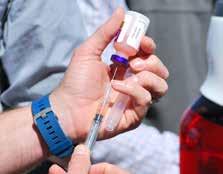
Courtesy Merck Animal Health
W
hen it comes to vaccination, communication is key to reinforcing this critical service with clients who are bombarded with consumer opinions and online information. Help clients keep perspective by reminding them that, while vaccination comes with inherent risks—as do all medical procedures—the overall incidence of vaccine-associated adverse events (VAAEs) is very low. Bolster clients’ vaccination knowledge—and confidence—by taking them through a three-part conversation.
of how vaccines affect the equine immune system. Stick to the basics of natural (innate) and adaptive (acquired) immunity, focusing on the differences between killed and modified-live vaccines. The primary message is that each type of vaccine stimulates the immune system differently.
response to a vaccine. This is what makes it so difficult to identify horses predisposed to allergic reactions and is why VAAEs are not generally due to a faulty product. What’s more, environment may play a role in VAAEs, making it difficult to predict reactions without a previous or repeated episode. Caution must be exercised when vaccinating any horse previously reported to have had a VAAE, especially if the clinical signs suggested an allergic type of vaccine reaction.
PART 2: EMPHASIZE CUSTOM PROTOCOLS. Make sure clients understand the
PART 3: ACKNOWLEDGE POTENTIAL VAAES. VAAEs can vary in type and sever-
PART 1: EXPLAIN HORSES’ IMMUNE RESPONSE. Give clients a quick rundown
American Association of Equine Practitioners’ core and risk-based vaccination guidelines are taken into consideration along with your personal experience with the horse, including the individual’s age, medical conditions, lifestyle and history of VAAEs. Just as some people are allergic to certain medications or foods, vaccine reactions in horses may be based upon their unique immunological makeup and
ity, and horses rarely experience severe reactions. For perspective, let clients know that most reactions are similar to what a person might experience after a flu shot. Still, make sure clients understand these three types of VAAEs, and remind them it’s best for the veterinarian to administer vaccinations so you’re present if a VAAE occurs. • Local injection site reactions. By far the
ABOUT THE AUTHOR Duane E. Chappell, DVM, Associate Director, Equine Pharmacovigilance and Professional Services at Merck Animal Health, has a background in academia, equine research and private practice. Dr. Chappell and the pharmacovigilance team answer calls daily from veterinarians on immunology and questions associated with Merck Animal Health products, including potential VAAEs. SPONSORED BY MERCK ANIMAL HEALTH
most common type of VAAE. Usually occur within 24 hours of vaccination and resolve shortly thereafter. Signs include: • Localized swelling of varying degree, injection-site tenderness, muscle soreness • Decreased range of motion of the limb or head and neck less commonly • Abscesses on very rare occasions • Mild systemic reactions. Usually resolve within 24 to 48 hours without treatment. Signs include: • Low grade fever (less than 102°F), lethargy, lack of appetite • Allergic systemic reactions. Require veterinary attention. Signs include: • Sweating, elevated heart rate, respiratory distress, colic or hives • Anaphylaxis, extremely rare and most severe VAAE Setting realistic expectations with clients about what to expect after vaccinations, including potential VAAEs, goes a long way to easing their minds, building a strong relationship and reinforcing your credibility along with the importance of veterinarian-administered vaccines. By working together, you and your clients form a formidable team in keeping horses protected against disease.
WANT TO ASK A QUESTION? EMAIL THE EDITOR. For more immunology-related information, visit merck-animal-health-usa.com/species/equine
ModernEquineVet.com | Issue 4/2020
7
ORTHOPEDICS
Translating These Concepts to Equine Rehab Looking at the biomechanics of an injury can help individualize equine rehabilitation, according to Sheila Schils, PhD, owner of the EquiNew Therapy in River Falls, Wis and former Professor of Equine Science at the University of Wisconsin. Consider these concepts when making corrections. EXERCISES TO LOWER PEAK FORCES INCLUDE: • slowing the speed of the gait to increase time on the ground and then to increase joint flexion, • encouraging elevated carpal and hock action during jumping, • tightening the distances between trotting poles or fences to help slow the horse or • using 1 strides while choosing the appropriate height of the fence to obtain the desired joint flection.
Exercises to Improve Symmetrical Limb Placement • Handlers can lead the horse from the side that improves the symmetry of limb weight bear. Teaching the horse to lead from the right side can dramatically improve limb placement. • Being aware of symmetrical limb placement during dry or water treadmill work. • Riders can reposition the hindquarters so that the hindquarters step into the prints of the front feet on a straight or curved line. • Riders can reposition the shoulders so that the hindquarters step into the prints of the front feet on a straight or curved line. • Repositioning the hindquarter and the forehand are 2 distinctly different muscular activities and both must be evaluated as which is the ideal means to improve the symmetry of weight bearing • Straight lines should be emphasized and turns should look more like an octagon than a curve.
these muscles are different muscles than the decelerating muscles, the decelerating muscles are not overworked and this reduces injury.” In addition, this improved deceleration ability will reduce impact forces. “It’s like buying 1 and getting 1 for free.” Think of the human or equine hamstring, this is a common muscle which is used to decelerate the leg. In addition, the biarticular function of some muscles—for example the coaction of both the hip and knee joint—also increase the risk of injury, Dr. Schils explained. When hip flexion and knee exten-
sion occurs, the hamstrings are placed in a maximally lengthened state, which is predisposing them to injury. Biomechanical studies have shown that for a limb to support the weight of the body on a straight line, the limb must be placed under the center of gravity of the body, which means the limb must swing slightly medial to “find the belly button.” However, if this medial placement is asymmetrical—where 1 limb— is less medially placed than the opposite limb this can indicate a lack of willingness to bear weight on that limb. “For me—during a movement evaluation—this is the first thing I look for as the horse walks and trots on a straight line to me and away from me to determine if the horse has a right or left hind or front limb preference. Then the horse is moved on a circle and if this excessive medial limb placement is seen regardless of the direction of movement, the horse is indicating a strong preferential limb,” she told Modern Equine Vet. And to make matters worse, the limb that is asymmetrically medially placed is almost always the limb accepting more weight, which could increase injuries, she added. In addition, the angulation of the swing phase of the excessively placed limb requires that limb to move with a faster and longer stride than the opposite limb, unbalancing the gait. This deviation may make this limb appear to step faster and not stay on the ground as long as the opposite hind. There are 4 possible reasons why horses place one limb further under their body than the opposite limb, she suggested: 1. That limb is the stonger of the 2 limbs. 2. That limb is the more coordinated of the 2 limbs. 3. That limb is both the stronger and the more coordinated limb. 4. The non medially placed limb is painful and therefore the horse resist weightbearing on that limb. Evaluating the biomechanics of the entire horse cannot only help when developing a rehabilitation plan, but it could help develop recommendations that could prevent injuries in the first place, according to Dr. Schils. “Biomechanics is a complex field of study due to all the interrelationships, you can’t just look at the trees, you have to really sit down and understand the function of the forest first. It can require a different way of looking.” MeV
For more information: Schils S, Ober TR, Butcher MT. Review of the biomechanics of injury in the equine athlete: From research to clinical practice. AAEP Proceedings 2019;65:273-280. 8
Issue 4/2020 | ModernEquineVet.com
There’s nothing else like it. Over the past 30 years, Adequan® i.m. (polysulfated glycosaminoglycan) has been administered millions of times1 to treat degenerative joint disease, and with good reason. From day one, it’s been 2, 3 the only FDA-Approved equine PSGAG joint treatment available, and the only one proven to. Reduce inflammation Restore synovial joint lubrication Repair joint cartilage Reverse the disease cycle When you start with it early and stay with it as needed, horses may enjoy greater mobility 2, 4, 5 over a lifetime. Discover if Adequan is the right choice. Talk to your American Regent Animal Health sales representative or call (800) 458-0163 to order. BRIEF SUMMARY: Prior to use please consult the product insert, a summary of which follows: CAUTION: Federal law restricts this drug to use by or on the order of a licensed veterinarian. INDICATIONS: Adequan® i.m. is recommended for the intramuscular treatment of non-infectious degenerative and/or traumatic joint dysfunction and associated lameness of the carpal and hock joints in horses. CONTRAINDICATIONS: There are no known contraindications to the use of intramuscular Polysulfated Glycosaminoglycan. WARNINGS: Do not use in horses intended for human consumption. Not for use in humans. Keep this and all medications out of the reach of children. PRECAUTIONS: The safe use of Adequan® i.m. in horses used for breeding purposes, during pregnancy, or in lactating mares has not been evaluated. For customer care, or to obtain product information, visit www.adequan.com. To report an adverse event please contact American Regent, Inc. at (800) 734-9236 or email pv@americanregent.com. Please see Full Prescribing Information at www.adequan.com.
www.adequan.com 1 Data on file. 2 Adequan® i.m. Package Insert, Rev 1/19. 3 Burba DJ, Collier MA, DeBault LE, Hanson-Painton O, Thompson HC, Holder CL: In vivo kinetic study on uptake and distribution of intramuscular tritium-labeled polysulfated glycosaminoglycan in equine body fluid compartments and articular cartilage in an osteochondral defect model. J Equine Vet Sci 1993; 13: 696-703. 4 Kim DY, Taylor HW, Moore RM, Paulsen DB, Cho DY. Articular chondrocyte apoptosis in equine osteoarthritis. The Veterinary Journal 2003; 166: 52-57. 5 McIlwraith CW, Frisbie DD, Kawcak CE, van Weeren PR. Joint Disease in the Horse.St. Louis, MO: Elsevier, 2016; 33-48. All trademarks are the property of American Regent, Inc. © 2020, American Regent, Inc. PP-AI-US-0372 02/2020
DERMATOLOGY
Topical CBD A d a m
M a r c u s
Treatment with a topical cannabinoid ap-
Source: Dr. Andrew Dart
pears to promote wound healing in horses, according to a preliminary study. Researchers in Australia found that a 1% solution of cannabidiol (CBD) suspended in Unique Manuka Factor (UMF) 5 honey altered the normal healing process of surgically created limb wounds. Although the mechanism of action of the CBD in wound healing is unclear, the researchers said the effects may reflect antiinflammatory and antibacterial properties of CBD.
Wounds before and after application of the CBD and honey combination.
10
Issue 4/2020 | ModernEquineVet.com
“This is a preliminary study and the first study investigating the effects of CBD on wound healing in the horse. The very few studies investigating the effects of cannabinoids in wound healing in animals have focused on rodents. So the first study using a new model commonly raises more questions than answers,” said Andrew Dart, BVSc, PhD, DACVS, an expert in topical wound care at the University of Sydney, in Australia, who led the research. “It is too early to make any recommendations. However, the reported anti-inflammatory and antibacterial effects of the CBD used topically in this study appear to have had a systemic effect and modulated healing in all the wounds irrespective of treatment. On this basis the effects of CBD on second intention wound healing in the horse warrants further study.” For the trial, Dr. Dart and his colleagues created 5 2.5 cm x 2.5 cm skin wounds on the metacarpi of 6 horses, then contaminated the lesions with 3. fresh horse feces for 24 hours to replicated naturally occurring distal limb wounds. The wounds then received 5 treatments: CBD/UMF 5 honey; UMF 5 honey alone; UMF 20 honey or a saline solution. Bandages were applied daily for 12 days to promote the early signs of dysregulated healing. The researchers applied the treatments every day for 42 days, assessing the size of the wounds weekly, the healing rate and the total time to heal. Based on previous studies performed by the authors, wounds were expected to retract for the first 7 days. Wounds treated with UMF 20 have been repeatedly shown to reduce this wound retraction and enhance healing compared with wounds treated with
Shutterstock/NIKCOA
B y
May Stimulate Wound Healing in Horses
Intraoral
•
DIGI
HY
• T
AL
E DENT
•
U IN
AL
EQ
In tra ora
AP
Equ
in e
R
NEW
lD
•
Dental DR
R ST
THE
LD WOR ’S FI
RADIOGR
Picture this… Denti Pod™ is the world’s first intraoral equine dental digital radiography detector. The elusive advantages of dental diagnostics are now readily available to you.
Denti Pod Intraoral Digital Radiography Detector 100mm x 150mm Imaging Area
You cannot practice a veterinary level of equine dentistry without dental radiology. Period. The average equine or mixed animal practice that is doing equine dentistry could, and should, easily incorporate dental radiology. If not, then I guarantee they are missing things. It is really quick, it is really easy, it is good for the bottom line, it is good for patient care, and it sets the general practitioner apart… doing better dentistry on fewer horses, while increasing practice revenue. In my opinion, dental equipment, including radiology, is the single greatest return on equipment investment in the clinical equine practice. Dentistry is a huge profit center. The initial purchase of the equipment is minor compared to what you generate long term. Jon M. GIECHE, D.V.M., FAVD EQ, Diplomate AVDC EQ (One of only eight AVD-EQ Fellows in North America!)
Introducing Denti Pod, the latest advancement in equine dental care.
As part of the revolutionary new Cuattro Hub™ portable multi-modality wireless diagnostic system, Denti Pod brings intraoral Dental DR anywhere you need it. Switch between acquisition of Dental DR, Wireless Ultrasound and Large Animal DR, all on one device, at the press of a button. Call today to schedule a demo.
www.heska.com 800.464.3752
© 2019 Heska Corporation. All rights reserved. HESKA is a registered trademark and Cuattro Hub and Denti Pod are trademarks of Heska Corporation in the United States and in other countries. Specifications are subject to change without notice. Pricing dependent on final configuration. US19AD0501
Imaging the Possibilities
DERMATOLOGY
feces, saline or UMF 5 honey. Wounds treated with feces retract more than saline and UMF 5 treated wounds. “However, we found all wounds, irrespective of treatment, did not retract in the first 7 days. … All wounds behaved similarly,” Dr. Dart told Modern Equine Vet. “Based on the previous studies we have performed, we would have expected that at least the wounds treated with feces, saline, UMF 5 honey would have retracted. So, in the current study there seemed to be some underlying effect on all the wounds irrespective of treatment. “That effect could reflect the anti-inflammatory effects of CBD which is what we might expect if there
was a systemic absorption delivering local anti-inflammatory effects.” Dr. Dart said the dose of CBD used in this study was based on a limited number of previous rodent studies and was not expected to deliver a systemic effect. “Our study was designed to evaluate topical effects of CBD on open wounds and detect if there was any local effect on adjacent wounds. It was not designed to detect a systemic benefit. Given only cannabidiol could have a systemic effect, the only explanation was that we saw possible was that there was systemic absorption that delivered a local effect on all wounds. … There seems to be no other explanation.” MeV
For more information: McIver, Tsang AS, Symonds NE. Effects of topical treatment of cannabidiol extract in a unique Manuka factor 5 manuka honey carrier on second intention wound healing on equine distal limb wounds: a preliminary study. Aust Vet J. 2020 Feb 24. doi: 10.1111/avj.12932 (Epub ahead of print). https://onlinelibrary.wiley.com/doi/abs/10.1111/avj.12932
FDA Approves EquiHaler for Severe Equine Asthma The Center for Veterinary Medicine approved the prodrug ciclesonide inhalation spray (Aservo EquiHaler, Boehringer Ingelheim Animal Health) to manage the clinical signs associated with severe equine asthma in horses. Ciclesonide is a corticosteroid. This is the first corticosteroid-inhaled therapy indicated for horses with severe equine asthma, which causes exercise intolerance, labored breathing at rest, wheezing and coughing. Severe equine asthma is associated with exposure to specific environmental triggers, such as dust and hay. Affected horses can also have a familial history of equine asthma. Aservo EquiHaler is a non-pressurized metereddose inhaler and drug cartridge combination that includes an ergonomic handle and dosing lever for easier handling, and a nostril adaptor that fits gently inside the nostril of the horse, allowing the horses to easily inhale the medicated mist. Once the inhaler has been activated, it must be used within 12 days. The safety and efficacy of the medication in horses with severe equine asthma were shown in a multisite field study involving 26 U.S. veterinary clinics. The data demonstrated that ciclesonide, when used according to the label, is safe and effective for management of clinical signs associated with severe equine asthma in horses. The most common adverse effects were coughing and nasal discharge. A small number of horses involved in the studies also experienced an elevated leukocytosis and/or neutrophilia, sneezing, nasal ir12
Issue 4/2020 | ModernEquineVet.com
ritation or bleeding and laminitis. In 1 study that compared the drug against both a placebo and inhaled dexamethazone, 16 horses were enrolled in 3 subsequent dose-titration studies (8 horses/ study) to investigate the effects of inhaled ciclesonide given for 2 weeks at doses ranging from 450 to 2,700 μg twice daily or 3,712.5 μg once daily. The asthma exacerbations were induced. Veterinarians who were blinded to the treatment assessed lung function and clinical scores taken before and after 7 and 14 days of treatment. Serum cortisol was measured before, several times during, and after treatment. After 7 days, they found that both the ciclesonide and dexamethasone significantly improved pulmonary function over placebo. However, only dexamethazone suppressed serum cortisol suppression. Placebo did not exert any significant beneficial effect. The Aservo EquiHaler, which is only available by prescription, has not been evaluated for safety and effectiveness in other equids, such as donkeys or mules. MeV
For more information: Lavioe JP, Bullone M, Rodrigues N., et al. Effect of different doses of inhaled ciclesonide on lung function, clinical signs related to airflow limitation and serum cortisol levels in horses with experimentally induced mild-to-severe airway obstruction. Equine Vet J. 2019;51(6):779-786. https://www.ncbi.nlm.nih.gov/pubmed/30854685
INFECTIOUS DISEASES
Not Just Humans: Study IDs Key Signs of Coronavirus in Horses The coronavirus infection that has killed more than 161,000 people, crippled the world's economy and brought normal life to a halt, may be new to epidemiologists, but the family of viruses is becoming increasingly familiar to veterinarians. In a presentation at the 65th Annual Convention of the American Association of Equine Practitioners, Emily Berryhill, DVM, DACVIM, described some of the clinical features of infection with the equine coronavirus (ECoV). Dr. Berryhill, an assistant professor of medicine and epidemiology at the University of California, Davis, School of Veterinary Medicine, and her colleagues looked at case histories of 33 horses hospitalized with a confirmed diagnosis of ECoV and 33 control animals hospitalized for other infections. Three of the horses with ECoV also were infected with other microbes, including Salmonella. For 20 horses for whom data were available, 19 (95%) came from a multi-horse facility and nearly one-quarter had traveled to a horse show within the past 3 weeks, Dr. Berryhill said. Nearly 20% had been linked to other confirmed cases of ECoV or fevers of unknown origin at their facilities. Symptoms of ECoV infection range widely and often are similar to those associated with other pathogens, Dr. Berryhill said. Some horses are mildly affected while others can become critically ill and require intensive medical care. Electrolyte imbalances are common, but not specific to ECoV infection. But some differences did emerge. More than 80% of horses with ECoV infection and 100% of animals with a co-infection had a historic fever (median, 104°F), compared with 55% of the control animals (median, 102.7°F). Anorexia also was a common feature of ECoV infection, occurring in 47% of animals, compared with none of those without the virus. Colic and lethargy occurred in 43% and 27% of horses with ECoV, rates that weren’t significantly different from those in the control group. “ECV should of course be considered as a differential for horses that have fever, colic and anorexia—but
Image courtesy of Emily Berryhill, DVM, DACVIM
By Adam Marcus
diarrhea is not required—and these signs can have varying degrees of severity,” Dr. Berryhill said. However, ECoV was associated with discrete differences in blood cell markers, being more prone to abnormally low total white blood cell count, lymphopenia, leukopenia and neutropenia than horses in the control group, Dr. Berryhill reported. Equine coronavirus is commonly diagnosed by qPCR on fecal samples. “In our hospital, qPCRs are commonly run when horses have signs of leukopenia or fever, or localizing GI or respiratory signs,” Dr. Berryhill said. “The PCRs are relatively quick and are a great diagnostic both for the horse itself and for infectious disease control within the clinic.” Follow-up qPCR tests were available for 15 of the 33 horses, 7 of which were positive for ECoV a median of 7 days after initial diagnosis, Dr. Berryhill said. “I think this is particularly important for client education because you know that you can have a horse that is looking totally normal but can still be shedding the virus and so infectious disease control—especially if the horse is housed at home—needs to be vigilant and long-standing.” As for long-term consequences of infection, Dr. Berryhill said animals that survive coronavirus infection appear to regain full health. “Their GI tract, if you can get them through the insult, seems to recover pretty well,” she said. MeV ModernEquineVet.com | Issue 4/2020
13
TECHNICIAN UPDATE
Critical Care of an Intensive Post-Op Colic Patient By Molly Cripe Birt, BS-RVT, VTS-EVN
Image courtesy Molly Cripe Birt
A 13-year-old chestnut paint mare weighing 624 kg presented to Purdue University Large Animal Hospital (PULAH) for colic lasting 12 hours. Although she had a stoic attitude, her general appearance indicated that she had been rolling. She had a rectal temperature of 101.7° F. Her heart rate was initially 40 beats per minute (bpm) but elevated to 93 bpm. She was tachypneic at 93 breaths per minute (brpm) with dark pink mucus membranes (MM) and a capillary refill time (CRT) of 3 seconds. Borborygmi were absent in all 4 quadrants of the abdomen, and the owners reported a decrease in fecal output. The packed cell volume (PCV) was 52% and the total protein (TP) was 8 g/dL, indicating dehydration. The lactate was 4.5 mmol/L, indicating poor tissue perfusion and possible tissue ischemia or necrosis. The blood gas analysis yielded normal results, although the complete blood count (CBC) and chemistry panel showed an elevated total bilirubin at 5.90 mg/dL and an elevated creatine kinase (CK) at 2,053 IU/L, consistent with hepatic obstruction and muscle exertion typical in colic patients. A 14-gauge 5½" long-term catheter was aseptically placed in the right jugular vein. Because of dehydration, elevated lactate and CK, 4 mL/kg (2 L) hypertonic saline was given intravenously, followed by an open bolus of crystalloids (Plasmalyte).
A nasogastric tube was placed and 8.5 L reflux was obtained. Rectal palpation identified a large colon displacement and many distended loops of small intestine. Abdominal ultrasound showed dilated, nonmotile loops of small intestine. An abdominocentesis was performed and the peritoneal fluid lactate was 8.3 mmol/L and TP 5.4 g/dL, which was consistent with bowel ischemia, necrosis and tissue devitalization. Due to abnormal rectal and ultrasound findings with unstable vitals, the owners authorized an abdominal exploratory. She was administered 22,000 IU/kg (13,728,000 IU) IV potassium penicillin and 6.6 mg/kg (4,118 mg) IV gentamicin. She was sedated, induced and positioned into dorsal recumbency. The abdomen was clipped and aseptically prepared for surgery. A ventral midline incision was made and distended small intestine was seen on entry into the abdomen. An abnormally positioned large colon confirmed a suspected right dorsal displacement. The large colon, small colon and cecum were replaced back into the abdomen, and the small intestine was examined further to discover a pedunculated lipoma obstructing mesenteric blood flow of the mid-jejunum. The affected bowel was dark purple to black, requiring a 6-foot resection anastomosis. Once the resection was complete, the abdomen was thoroughly lavaged and carboxymethylcellulose was infused before closure of the incision. She was transported to recovery where she recovered without complications. Twice maintenance IV fluids (120 mL/kg/day) were delivered after she was returned to her stall.
A nasogastric tube was placed and 8.5 L of reflux was obtained.
14
Issue 4/2020 | ModernEquineVet.com
The mare received IV fluids and medications. Although stoic, her general appearance indicated she had been rolling.
Image courtesy Molly Cripe Birt.
TECHNICIAN UPDATE
A distended small intestine was seen on entry into the abdomen. An abnormally positioned large colon confirmed a suspected right dorsal displacement.
Because of hypocalcemia that developed under general anesthesia, 25 mL/L 23% calcium gluconate was added to the crystalloids. A supportive belly band was placed to protect the incision. Ice boots were placed on all four hooves to prevent laminitis. For the next 13 days, potassium penicillin was given every 6 hours, IV gentamicin once daily and 1.1 mg/kg (686 mg) flunixin meglumine every 12 hours. Additionally, IV polymyxin B was given for 1 day only, and 1 pint dimethylsulfoxide (DMSO) was added to a 5L bag of crystalloids and given intravenously once daily for 2 days. The following morning, the mare was slightly febrile, tachycardic at 72 bpm and tachypneic at 40 brpm. Although she was standing quietly, it was clear she was uncomfortable, so a NGT was placed and 8 L reflux was obtained, dropping her heart and respiratory rates. She was refluxed every 2 hours. The net total was quantified to monitor for losses, and PCV/ TP/lactate and blood gas analysis values were checked twice daily for hydration and electrolyte balance. This practice was continued for 7 days, as the mare developed a significant postoperative ileus (POI) and was likely endotoxic. The volume of reflux obtained was exorbitant, averaging 40–50 L/day. POI commonly occurs in patients recovering from small intestinal surgery and accounts for a 13% to 86% of postoperative death. POI can be caused by bowel tissue inflammation from the original insult and rough handling of 16
Issue 4/2020 | ModernEquineVet.com
The small intestine was examined further and a pedunculated lipoma obstructing mesenteric blood flow of the mid-jejunum (right) was found.
the bowel during surgery. Endotoxemia and the resulting electrolyte imbalances can also contribute to POI. Endotoxemia is the release of gram-negative bacterial components into the bloodstream, causing tachycardia, tachypnea, dehydration and toxic symptoms. Treatment for POI included the decompression of her stomach from reflux via a NGT, IV fluid therapy and the use of prokinetic and anti-endotoxic drugs. Blood gas analysis results were unremarkable, but it was decided to add potassium chloride and continue 25 mL/L 23% calcium gluconate in the crystalloids for electrolyte support. Although her PCV/TP/lactate levels were acceptable, there were concerns that her losses would overtake the volume of fluids delivered. Therefore, her fluid rate was adjusted to account for maintenance and losses, sometimes being delivered at 2.5 to 3 times maintenance (150–180 mL/kg/day). Lidocaine is a commonly used drug for POI, as it is regarded by some practitioners as a prokinetic drug that has anti-inflammatory properties and stimulates peristaltic movement. A loading dose of 2% lidocaine was given IV over 15 minutes, followed by a constant rate infusion of 2% lidocaine. A positive effect should be seen within 12 to 24 hours. Other prokinetic drugs could have been used, such as metoclopramide or ranitidine. Antimicrobials, anti-inflammatories and other drugs can also be applied as an anti-endotoxic drug.
Icing to prevent laminitis
For this horse, the following was included: flunixin meglumine, which inhibits cyclooxygenase-2 responsible for inflammation associated with endotoxemia; polymyxin B, which is a broad-spectrum cyclic peptide antibiotic that binds to endotoxins; and DMSO, a scavenger of hydroxyl radicals responsible for reperfusion injury complications. Although she tolerated the lidocaine CRI, it did not seem to have an effect on her POI, and it became clear that there was another underlying problem. On day 5, she became less interested in PULAH staff. On physical exam, her heart rate would rest around 48 bpm but rise to 70 bpm intermittently; her respiratory rate was consistently elevated, although her lung sounds were clear. Borborygmi were decreased in all four abdominal quadrants and her hooves were warm to the touch. Butorphanol was given intramuscularly for pain management as needed. She became hyperproteinemic at 8.8 mg/dL, but her PCV remained normal despite the difficulties keeping ahead of fluid losses. The attending veterinarian was concerned that she did not have POI but some form of obstruction. He recommended a repeat laparotomy or humane euthanasia. While initially hesitant, the owners agreed for relaparotomy. On day 9 of hospitalization, surgery was performed, revealing irritated but healthy bowels with an adhesion between the jejunal anastomosis stump and omental remnant. This adhesion caused a mechanical obstruction. The adhesion was ligated and
broken down using blunt Mayo scissors. No further abnormalities being found, the abdomen was lavaged and closed. She recovered from anesthesia without complication and her belly band was reapplied. The mare’s physical exam began improving. The treatment plan remained the same, although the volume of reflux began to decrease significantly after the second surgery. Her PCV trended at 27% and TP at 8.2 g/dL. Because her fluid losses were not as great, her fluid rate was tapered to maintenance and she was offered small amounts of water. The 2% lidocaine CRI was discontinued. Because she had been off feed for 8 days and lost nearly 45 kg, there was concern about a negative energy balance. Triglyceride levels were 2,100 mg/dL, indicating a hypertriglyceridemia (>500 mg/dL). Her decreased caloric intake caused fat mobilization from adipose tissue. Her over-conditioning allowed more fat to circulate systemically, which is why her triglyceride levels were so elevated. Circulating fat accumulated in the liver, causing hepatic lipidosis, of which she never showed clinical signs. Partial IV nutrition support was initiated by adding 20 mL/L 50% dextrose to the crystalloids creating a 1% solution; 23% calcium gluconate and potassium chloride were continued as additives to compensate for electrolyte losses. In addition to reading her PCV/TP twice daily, the mare's blood glucose (BG) levels were monitored to ensure she did not become hyperglyceModernEquineVet.com | Issue 4/2020
17
TECHNICIAN UPDATE
Shutterstock/nelelena
Teaching Points Due to the extent of her complications, attentive monitoring and rigorous medical intervention was required. We anticipate critical postoperative colics to have complications and deteriorate within 48 to 72 hours; in this mare’s case, it was within hours of standing from the initial surgery. As an older, overweight horse, she was particularly prone to develop any number of complications. Although never diagnosed, it was likely she may have equine metabolic syndrome or pituitary pars intermedia dysfunction, both of which prolong healing time, increase the likelihood of developing laminitis and developing metabolic imbalances. A technician responsible for monitoring colic patients must be able to pass NGTs and successfully siphon reflux. In this case, there were many instances where I had to adjust the NGT to find the “sweet spot” where the reflux was pooling in the stomach. More importantly, as the amount of reflux increased, I needed to monitor her fluid losses through urination and refluxing. This included estimating urine amounts and quantifying net reflux, but also carefully monitoring her heart and respiratory rates, mucous membrane color and moisture, CRT, skin turgor and PCV/ TP. Anticipating a large amount of loss, I acted aggressively to stay ahead by adjusting the patient’s fluid rate as necessary. For instance, when I refluxed 16 L from the mare on day 2, I discussed my concerns with the attending veterinarian, ran a PCV/TP and blood gas analysis. Although there was no deficit, I calculated a 2½ to 3 times maintenance rate based on her losses. I calculated and added 23% calcium gluconate and potassium chloride to cyrstalloids as a supportive measure against electrolyte loss. As a technician caring for critical postoperative and medical cases, I must be able to complete a thorough physical exam, and anticipate and identify any abnormalities. By being able to recognize early symptoms, the veterinary team can quickly adjust the treatment plan to avoid the worsening of the patient’s status. This mare’s case was not atypical of a postoperative colic, but I recognize that her complications could have been different and much worse in the first 72 hours from the initial surgery. So, although her postoperative period was complex, I was always monitoring for worsening of her condition.
mic due to insulin resistance as a result of a metabolic condition. The first reading of the BG was 126 mg/dL and then, 6 hours later, 119 mg/dL; these levels were acceptable and the concentration of 50% dextrose and fluid rate were unchanged. Although partial or total parenteral nutrition was available, the 1% dextrose solution worked well for this horse. Her NGT was pulled on day 10, and she was slowly reintroduced to small amounts of hay. Her PCV/TP/ BG results were within normal limits, and the fluid rate was decreased to half maintenance. However, she began to spike fevers of 103° F or higher, and a small amount of serosanguinous discharge was present on the cranial aspect of the incision when the belly band was removed. The amount of drainage increased and became purulent over 24 hours. Ultrasound of the lungs and abdomen were unremarkable, but there were pockets of flocculant fluid along the subcutaneous line with an intact body wall. Several staples were removed at the cranial inci18
Issue 4/2020 | ModernEquineVet.com
sion to allow for better drainage. The belly band was changed twice daily. While postoperative incisional infections are prevalent in 80% of re-laparotomies, the aggressiveness of the bacteria was concerning. The attending veterinarian suspected that the infection was from an aerobic, environmental source, therefore potassium penicillin and gentamicin were discontinued. Diluted in 60 mL of sterile water, IV enrofloxacin was given once daily for several days before being given orally. Enrofloxacin is a bactericidal fluoroquinolone effective against gram-negative and gram-positive bacteria, including Klebsiella pneumoniae cultured from the incision. As the mare was eating and drinking well, her triglyceride levels were significantly decreased at 527 mg/dL. On day 15, her fluids were discontinued, the catheter pulled and enrofloxacin was given orally. Flunixin meglumine was discontinued and replaced with oral phenylbutazone. On day 17, she became tachypneic at 100 bpm and
with a booming arrhythmia. A NGT was placed with no reflux; she was not in pain. An electrocardiogram (ECG) was applied, revealing sustained abnormal QRS complexes, absent P waves and abnormal T waves. This reading was consistent with ventricular tachycardia. Ventricular tachycardia is the rapid rhythm of the ventricles in the heart that occurs with myocarditis, endocarditis, electrolyte and metabolic disturbances, endotoxemia and other unknown causes. S. was asymptomatic, but could have developed syncope, respiratory distress, pulmonary edema or congestive heart failure. A CBC and chemistry panel was submitted to check for causes. It revealed a neutrophilia at 13.9 K/ uL with banded neutrophils and hyperglobulinemia of 45 g/dL, both typical of a postoperative colic. The alkaline phosphatase and gamma-glutamyl transferase were increased at 887 IU/L and 8 IU/L, respectively. These values become elevated in the presence of hepatic lipidosis and, were unremarkable given her hypertriglyceridemia. The magnesium was low at 4.5 1.3mg/ dL, perhaps because of anorexia. But a hypomagnesemia with a hyperkalemia of 5.8 mmol/L and a hyponatremia at 127 mmol/L caused concerns about renal damage. The kidneys could have been damaged with the extended period of aminoglycoside and flunixin meglumine use, causing sodium loss and decreased potassium excretion. Therefore, urine was collected via urinary catheterization for specific gravity and urinalysis. Results showed cloudy urine with a normal concentration, evidence of bacteria and no tubular casts. This was more consistent with an asymptomatic cystitis and do not reveal cause for renal abnormalities. A Holter ECG monitor was applied for continuous
heart rate and rhythm monitoring. A 14-gauge 5½” long-term IV catheter was aseptically placed in the left jugular vein for delivery of 2% lidocaine as an antiarrhythmic drug. A bolus of 1.3 mg/kg (35 mL) 2% lidocaine was given intravenously over 10 minutes and repeated 1 hour later. A 2% lidocaine CRI was started at 2.6 mg/kg/hr (75 mL/hr) and the ECG was monitored for changes. Corn syrup was given by mouth to ignite potassium uptake into cells. The ECG was monitored and venous blood gas analyzed for changes. A normal sinus rhythm was occasionally seen, and 12 hours later the rhythm converted completely to a sinus rhythm. The 2% lidocaine CRI was discontinued after 24 hours, and the ECG readings were normal over the next 48 hours. The IV catheter was pulled. The mare was discharged to the referring veterinarian after 25 days of hospitalization. The mare’s prognosis was fair to good providing no further complications arose. The belly band was to be changed every 2 days until the incision healed completely, and the remaining skin staples could be removed within 7 days. We were pleased to hear that she did well post discharge. MeV
About the Author
Molly Cripe Birt, RVT, VTS-EVN LA, is a surgery technician at the Veterinary Teaching Hospital, Purdue University, College of Veterinary Medicine in West Lafayette, Ind. She graduated with her Bachelor’s degree in Veterinary Technology in 2006 and earned her Veterinary Technician Specialty in Equine Veterinary Nursing in 2015. She is currently the president of the Academy of Equine Veterinary Nursing Technicians.
What Does the COVID-19 Stimulus Package Do for You? The American Veterinary Medical Association (AVMA) is developing resources to help veterinarians learn more about the expanded benefits and programs available because of COVID-19. The programs include emergency medical leave, paid sick leave, expanded unemployment insurance, suspension of federal student loan payments and interest accrual, tax credits for small businesses, and new loan programs to assist small businesses, independent contractors and the self-employed who are coping with the financial impacts of COVID-19. To help veterinarians understand and access the new benefits and relief measures available to them, the AVMA has developed several resources available
on AVMA's website, with more in development, including free, on-demand AVMA focusing on relief provisions and important resources for veterinarians; details about new small business loan programs; information on small business exemptions from new paid leave requirements; updates and resources from government agencies; addressing contractual obligations during COVID-19; tax provisions for veterinary small businesses; and new paid leave requirements. MeV FOR MORE INFORMATION, check the AVMA's COVID-19 webapage https://www.avma.org/resourcestools/animal-health-and-welfare/covid-19. ModernEquineVet.com | Issue 4/2020
19
The Modern
Equine Vet
Reach your veterinarians wherever they are, whenever they want. FOR ADVERTISING RATES AND INFORMATION, EMAIL Matthew Todd or Lillie Collett