
5 minute read
SPORTS MEDICINE

Advanced Imaging Can Guide Rehab Protocols and Monitor Progress
Marie Rosenthal, MS
The goals of treating musculoskeletal injuries in Signs of a problem in medium level dressage horse in right the sporting horse are to stop the acute inflammacanter: Ears back; intense stare; tion, minimize inflammation after the acute phase, and the bit is pulled through to the right. prevent reinjury and help the horse return to work.
Advanced imaging is not just useful in diagnosing an injury, but also in guiding rehab protocols, monitoring and assessing progress and prognosis, as well as determining the likelihood of return to work, explained Lauren V. Schnabel, DVM, PhD, DACVS, DACVSMR, associate professor of Equine Orthopedic Surgery and associate director, Comparative Medicine Institute, North Carolina State University.
The rehabilitation protocols must be tailored to each horse, even for “simple” tendon and ligament lesions. “‘Simple’ is in quotes because nothing is simple. Any injury can become complicated,” said Dr. Schnabel, who described simple lesions as soft tissue injuries of the distal limb outside the hoof capsule with overt fiber pattern disruption.
“Your rehab plan really is case specific,” she said at the AAEP 2020 Virtual Annual Convention and Trade Show, but offered some pointers in developing a plan.
To monitor the healing and rehabilitation, they frequently rely on Doppler ultrasonography to evaluate for that potential return of vascularity—and depending on where the lesion location—elastography to assess for softening of the tendon or ligament that may proceed any changes in fiber pattern.
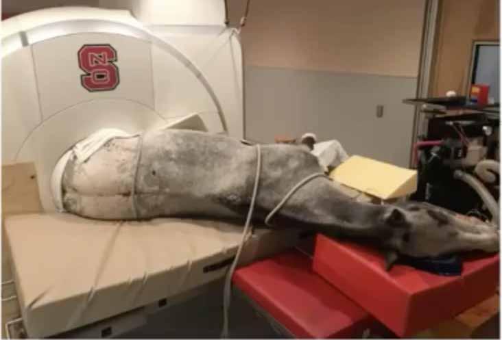
Because of the need for general anesthesia, high-field MRI is used more for diagnosis than monitoring. Quite a bit of manpower and positioning is needed, and how much can be seen frequently depends on the configuration of the horse. Used with permission of the AAEP. First presented at the 2020 AAEP Annual Convention and Trade Show.
General Rehabilitation Plan
Although plans are tailored to each horse and its injury, there are some general points that they often consider, she said.
“Typically, we monitor these horses every 2 to 4 weeks initially during the acute phase, and then every 4 to 8 weeks until they’re healed,” she said. “Our imaging findings are assessed in combination with a physical and lameness evaluation, and the results of these examinations together guide our decisionmaking on rehab protocols that aim for an eventual return to full work.”
Increasing exercise intensity to the next stage of the rehab plan depends on a favorable lameness exam with improvement in healing and imaging over the previous examination. Because it depends on the progress made, “our exercise instructions are very specific only until the next recheck appointment to guarantee that they come back,” she said. “We might need to tweak things.”
Always keep in mind the horse’s work when intensifying the regimen, Dr. Schnabel suggested. “We want to be careful about what’s specific to that horse’s discipline, as we go up to the next levels of our rehab intensity,” she said.
Foot balance and the need for therapeutic shoeing are assessed for every case to assure the best support for the horse’s limbs. “It’s just always sort of mind boggling how many of even these high-performance horses will come in with really poor foot balance. So, we want to address that certainly prior to starting a rehab program, to put the horse in the best position to succeed.”
All owners and trainers are also counseled on the importance of maintaining overall fitness, even during rest and rehab with mobility and strength-building exercises, as well as core stability exercises. These exercises keep horses in shape during rehab and in a much better position to return to full work.
Dr. Schnabel’s team likes to specify an overall expectation for the time to return to full work— provided the lesions heal. They also advise owners and trainers to make sure the horse stays calm, and maintains stall rest with controlled exercise. “For that reason, we often do provide long-term sedatives, such as reserpine or trazodone for these horses,” she said, adding that she probably uses more trazodone than reserpine, because there are fewer gastrointestinal side effects and trazodone is fast acting.
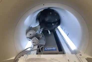

High-field MRI is commonly used to diagnose injuries localized to the carpus, tarsus or proximal suspensory region because of the need for general anesthesia to avoid excessive movement. Image courtesy of the AAEP, presented at the 2020 Virtual Annual Convention and Trade Show.
STEP 1: Walking
Following strict stall rest, the plan usually starts with controlled daily hand walking, gradually increasing the time building up to about 40 minutes by the end of an 8-week period.
She said that the exercise should be done under saddle rather than in hand when appropriate for the horse. “Most of our disasters during rehab happen while people are in-hand walking or hand grazing the horse out in the open. The horse gets away and gallops around for a few minutes or it’s just completely wild and slips, etc.
“So, we seem to do much better with these horses walking under tack than we do with hand walking,” Dr. Schnabel said. “If they have to be hand walked or are unsafe to ride for whatever reason, then we certainly recommend doing that walking within an enclosed paddock or ring in case the horse gets loose.”
STEP 2: Trotting
If there is improvement and no increase in lesion severity at that 8-week recheck, they introduce very short, straight, trot sets with walking in between, gradually increasing to 30–35 minutes of trot work with walk breaks by week 16.
“We also gradually introduced ground poles or cavallettis during that trotting period to improve strength and proprioception and just to keep the horse's brain engaged in its work,” she said.
“It’s certainly been our experience that trot work is often required to provide the proper biomechanical stimulus to the tendon or ligament for healing,” Dr. Schnabel said.
So after 8 weeks of trotting, they look for definitive improvement in both the fiber pattern and regression of vasculature on Doppler ultrasonography before increasing work intensity or starting canter work.
“At this stage, persistent hypervascularity—even with return to normal fiber pattern on gray scale—is really worrisome to us for the horse's prognosis and particularly to increase the work without possible further injuries,” she warned. “If they’ve gone through their 8 weeks of trotting and they're still persistently hypervascular, we’re going to be worried that they’re not ready to go on further, and we’re going to back things down.”
During the reevaluation, make sure that owners and trainers are compliant with the plan, but remember adding or stopping some modality might also be warranted.
STEP 3: Canter
The preference is for the horse to canter under saddle for about 4 weeks prior to any turnout in a sizeable paddock. She wants to see the animal as fit as possible and cantering under saddle before they have free reign around a pasture.
“After that point, the increase in work intensity is very disciplined and horse-specific but once a horse has returned to full work, it’s common for us to reassess the old injury site approximately every 3 to 6 months or sooner—depending on the horse—to monitor for subtle signs of re-injury and adjust work level or rehab accordingly,” she said.
Adjuncts
There are many options for enhancing these rehabilitation exercises, including the underwater treadmill, saltwater spa, vibration plates, laser shockwave, therapeutic ultrasound and stem cells that can be used depending on the case and the owner’s desire and financial ability.


