Is That Lameness All In His Head?
Fixing the 'Hypos' in Critical Neonates
Technician Update: Standing Surgery for Bladder Stones
Testing for Bisphosphonate Use

Vol 13 Issue 8 2023 www.modernequinevet.com
ASK THE INFECTIOUS DISEASE EXPERT Equine Vet The Modern
SALES: ModernEquineVet@gmail.com
EDITOR: Marie Rosenthal
ART DIRECTOR: Jennifer Barlow
CONTRIBUTING WRITERS: Paul Basilio
COPY EDITOR: Patty Wall
• Rob Warren
LEGAL DISCLAIMER: The content in this digital issue is for general informational purposes only. PercyBo Publishing Media LLC makes no representations or warranties of any kind about the completeness, accuracy, timeliness, reliability or suitability of any of the information, including content or advertisements, contained in any of its digital content and expressly disclaims liability of any errors or omissions that may be presented within its content. PercyBo Publishing Media LLC reserves the right to alter or correct any content without any obligations. Furthermore, PercyBo disclaims any and all liability for any direct, indirect, or other damages arising from the use or misuse of the information presented in its digital content. The views expressed in its digital content are those of sources and authors and do not necessarily reflect the opinion or policy of PercyBo. The content is for veterinary professionals. ALL RIGHTS RESERVED. Reproduction in whole or in part without permission is prohibited.
2 Issue 8/2032 | ModernEquineVet.com TABLE OF CONTENTS
Published by PERCY BO media publishing PO Box 935 • Morrisville, PA 19067 Marie Rosenthal and Jennifer Barlow, Publishers
The Modern NEONATOLOGY How to Fix the ‘Hypos’ in Critically Ill Neonates 8 TECHNICIAN UPDATE Standing Perineal Surgery for Cystolith Removal in a Stock Horse Gelding .................................................................................................................. 18 NEWS NOTES Genetics Driving Speed in Racehorses 16 Hair Samples Useful for Detecting Long-term Bisphosphonate Use in Horses 17 UC Davis Efforts Contribute to Continued Decline in California Racehorse Fatalities .................................................................................................... 23 How Common Is Sudden Cardiac Death?................................................................................. 23 SPONSORED EDITORIAL What are the Trends in West Nile Virus Activity and the Risk to U.S. Horses? 7 Maybe That Lameness is All in the Horse’s Head COVER STORY 4 Cover: Shutterstock/Callipso88 ADVERTISERS Arenus Animal Health/Aleira-Releira 3 Merck Sponsored Editorial 7 Arenus Animal Health/AssureGuard Gold .......................................... 9 CareCredit Advertorial 10 American Regent/Adequan 13 Merck Animal Health/Prestige 15 Equine Veterinary Nursing Technician 21
Equine Vet

Maybe That
4 Issue 8/2022 | ModernEquineVet.com
LAMENESS
Lameness is ALL IN THE
HORSE’S HEAD
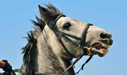
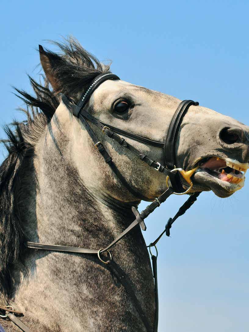
It might seem a bit out there, but inflammation of the equine temporomandibular joint (TMJ) may play a role in lameness and poor performance of the horse, admitted James L. Carmalt, MA, VetMB, MVetSc, PhD, FRCVS, DABVP(Eq), DAVDC(Eq), DACVSMR(Eq), DACVS, a professor of Equine Surgery, Dentistry, Sports Medicine & Rehabilitation, in the Department of Large Animal Clinical Sciences, Western College of Veterinary Medicine, University of Saskatchewan.
“This is not your classic lameness with the head ‘bobbiness’,” he said, “and we may end up opening a can of worms as we get into this.”
However, the work of he and his colleagues point to a link between TMJ inflammation and lameness.
Until 10 or 15 years ago, no one paid much attention to the TMJ, but they came to realize that the joint is clearly dynamic, and just as other joints do, there are changes associated with aging and use. Additionally, some young horses have developmental abnormalities in the joint, which can also affect mastication if inflamed.
Dr. Carmalt did come across a horse, which kept throwing its rider. The team noted a mild left hindlimb lameness, which was confirmed by using the Lameness Locator by Equinosis. Interestingly, they noticed that the lameness worsened when the side-reins (attached to a surcingle) were shortened. When they blocked the TMJ, the baseline lameness remained, but it did not worsen when the reins were shortened.
“We had not touched this horse in any way, other than to block the temporomandibular joints,” Dr. Car-
malt said, “and it fundamentally changed the movement of the horse. Not only subjectively—but because we were thinking about publishing it—we got objective data, and the objective data supported that subjective assessment.”
They treated the horse successfully, and it was better for about 9 months before the behavior became a problem again.

Prior to this first clinical case, they had been pretty sure that TMJ inflammation could result in a performance problem but were not sure how, or to what degree. At first glance, the joint is not under a heavy load, but consider it a moment, and it’s easy to see that the bit is in the mouth and under the control of the rider, so there could be a link between pain or abnormalities of the joint and the horse’s movement, he said.
Taking the Reins
Part of the problem of rein lameness is its definition, he explained. “The definition of rein lameness is difficult because it's a lameness that can be appreciated [felt or seen] only when the horse is being exercised under saddle or wearing a bridle and being exercised.
“And we've all had these cases where the client comes and says, ‘I feel my horse is off.’
“These riders are feeling something. You look at it as a veterinarian without any person on the horse. You trot it, you lunge it, you can't see it. I'm thinking, if I can't see it, how the heck, do you expect me to work this out?
“Now you put some tack on the horse, with or with-
By Marie Rosenthal MS
ModernEquineVet.com | Issue 8/2022 5 Shutterstock/pirita
out the rider, and start moving the horse. That changes everything, and now, you can begin to see what you're talking about,” Dr. Carmalt told the Modern Equine Vet.
Dr. Carmalt and his colleagues set out to see if there was a measurable link between inflammation of the TMJ, lameness and performance. The objective of the study was to determine the effect of temporomandibular joint inflammation on rein tension with the hypothesis being TMJ inflammation would alter the response to rein input on horses moving on a treadmill.
They trained 5 horses to walk and trot on a treadmill wearing long-reining equipment with rein-tension devices and 28 reflective optical tracking markers. They assessed the horse’s movement during a free walk and trot without rein-tension and again at the same gaits with rein-tension. Data were collected continually from both sides of the horse using the rein-tension devices and by using 12 kinematic cameras focused on the optical tracking markers. After collecting the initial data, one TMJ was randomly assigned to receive an injection of a substance that causes mild irritation but was self-limiting. Six hours later the study was repeated. The horses got 10 days off after the injection, before the study was repeated, and this time the opposite TMJ was injected.
Throughout the study, the highly experienced horse handler and research collaborator, Nathalie Reisbig, MedVet MS, PhD, had no idea which joint was injected.
“Despite that, she could feel it in her hand. If we injected the dominant side, the horse switched, and the dominance went to the originally non-dominant side. And if we injected the non-dominant side, the horse became very heavy in the hand on the previous dominant side.
“If, for example, we injected the left side and then she needed a little bit of left rein to keep the horse in the middle of the treadmill, this horse would overcompensate and go really left,” he explained.
“This makes sense,” Dr. Carmalt said, “The horse is trying to avoid the pain.”
One interesting finding, he said, was that they did not see large changes in horse movement when they looked at the kinematic findings. The one significant finding was that after injection, horses had a more vertical head position at trot. “Again, this makes sense. The horse has pain that is being exacerbated by rein tension. If the horse stays “behind the bit” the pressure exerted on the TMJs by the handler is reduced.
Overall, the lack of other significant differences may have been because the “horse handler was working substantially harder to keep that horse straight and moving correctly. So, the handler themselves probably had an important effect on the negative result of the kinematics,” he said.
Listen to the Riders
They have blocked the temporomandibular joints on several other client horses, and the riders tell him that the horses are moving substantially better.
He said the moral of the story is just because a veterinarian cannot see the problem, when a rider or client comes in saying that they are “feeling” something is different when they ride the horse, consider blocking the TMJ.
“If you've got TMJ inflammation, horses can have dramatic responses to rein input without being classically lame,” he said.
“The riders are not wrong. They are feeling something that is different, and our job is therefore to go after what is different.”
Despite the current interest in TMJ disease, it is important to rule out the more common causes of lameness, such as foot pain, or bone spavin before looking at TMJ problems, said Dr. Carmalt, who spoke about the study and the issue during the 68th Annual AAEP Convention, held in San Antonio. MeV For
6 Issue 8/2032 | ModernEquineVet.com LAMENESS
Reisbig
effect
temporomandibular joint inflammation on response to rein-tension
https://www.frontiersin.org/articles/10.3389/fvets.2023.1213423/full Also check out Dr. Carmalt's facebook page: https://www.facebook.com/groups/625206024604903
more information:
NA, Pifko J, Lanovaz JL, et al. The
of acute equine
and kinematics. Front Vet Sci. 2023;19(10):1213423.
“If you have TMJ inflammation, horses can have dramatic responses to rein input without being classically lame.”
Dr. James L. Carmalt
Infectious Disease Expert
“What are the best disinfecting practices to help mitigate disease spread?
Arguably more important than managing a disease outbreak is minimizing the chance that one occurs in the first place. When implemented in conjunction with your customized vaccination recommendations, this disinfection protocol helps clients reduce the risk of infectious diseases in barns, boarding facilities, transport trailers and more.
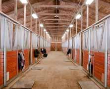
STEP 1. CLEAN
• Remove organic debris, buckets, feed tubs and bedding from the area.
• Clean the area using standard procedures with a liquid detergent solution to soften and remove dried organic material. If cleaning stalls, rinse them as a final step, beginning at the top of walls and working toward the drain.
•Allow surfaces to dry before applying disinfectant.
STEP 2. DISINFECT
• Disinfect environmental surfaces. Disinfectant solutions require appropriate surface contact time and should be applied according to product label directions.
• Choose disinfectants that are EPA-registered, have documented effectiveness in the presence of organic matter and are safe to use around horses and humans. Read all disinfectant labels and implement necessary protective equipment prior to disinfection.
• For complete surface coverage, use an electrostatic sprayer to apply the disinfec-


ABOUT THE AUTHOR
tant (see the recommended products).
• To disinfect, use a continuous path through the space, working from low to high areas in a sweeping motion until
surfaces are thoroughly wet.
• Do not wipe surfaces following electrostatic disinfection and allow surfaces to dry thoroughly before applying an antimicrobial solution.
STEP 3. PROTECT
• Using a spray-on antimicrobial solution in conjunction with cleaning and disinfecting protocols provides a protective coating against the growth of bacteria, fungi, mold and algae.
Repeat this protocol as needed based on the duration of efficacy noted on the product labels of your chosen disinfectants and antimicrobial solutions.
PARTNERING TO IMPROVE ENVIRONMENTAL INFECTION CONTROL

Driven by an Unconditional commitment to equine health and welfare, Merck Animal Health is collaborating with ByoPlanet® to curate a disinfectant product suite that is safe for use around horses and people. Together, Merck Animal Health and ByoPlanet are partnering with The United States Equestrian Federation (USEF) to reduce the risk of disease introduction and spread on USEF competition grounds and to help protect the health of USEF equine athletes. ByoPlanet’s patented technologies paired with Merck Animal Health’s leading antimicrobic products have the potential to revolutionize pathogen control in the equine industry by providing enhanced protection for horses and giving peace of mind to their caretakers.
RECOMMENDED PRODUCTS
• Clean Republic Multi-Purpose Disinfectant. With an EPA Category IV toxicity rating (the lowest available level of toxicity), this noncorrosive, non-bleaching pH-neutral product kills 99.9% of bacteria and is made with only salt, water and electricity. It contains no fragrances or harsh fumes.

• ByoPlanet® electrostatic sprayers. Applying disinfectant with ByoPlanet electrostatic sprayers provides 360° of surface coverage with less waste, quicker re-entry into the environment and better efficacy against pathogens in hidden areas.
• Armatrex spray-on antimicrobial solution. Safe for use in many live-animal settings and on a variety of surfaces, such as stalls and cross ties, Armatrex provides up to 90 days of protection. ByoPlanet electrostatic sprayers are the recommended application system.

Chrissie Schneider, DVM, MS, DABVP (equine practice), cVMA, is a senior equine professional services veterinarian at Merck Animal Health. Prior to joining Merck Animal Health in 2020, Dr. Schneider was in clinical equine practice for 11 years – including an equine internship, an Equine Field Service residency at The Ohio State University and six years as an associate at an equine-exclusive ambulatory practice in central Ohio.
WANT TO ASK A QUESTION?
EMAIL THE EDITOR.
For more information, visit the Merck Animal Health Armatrex webpage.
ASK
Copyright © 2023 Merck & Co., Inc., Rahway, NJ, USA and its affiliates. All rights reserved
This column, brought to you by Merck Animal Health, features insightful answers from leading minds.
THE
ModernEquineVet.com | Issue 8/2023 7
How to
Fix the ‘Hypos’
in Critically Ill Neonates
Neonatal calls can produce significant anxiety in even the most experienced equine practitioners. Even before a primary diagnosis can be formulated, triage and stabilization are crucial to helping these fragile patients.

Tiffany L. Hall, DVM, DACVIM, DACVECC, a critical care specialist at the Equine Medical Center of Ocala, in Florida, has plenty of first-hand experience with critical neonates. To arm practitioners with the confidence needed to take care of patients, she recently shared a wealth of information collected from clinical experience, various personal mentors, and the wisdom of other equine neonatologists.
The 7 ‘Hypos’

The field of medicine is awash in acronyms and mnemonic devices to help clinicians keep the important things straight, so it’s no surprise that critically ill equine neonates have their own catchy system.
“The 7 ‘hypos’ are generalized conditions seen in many that result from a variety of underlying pathophysiologies,” Dr. Hall said during a presentation at the 68th AAEP Annual Convention in San Antonio. “This approach to evaluation allows for systemic and goal-directed therapy in emergency situations. After assessing and stabilizing the equine neonate, then you can go back and decide what further diagnostics you
By Paul Basilio
NEONATOLOGY
8 Issue 8/2032 | ModernEquineVet.com Shutterstock/pfluegler-photo
THE

Arenus Animal Health | 866-791-3344 | www.arenus.com SO INNOVATIVE...
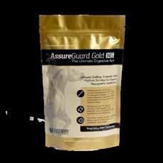
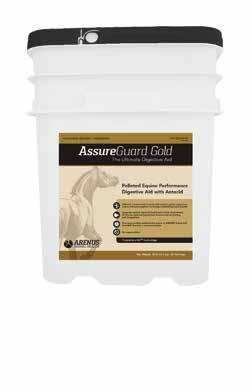
Replace your mineral oil, electrolytes, adsorbents, or other treatment options with Assure Guard Gold-NG the only effective and easy to administer slow gelling quick relief formulation including 2 cups of ultra pure psyllium, 72 billion CFU of probiotics, prebiotics, antacids, L-glutamine, electrolytes and energy. For continued support, consider a 10 day supply of Assure Guard Gold after treatment. IT’S LIKE MAGIC Ask your Arenus Veterinary Solution Specialist how Assure Guard Gold-NG and Assure Guard Gold can help your equine patients quickly and effectively recover from the digestive upsets you treat daily.
® Assure Guard
Together, Assure Guard Gold-NG And Assure Guard Gold Create A Powerhouse Against Your Most Challenging Digestive Cases. Use Assure Guard Gold-NG For Fast Relief And Maintain Excellent Digestive Health With Assure Guard Gold.
REAL MAGIC IS IN THE RESULTS
The Ultimate Digestive Aid
Gold
What makes a horse owner’s pulse race?


Over half of horse owners are stressed by a bill less than $1,500
A year’s worth of horse care can cost as much as $36,851
Horse owners underestimate their yearly costs by up to


363%


Equine costs can trigger stress in horse owners. The Challenge:
It’s no secret that horses hold a special place in their owners’ hearts. In fact, the Synchrony Equine Lifetime of Care study revealed that more than half of horse owners consider their horse a member of the family. That being said, stress can come with any relationship—even more so when nances come into play, and many horse owners aren’t aware of the cost that comes with a lifetime of equine care.
Understanding the emotional, psychological and nancial factors associated with horse ownership can allow you to better empathize with clients and help prioritize their needs—and the needs of their horse. Read the whole study to learn how clients might be feeling before, during and after veterinary appointments at equinelifetimeofcare.com.
The CareCredit health and animal care credit card helps give clients a way to pay over time for the services and products you o er. They can use it for wellness exams, vaccinations, emergency care and more—and your practice receives payment in two business days. It’s a nancing solution that can ease horse owners’ anxiety around payment and empower you to have stress-free cost conversations so you can both focus on giving your patient a lifetime of care.






exible nancing can help clients be prepared in a heartbeat.
Friendly,
The Solution:
Horse owners who don’t know what nancing options their veterinarian provides: 54%
a recreational horse’s lifetime, expenses
Source: CareCredit Equine Lifetime of Care Study, 2023. ©2023 Synchrony Bank MEV0823VA Scan to go to the Equine Lifetime of Care study. Get started for free by calling 844-812-8111 and mention code MEV0823VA.
Horse owners who will keep their horse for a lifetime: 90% Throughout
can add up to $412,001
• Hypoperfusion
• Hypoglycemia
• Hypothermia
• Hypoxemia
• Hypoventilation
• Hypo-base
• Hypo-immunity
need, how to determine the underlying problem, and whether you need to refer the patient.”
Hypoperfusion
Hypoperfusion occurs when the delivery of oxygen and nutrients to tissues is inadequate to meet the body’s demands. In neonates, this is typically due to low blood volume, inappropriate vascular tone, or poor myocardial contractility.
“Perfusion is best assessed through the physical examination,” Dr. Hall said. “Pay particular attention to the perfusion parameters of mentation, distal extremity temperature, metatarsal artery pulse quality, and urine production.”
She added that a hand-held lactatometer can obtain serial lactate measurements to help guide therapy and give an idea of disease severity.
Hypoglycemia
Anything that prevents a foal from nursing within a few hours of birth can lead to rapid hypoglycemia. A standard human glucometer can readily and easily determine glucose concentrations stall-side.
“Most equine neonatologists no longer recommend carte blanche administration of 5% dextrose in the neonatal foal,” she added. “This is based on research in human critical patients, which has demonstrated that rapid changes in glucose concentration can be associated with poorer outcomes.”
Hypothermia
All neonates are particularly susceptible to hypothermia, but critically ill neonates are incapable of proper thermal regulation due to a lower metabolic rate relative to their surface area.
“It’s important to note that modest hypothermia is actually considered beneficial, particularly in human critically ill patients undergoing CPR, or in human neonates undergoing treatment for hypoxic ischemic encephalopathy,” Dr. Hall said. “We are unable to actively cool our patients, so instead we allow them to be mildly hypothermic as long as their body temperature is above 96° and they have a normal heart rate.”
Hypoxemia
Hypoxemia is typically the result of 3 underlying conditions: hypoventilation, a ventilator-perfusion (V/Q) mismatch, and diffusion impairment due to meconium aspiration or severe respiratory disease.
“Modest hypoxemia may be addressed by placing the foal in sternal recumbency,” she explained. “You can also administer intranasal oxygen therapy if it’s available, but it should be used in the short-term in the field unless you have a humidifier.”
Hypoventilation
Hypoventilation is defined as elevated PaCO2 levels >55 mm Hg. It typically results in respiratory acidosis, associated metabolic responses, cerebral vasodilation and increased intracranial pressure. Its origins may be neurologic (eg, hypoxic ischemic encephalopathy, neonatal maladjustment syndrome) or neuromuscular (eg, progressive paralysis or fatigue), or it may be associated with musculoskeletal pain or a space-occupying mass.
“It can also be pulmonary in origin,” Dr. Hall explained. “When diffusion impairment becomes so severe in acute respiratory distress syndrome, we do allow a permissive hypercapnia, as long as their mentation is appropriate or the blood pH is >7.25.”
Hypobase and Hypoimmunity
In neonates, hypobase—or acidemia—is generally improved by addressing the other hypos.
For hypoimmunity, there are a multitude of peripartum stressors that could affect colostrum quality
12 Issue 8/2022 | ModernEquineVet.com NEONATOLOGY
7
‘HYPOS’ OF EQUINE NEONATES
Even before a diagnosis can be made, triage and stabilization are crucial to helping these fragile patients.
nothing else like it.
For more than 30 years, Adequan® i.m. (polysulfated glycosaminoglycan) has been administered millions of times1 to treat degenerative joint disease, and with good reason. From day one, it’s been the only FDA-Approved equine PSGAG joint treatment available, and the only one proven to.2, 3
Reduce inflammation
Restore synovial joint lubrication
Repair joint cartilage
Reverse the disease cycle
When you start with it early and stay with it as needed, horses may enjoy greater mobility over a lifetime.2, 4, 5 Discover if Adequan is the right choice. Visit adequan.com/Ordering-Information to find a distributor and place an order today.
BRIEF SUMMARY: Prior to use please consult the product insert, a summary of which follows: CAUTION: Federal law restricts this drug to use by or on the order of a licensed veterinarian. INDICATIONS: Adequan® i.m. is recommended for the intramuscular treatment of non-infectious degenerative and/or traumatic joint dysfunction and associated lameness of the carpal and hock joints in horses. CONTRAINDICATIONS: There are no known contraindications to the use of intramuscular Polysulfated Glycosaminoglycan. WARNINGS: Do not use in horses intended for human consumption. Not for use in humans. Keep this and all medications out of the reach of children. PRECAUTIONS: The safe use of Adequan® i.m. in horses used for breeding purposes, during pregnancy, or in lactating mares has not been evaluated. For customer care, or to obtain product information, visit www.adequan.com. To report an adverse event please contact American Regent, Inc. at 1-888-354-4857 or email pv@americanregent.com.
Please see Full Prescribing Information at www.adequan.com
1 Data on file.
2 Adequan® i.m. Package Insert, Rev 1/19.
3 Burba DJ, Collier MA, DeBault LE, Hanson-Painton O, Thompson HC, Holder CL: In vivo kinetic study on uptake and distribution of intramuscular tritium-labeled polysulfated glycosaminoglycan in equine body fluid compartments and articular cartilage in an osteochondral defect model. J Equine Vet Sci 1993; 13: 696-703.
4 Kim DY, Taylor HW, Moore RM, Paulsen DB, Cho DY. Articular chondrocyte apoptosis in equine osteoarthritis. The Veterinary Journal 2003; 166: 52-57.
5 McIlwraith CW, Frisbie DD, Kawcak CE, van Weeren PR. Joint Disease in the Horse.St. Louis, MO: Elsevier, 2016; 33-48.
All trademarks are the property of American Regent, Inc.
© 2021, American Regent, Inc.
PP-AI-US-0629 05/2021
There’s
www.adequan.com
and prevent absorption and ingestion of immunoglobulins.
“How we address inadequate passive transfer in a healthy foal depends on a number of factors, including how many foals are on the property, whether the property is a clean environment, and the history of the farm,” Dr. Hall said.
How to fix them
“In the field, you can quickly address many ‘hypos’ by administering antibiotics and plasma, restoring the circulating volume, optimizing ventilation and oxygenation by warming the foal passively, and providing a glucose substitute,” Dr. Hall explained. “It’s been demonstrated in human studies that the number one factor that improves outcomes in people presenting to hospitals with sepsis is early administration of antimicrobials.”
Many equine neonatologists agree that antimicrobials should be administered in the field prior to referral, although this wasn’t always the case. In the past, a practitioner might hesitate in administering antibiotics before the referral center could obtain a blood culture.
“We no longer worry about that,” she said. “We’d rather have the antibiotics on board. We can still get our blood cultures—there are special bottles that can [do that], even if the horse has received antibiotics.”
The combination of beta-lactam and an aminoglycoside is the gold standard, with ampicillin and amikacin the top preferences. Procaine penicillin G can be swapped for ampicillin if needed, and cephalosporin is a second-tier option.
Most foals are also going to require intravascular fluids. Dr. Hall recommends a crystalloid administered over a period of about 20 minutes.
FIELD STABILIZATION LIST
“Critically ill foals are at an exceptionally high risk of fluid overload, so I recommend that you don’t exceed 40 mL/kg in the first hour of administration,” she explained. “If you are giving plasma, always remember to include that in your total volume.”
For a glucose substitute, the easiest way is to administer mare’s milk through a tube if the foal is not nursing.
“A critically ill foal’s GI tract might not be functioning normally, so you may also administer 3 to 5 mL of 50% dextrose orally over the mucus membranes while you gain IV access,” Dr. Hall said. “Frequent fluctuations in glucose negatively impact outcomes in humans, so I recommend that you give 1% dextrose solutions—or at least do not exceed 2% dextrose—as a single bolus over 20 minutes.”
Finally, optimization of ventilation and oxygen can be achieved by placing the foal in sternal recumbency. Ventilation is typically addressed with chemical or mechanical ventilation in the field.
“The easiest way to do this is to administer caffeine at 10 mg/kg orally once,” she said. “That will help stimulate the central respiratory center and increase respiratory rate.”
Dr. Hall noted that caffeine administration is not recommended for all foals. The best candidates are foals who pause anywhere from 10 to 20 seconds between breaths.
“After you’ve completed your initial assessment and performed some of these therapies to stabilize the foal, you can circle back and do a more in-depth investigation to determine the underlying condition and determine whether the foal should be referred,” Dr. Hall said. MeV
14 Issue 8/2032 | ModernEquineVet.com
NEONATOLOGY
CIRCULATING VOLUME • 20 mL/kg crystalloid IV (1 L) • Plasma (1-2 L) • ≤ 2 L/50 kg in 1 hour ANTIMICROBIALS • Ampicillin 1-2 g IV (or PPG 3.5 mL IM) • Amikacin: 25 mg/kg IV/IM (5 mL/50 kg) • Ceftiofur 5 mg/kg IV/IM/ SQ (5 mL/50 kg) VENTILATION • Sternal recumbency • Caffeine 10 mg/kg PO once • Blanket or heating lamp GLUCOSE REPLACEMENT • Tube with mare’s milk • 2% dextrose in fluids (40 mL/1 L) • 3.5 mL dextrose PO
Many neonatalogists agree that antimicrobials should be administered prior to a referral.
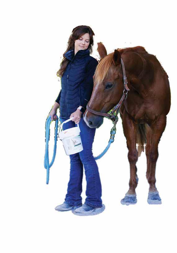
Get the scoop on the #1 TREATMENT FOR EPM 1
PROTAZIL® (1.56% diclazuril)
Antiprotozoal Pellets:
• Starts working fast (within 12 hours) — no loading dose required 2
• The only FDA-approved alfalfa-based top-dress treatment for EPM
• Easy to administer
Ask
IMPORTANT SAFETY INFORMATION: Use of PROTAZIL® is contraindicated in horses with known hypersensitivity to diclazuril. The safety of Protazil in horses used for breeding purposes, during pregnancy, or in lactating mares, and use with concomitant therapies in horses has not been evaluated. Do not use in horses intended for human consumption. Not for human use. For complete safety information, refer to the product label.
1 Equine Mega Study, Brakke Consulting, 2023.
2 Hunyadi L, Papich MG, Pusterla N. Pharmacokinetics of a low- dose and DA-labeled dose of diclazuril administered orally as a pelleted top dressing in adult horses. J of Vet Pharmacology and Therapeutics (accepted) 2014, doi: 10.111/jvp.12176. The correlation between pharmacokinetic data and clinical effectiveness is unknown.
“Protazil is very safe, it works, it’s cost effective and it’s super easy to give.”
Jennifer Groon, VMD The Feiner Equine
A Scary and Difficult Diagnosis. An Easy and Effective Treatment.
your Merck Animal Health Equine representative about PROTAZIL® or call 800-521-5767.
Copyright © 2023 Merck & Co., Inc., Rahway, NJ, USA and its affiliates. All Rights Reserved. US-PRA-230500004.
For Oral Use In Horses Only
CAUTION Federal (U.S.A.) law restricts this drug to use by or on the order of a licensed veterinarian.
NADA #141-268 Approved by FDA
DESCRIPTION
Diclazuril, (±)-2,6-dichloro-α-(4-chlorophenyl)-4-(4,5-dihydro-3,5-dioxo-1,2,4-triazin-2(3H)-yl)benzeneacetonitrile, has a molecular formula of C17H9CI3N4O2, a molecular weight of 407.64, and a molecular structure as follows:
Genetics Driving Speed in Racehorses
By Duncan Sandes University of Exeter
Diclazuril is an anticoccidial (antiprotozoal) compound with activity against several genera of the phylum
Apicomplexa. PROTAZIL® (diclazuril) is supplied as oral pellets containing 1.56% diclazuril to be mixed as a top-dress in feed. Inert ingredients include dehydrated alfalfa meal, wheat middlings, cane molasses and propionic acid (preservative).
INDICATIONS
PROTAZIL® (1.56% diclazuril) Antiprotozoal Pellets are indicated for the treatment of equine protozoal myeloencephalitis (EPM) caused by Sarcocystis neurona in horses.
DOSAGE AND ADMINISTRATION
Dosage PROTAZIL® (1.56% diclazuril) is administered as a top dress in the horse’s daily grain ration at a rate of 1 mg diclazuril per kg (0.45 mg diclazuril/lb) of body weight for 28 days. The quantity of PROTAZIL® necessary to deliver this dose is 64 mg pellets per kg (29 mg pellets/lb) of body weight.
Administration: To achieve this dose, weigh the horse (or use a weigh tape)). Scoop up PROTAZIL® to the level (cup mark) corresponding to the dose for the horse’s body weight using the following chart:
Range
Horse (lb)
of Pellets Weight Range of Horse (lb) mLs of Pellets
- 524 20 1275 - 1524 60
- 774 30 1525 - 1774
- 1024 40 1775 - 2074 80 1025 - 1274 50 - -
One 2.4-lb bucket of PROTAZIL® will treat one 1274-lb horse for 28 days. One 10-lb bucket of PROTAZIL® will treat five 1100-lb horses for 28 days.
CONTRAINDICATIONS
Use of PROTAZIL® (1.56% diclazuril) Antiprotozoal Pellets is contraindicated in horses with known hypersensitivity to diclazuril.
WARNINGS
For use in horses only. Do not use in horses intended for human consumption. Not for human use. Keep out of reach of children.
PRECAUTIONS
The safe use of PROTAZIL® (1.56% diclazuril) Antiprotozoal Pellets in horses used for breeding purposes, during pregnancy, or in lactating mares has not been evaluated. The safety of PROTAZIL® (1.56% diclazuril)
Antiprotozoal Pellets with concomitant therapies in horses has not been evaluated.
ADVERSE REACTIONS
There were no adverse effects noted in the field study which could be ascribed to diclazuril. To report suspected adverse reactions, to obtain a MSDS, or for technical assistance call
1-800-224-5318
CLINICAL PHARMACOLOGY
The effectiveness of diclazuril in inhibiting merozoite production of Sarcocystis neurona and S. falcatula in bovine turbinate cell cultures was studied by Lindsay and Dubey (2000).1 Diclazuril inhibited merozoite production by more than 80% in cultures of S. neurona or S. falcatula treated with 0.1 ng/mL diclazuril and greater than 95% inhibition of merozoite production (IC95) was observed when infected cultures were treated with 1.0 ng/mL diclazuril. The clinical relevance of the in vitro cell culture data has not been determined.
PHARMACOKINETICS IN THE HORSE
The oral bioavailability of diclazuril from the PROTAZIL® (1.56% diclazuril) Antiprotozoal Pellets at a 5 mg/kg dose rate is approximately 5%. Related diclazuril concentrations in the cerebrospinal fluid (CSF) range between 1% and 5% of the concentrations observed in the plasma. Nevertheless, based upon equine pilot study data, CSF concentrations are expected to substantially exceed the in vitro IC95 estimates for merozoite production (Dirikolu et al., 1999)2. Due to its long terminal elimination half-life in horses (approximately 43-65 hours), diclazuril accumulation occurs with once-daily dosing. Corresponding steady state blood levels are achieved by approximately Day 10 of administration.
EFFECTIVENESS
Two hundred and fourteen mares, stallions, and geldings of various breeds, ranging in age from 9.6 months to 30 years, were enrolled in a multi-center field study. All horses were confirmed EPM-positive based on the results of clinical examinations and laboratory testing, including CSF Western Blot analyses. Horses were administered PROTAZIL® (1.56% diclazuril) Antiprotozoal Pellets at doses of 1, 5, or 10 mg diclazuril/kg body weight as a topdress on their daily grain ration for 28 days. The horses were then evaluated for clinical changes via a modified Mayhew neurological scale on Day 48 as follows:
0. Normal, neurological deficits not detected.
1. Neurological deficits may be detectable at normal gaits; signs exacerbated with manipulative procedures (e.g., backing, turning in tight circles, walking with head elevation, truncal swaying, etc.).
2. Neurological deficit obvious at normal gaits or posture; signs exacerbated with manipulative procedures.
3. Neurological deficit very prominent at normal gaits: horses give the impression they may fall (but do not) and buckle or fall with manipulative procedures.
4. Neurological deficit is profound at normal gait: horse frequently stumbles or trips and may fall at normal gaits or when manipulative procedures were utilized.
5. Horse is recumbent, unable to rise.
Each horse’s response to treatment was compared to its pre-treatment values. Successful response to treatment was defined as clinical improvement of at least one grade by Day 48 ± conversion of CSF to Western Blot-negative status for S. neurona or achievement of Western Blot-negative CSF status without improvement of 1 ataxia grade.
Forty-two horses were initially evaluated for effectiveness and 214 horses were evaluated for safety. Clinical condition was evaluated by the clinical investigator’s subjective scoring and then corroborated by evaluation of the neurological examination videotapes by a masked panel of three equine veterinarians. Although 42 horses were evaluated for clinical effectiveness, corroboration of clinical effectiveness via videotape evaluation was not possible for one horse due to missing neurologic examination videotapes. Therefore, this horse was not included in the success rate calculation.
Based on the numbers of horses that seroconverted to negative Western Blot status, and the numbers of horses classified as successes by the clinical investigators, 28 of 42 horses (67%) at 1 mg/kg were considered successes. With regard to independent expert masked videotape assessments, 10 of 24 horses (42%) at 1 mg/ kg were considered successes. There was no clinical difference in effectiveness among the 1, 5, and 10 mg/ kg treatment group results.
Adverse events were reported for two of the 214 horses evaluated for safety. In the first case, a horse was enrolled showing severe neurologic signs. Within 24 hours of dosing, the horse was recumbent, biting, and exhibiting signs of dementia. The horse died, and no cause of death was determined. In the second case, the horse began walking stiffly approximately 13 days after the start of dosing. The referring veterinarian reported that the horse had been fed grass clippings and possibly had laminitis.
ANIMAL SAFETY PROTAZIL® (1.56% diclazuril) Antiprotozoal Pellets were administered to 30 horses (15 males and 15 females, ranging from 5 to 9 months of age) in a target animal safety study. Five groups of 6 horses each (3 males and 3 females) received 0, 5 (5X), 15 (15X), 25 (25X) or 50 (50X) mg diclazuril/kg (2.27mg/lb) body weight/day for 42 consecutive days as a top-dress on the grain ration of the horse. The variables measured during the study included: clinical and physical observations, body weights, food and water consumption, hematology, serum chemistry, urinalysis, fecal analysis, necropsy, organ weights, gross and histopathologic examinations.
The safety of diclazuril top-dress administered to horses at 1 mg/kg once daily cannot be determined based solely on this study because of the lack of an adequate control group (control horses tested positive for the test drug in plasma and CSF). However, possible findings associated with the drug were limited to elevations in BUN, creatinine, and SDH and less than anticipated weight gain. Definitive test article-related effects were decreased grain/top-dress consumption in horses in the 50 mg/kg group. In a second target animal safety study,
PROTAZIL® (1.56% diclazuril) Antiprotozoal Pellets were administered to 24 horses (12 males and 12 females, ranging from 2 to 8 years of age). Three groups of 4 horses/sex/group received 0, 1, or 5 mg diclazuril/kg body weight/day for 42 days as a top-dress on the grain ration of the horse. The variables measured during the study included physical examinations, body weights, food and water consumption, hematology, and serum chemistry.
There were no test article-related findings seen during the study.
STORAGE INFORMATION
Store between 15°C to 30°C (59°F to 86°F).
HOW SUPPLIED
PROTAZIL® (1.56 % diclazuril) Antiprotozoal Pellets are supplied in 2.4-lb (1.1 kg) and 10-lb (4.5 kg) buckets.
REFERENCES 1Lindsay, D. S., and Dubey, J. P. 2000. Determination of the activity of diclazuril against Sarcocystis neurona and Sarcocystis
Improvements in genetics appear to be a factor for increasing speeds among Britain’s Thoroughbreds, which contradicts earlier studies. However, the new research also showed the rates of genetic progress are relatively low, particularly over longdistance races.
Researchers analyzed a data set of 692,534 race times run by more than 76,000 horses born between 1995-2012. Over sprint, middle-distance and longdistance races, genetic improvement for Thoroughbred speed was found to be ongoing and statistically significant.
The study found that genetic improvement accounts for 60%, 55% and 17% of the total increase in speed observed over short, middle and long distances, respectively.
The research also showed that Thoroughbred speed is only weakly heritable across all distances in part explaining the slow rates of genetic improvement.

The researchers suggest the low rates of genetic improvement also raise the possibility that selection of racehorses for
breeding may be weaker than previously assumed, particularly over long distances, or some other constraint may be at play.
“There has been plenty of debate over the recent decades whether racehorses have reached a genetic limit to how fast they can run,” said Patrick Sharman, PhD, MSc, from the Centre for Ecology and Conservation at the University of Exeter’s Penryn Campus in Cornwall.
“Our analysis suggests that is not the case, and on the contrary, provides strong statistical evidence that racehorses continue to evolve to run faster in Great Britain. The question now is why are rates relatively low, particularly over long-distances?
“We estimated relatively low heritability for racehorse speed which goes some way to explaining it, but it would appear there is more to it. The selection of racehorses for breeding may be less accurate and weaker than generally assumed, particularly over long-distance races, but other factors could be constraining genetic improvement.”MeV
The article was originally posted here: https://bit.ly/47Ls2X3-MEV It was edited for style and length.
For more information:
Patrick Sharman, Alastair J. Wilson. Genetic improvement of speed across distance categories in thoroughbred racehorses in Great Britain. Heredity, 2023; DOI: 10.1038/s41437-023-00623-8
https://www.nature.com/articles/s41437-023-00623-8
16
| ModernEquineVet.com NEWS NOTES
Issue 8/2023
Weight
of
mLs
275
525
70 775
falcatula in cell cultures. J. Parasitology, 86(1):164–166. 2Dirikolu, L., Lehner, F., Nattrass, C., Bentz, B. G., Woods, W. E., Carter, W. E., Karpiesiuk, W. G., Jacobs, J., Boyles, J., Harkins, J. D., Granstrom, D. E. and Tobin, T. 1999. Diclazuril in the horse: Its identification and detection and preliminary pharmacokinetics. J. Vet. Pharmacol. Therap. 22:374–379. Intervet Inc d/b/a Merck Animal Health, 2 Giralda Farms, Madison, NJ 07940 Copyright © 2022 Merck & Co., Inc., Rahway, NJ, USA and its affiliates. All rights reserved. 07-2014, 211.x.3.0.3
Shutterstock/dikkenss
Hair Samples Useful for Detecting Long-term Bisphosphonate Use in Horses
By Amy Young UC Davis
Hair samples can be useful in detecting the long-term use of bisphosphonates in horses, according to a study by Heather
juries. The Horseracing Integrity and Safety Authority's Anti-Doping and Medication Control program has categorized bisphosphonates as banned substances. Horses that test positive under the program are subject to lifetime racing ineligibility.
Knych
, DVM, PhD, of the K.L. Maddy Equine Analytical Chemistry Lab's Pharmacology section and the Department of Molecular Biosciences, at U.C. Davis College of Veterinary medicine.
“From our previous research, we know that bisphosphonates remain in equine bone over long periods,” Dr. Knych said. “Hair sampling is non-invasive and has proven to be a reliable and reproducible matrix for equine drug testing. The ability to detect bisphosphonates long term in hair samples would benefit horse welfare and the integrity of the sport.”
The current study developed an assay and evaluated the use of hair for long-term detection of the bisphosphonate, clodronate. Results indicated that the drug was detectable in hair for up to 6 months in most of the horses studied. Further research is needed to better understand how long it takes clodronate to be incorporated into hair, as well as implications for widespread use of this testing method.
Bisphosphonates are molecules that strongly bind to bone material and prevent bone resorption and natural remodeling. Two bisphosphonates are approved for use in horses that are 4 years of age and older, clodronate disodium (Osphos) and tiludronate disodium (Tildren). They are safe and effective at relieving pain and lameness in horses with navicular syndrome.
However, off-label use to treat other equine bone conditions, especially in young horses, has raised concerns about long-term effects on bone, and weakened bones can put horses at risk for catastrophic in-
For more information:
Previous studies by Dr. Knych’s group reported that bisphosphonates can be detected in equine bone, blood and urine for extended periods. However, their results also showed that detection in blood and urine can be inconsistent. Compounds can generally be detected for much longer periods in hair than in blood or urine. As such, mane and tail hair samples can provide longterm historical records of drug exposures.
Important Tool
“Hair testing has become an increasingly important anti-doping matrix in horse racing, but not all prohibited substances are amenable to hair testing,” said Rick Arthur, DVM, a former Equine Medical Director and a co-author on the study.
“Regulators are keen to expand their understanding of which drugs of interest can be detected in hair and under what conditions. This study is an example of that effort. Anti-doping in horse racing not only addresses performance enhancing or other performance modifying substances, but horse health and welfare substances as well. Bisphosphonate misuse has been a major concern for horse safety, and now hair testing offers an alternative matrix to blood and urine,” Dr. Arthur said.
The study was supported by a special call for bisphosphonate research from the Grayson Jockey Club Foundation. MeV
The article was originally posted here: https://bit.ly/3KYtGuV-MEV It was edited for style and length.
Knych K, McKemie DS, Yim S, et al. Long-term monitoring of clodronate in equine hair using liquid chromatography-tandem mass spectrometry. J Chromatogr B Biomed Appl. 2023;1226: 123789. Published July 15. https://doi.org/10.1016/j.jchromb.2023.123789
https://www.sciencedirect.com/science/article/pii/S157002322300199X?via%3Dihub
ModernEquineVet.com | Issue 8/2023 17 NEWS NOTES
“Bisphosphonate misuse has been a major concern for horse safety.”
Dr. Rick Authur
Standing Perineal Surgery for Cystolith Removal in a Stock Horse Gelding
 By Rebecca Cameron AssocDAppSc (Equine), BVetTech, RVT, VTS -EVN
Cystoliths, more commonly known as “bladder stones,” are an uncommon condition in horses in which hard ‘stones’, typically made up of concentrated minerals, are formed in the bladder. Equine urine is typically alkaline and has a high mineral content providing a suitable environment for cystolith formation. Cystolith removal in horses is usually preformed under general anaesthesia.
A 13-year-old stock horse gelding, presented to the equine hospital for stranguria and pigmenturia after competing the previous day. The horse was seen 5 days previously by a referring veterinarian for the same complaints.
Examination
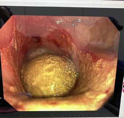
A physical examination was performed and all parameters were found to be within normal limits. The bladder was aseptically catheterized to obtain a urine sample for analysis. There was evidence of blood in the urine with no bacteria present. Endoscopy of the bladder was also performed and revealed
a large stone with a spiculated surface, approximately the size of a lemon. There was also evidence of mucosal irritation.
Surgery was booked for the following day because of the size of the cystolith and the degree of discomfort it was causing the horse. Due to the gelding’s calm temperament and the cost constraints of his owner, he was a good candidate for standing surgery. Standing cystolith removal has been performed more commonly in mares via the external opening of the urethra. In males a perineal urethrostomy (PU) is performed, which involves an incision below the anus into the urethra.
On the morning of the surgery, administration of pre-operative antimicrobial therapy including procaine penicillin intramuscularly, IV gentamicin and the non-steroidal anti-inflammatory drug (NSAID), IV phenylbutazone. The left jugular was aseptically prepared and catheterized with a 14 Ga angio catheter and sutured in place. The gelding was premedicated with acepromazine, methadone and detomidine, then walked into the standing surgical crush where he was started on a detomidine constant rate infusion (CRI) and a maintenance rate of Hartmanns.
The coccygeal area was clipped and aspectically prepared for the
18 Issue 8/2032 | ModernEquineVet.com
TECHNICIAN UPDATE
Equine urine is typically alkaline and has a high mineral content providing a suitable environment for cystolith formation.
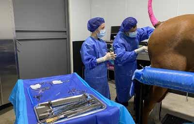
The stone was removed during a standing surgery. The cystolith was removed with endoscopic guidance through the PU site using a retrieval bag.
Images courtesy of Rebecca Cameron

CHECK OUT OUR NEW WEBSITE Equine Vet The Modern MODERNEQUINEVET.COM
administration of a caudal epidural between C1 and C2. Xylazine and lignocaine were given for the epidural. Epidurals can aid in desensitization of the anus, rectum, perineum, vulva, urethra and bladder.
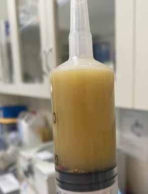
Administration of a caudal epidural is generally considered a safe procedure, although complications can occur, including infection and ataxia.
The surgical plane was assessed continuously with the aid of the Ghent standing sedation algorithm. The Ghent algorithm was developed to assess sedation depth, ataxia and surgical conditions and/or adjust the dose or administration rate of alpha-2-agonists in horses.
Once an adequate plane of anaesthesia was achieved, which in this case was sedated enough to not respond to outside noise or intervention. The tail was wrapped and secured to the side while a sterile urinary catheter was placed and the perineal area surgically prepared following aseptic protocol.
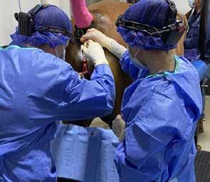
The cystolith was removed with endoscopic guidance through the PU site using a retrieval bag. The retrieval bag enclosed most of the stone, so it could
be broken up into smaller pieces and minimize any further mucosal irritation. The surgery took a long time due to the size and compacted consistency of the stone. Constant monitoring and top ups of analgesia were required as well as the use of local blocks with mepivacaine. After removal of the cystolith, the bladder was then thoroughly lavaged.
The following day, the bladder was lavaged under endoscopic guidance to ensure no further debris remained. This step is particularly important, as any small fragments have the potential to lodge in the urethra and cause complete blockage of the urinary tract. This procedure confirmed that the surgery was successful, and full removal of the cystolith and any debris in the bladder was confirmed.
The gelding remained in the hospital overnight where he was regularly monitored and had physical examinations conducted every 6 hours. Urine and fecal output also was recorded. The surgical incision was cleaned and monitored for any discharge, and petroleum jelly was applied around the incision to minimise urine scalding.
20 Issue 8/2032 | ModernEquineVet.com TECHNICIAN UPDATE
The retrieval bag enclosed most of the stone, so it could be broken up into smaller pieces and minimize any further mucosal irritation.
Administration of a caudal epidural is generally considered a safe procedure, although complications can occur.
Teaching Points
Patient selection and preparation are critical aspects for a successful standing procedure. Equine general anesthesia has been reported to have the highest complication rates of all domestic species with an anesthetic-related mortality of 1% for elective procedures and up to 10% for emergencies, along with the risk for myopathies and neuropathies due to the pressure of the large body weight on bony prominences.
Advances in sedative and local anesthetic techniques ensure many procedures that were once only performed under general anesthesia can now be performed safely while the patient is standing.
Discharge was the following day continuing with calesulpha-T paste (trimethoprim-sulfadimidine) orally twice daily (BID) for 14 days and the NSAID, flunixin meglumine BID for the first 3 days, and then orally once a day or another 4 days.
The PU site will naturally close on its own in a couple of weeks, and until this time intermittent urination may be observed from both the penis and the PU site. Until the site is closed completely, the area and down the back legs should be cleaned daily with warm water and mild soap followed by a light coating of petroleum jelly to prevent urine scalding, the owner was told. His tail was kept in a rectal sleeve to help keep the tail and surgical area clean until completely healed.
It was recommended to keep the gelding in a stall by himself with hand walking or turnout in a small yard so his attitude, appetite and behaviour could be monitored.
Performing surgeries standing is often more cost effective and associated with reduced healing time.
Alpha 2-agonists represent the main class of sedatives for standing surgery. The use of drug combination and CRIs are recommended to provide a bal-
ance and steady state of sedation and analgesia for the duration of the procedure. Anesthesia, sedation depth, sedation quality and degree of ataxia were all continuously recorded using the Ghent sedation algorithm as a guide.
The total length of surgery was 6 hours. It is important in these instances to consider the duration of action of the opioids used for analgesia. Methadone has a duration of action of 5 hours. There were noticeable changes in the geldings demeanor and comfort levels after 4 hours, which correlated with the duration of action of the methadone given as a premedication. As a result, he received a bolus of methadone, and the detomidine CRI was increased until he was comfortable again, then CRI was decreased back to the original dose.
Local blocks using mepivacaine were readministered to help desensitise the area. In reflection, an opioid also could have been used in the epidural to allow for longer duration. Urine output was monitored closely through the positioning of the sterile urinary catheter, and maintenance fluids were provided.
Ongoing Care
Horses who have formed calcium carbonate stones are at a higher risk for reforming them. Therefore, it is essential that diet is managed appropriately to prevent reformation of more cystoliths. Grassy hay was recommended instead of lucerne hay, which will decrease the amount of calcium in his urine. In addition to the diet adjustment, it was recommended that daily salt be added to his feed to encourage increased water intake. This gelding made a successful recovery from surgery, and was back to competing successfully a short time later. MeV
About the Author
Rebecca Cameron is a VTS-EVN working as a clinical anaesthetist at Scone Equine Hospital in Australia. She first studied applied science, majoring in Equine Science, and then completed a Bachelor of Veterinary Technology at the University of Queensland. Following graduation Rebecca had worked in many equine hospitals gaining new skills and developing a passion for equine anaesthesia. She loves foaling season and is also a dachshund fanatic with her much loved dachshund Aria at her side whenever she is not working.
22 Issue 8/2032 | ModernEquineVet.com
TECHNICIAN UPDATE
Shutterstock/nelelena
Once a horse has a calcium carbonate stone, they are at a higher risk for reforming the stones, so diet is essential.
UC Davis Efforts Contribute to Continued Decline in California Racehorse Fatalities
By Amy Young UC Davis
The number of racehorse fatalities in the 2022-23 fiscal year continues to decline, according to the California Horse Racing Board (CHRB).
Fatalities have been trending downward since 2005, with the recent report totaling 26 fatalities resulting from musculoskeletal injuries that occurred while racing or training at CHRB-regulated facilities (compared with 39 the previous year). According to the CHRB, 30,000 race starts and more than 100,000 workouts occur across 8,000 to 10,000 horses annually.
Long-standing efforts between the CHRB and the UC Davis School of Veterinary Medicine have built on past successes to benefit equine welfare and safety.
From research programs to testing facilities, UC Davis is integral to advances in racehorse health and injury prevention. The school is home to the J.D. Wheat Veterinary Orthopedic Research Laboratory, where Dr. Susan Stover’s group has characterized changes in proximal sesamoid bones that put race-
horses at risk for catastrophic fracture. These data, combined with standing equine positron emission tomography (PET) scanning, pioneered by at UC Davis by Dr. Mathieu Spriet, now allows at-risk horses to be identified and successfully rehabilitated for return to training and competition.
The California Animal Health and Food Safety Laboratory at UC Davis is home to the CHRB Post-mortem and the Kenneth L. Maddy Equine Chemistry Laboratory. The Post-mortem Examination Program provides necropsies (animal autopsies) for every horse that dies or is euthanized at CHRB racetracks and training facilities. The Maddy Laboratory is the CHRB’s primary drug-testing laboratory. The laboratory, which is part of the California Animal Health and Food Safety Laboratory System and is co-managed by professors Benjamin Moeller and Heather Knych, tests more than 40,000 samples from California racehorses annually.
In combination, these programs have provided valuable insights into the causes of racehorse injuries, as well as science-based approaches to improvements in injury detection and prevention. MeV
How Common Is Sudden Cardiac Death?
Premature depolarizations occur more often among horses during cross-country competition, and those in the upper divisions were at a higher risk, according to a recent study done my veterinarians from several U.S. facilities, including New Bolton Center, in Kennett Square, Pa., and Equine Sports Medicine Consultants, in Newark, Del.
They did a prospective, cross-sectional study to determine the frequency and types of rhythm disturbances that these elite athletes suffer. They also wanted to highlight the factors for premature depolarisations occurring during the cross-country phase of United States Eventing Association (USEA) and Fédération Equestre Internationale (FEI)-sanctioned events.
They obtained continuous electrocardiographic (ECG) recordings from 75 horses prior to, during and
For more information:
immediately following the cross-country competition and categorized them by complexity (singles, couplets, triplets, complex).
Premature depolarizations were identified in 42 of the 75 horses (56%) during the competition. The horses competing in the upper divisions of eventing had higher odds for having any premature depolarisations during the event compared with the lower divisions (OR = 17.5 [4.3, 72.01], P=0.006).
Another factor was the time the heart rate (HR) was >199 beats per minute, which was associated with more complex arrhythmias (triplets, salvos and tachyarrhythmias) (OR = 1.01 [1.0, 1.02], P=0.005), they said.
They also found that an irregular heartbeat at rest was associated with arrhythmia in the early recovery period (OR = 3.5 [1.1, 10.8], P=0.03). MeV
Durando MM, Slack J, Birks E, et al. Premature depolarisations in horses competing in United States Eventing Association and Fédération Equestre Internationale-sanctioned 3-day events. E Vet J. 2023 May 30. 2023 May 30. doi: 10.1111/evj.13948. https://beva.onlinelibrary.wiley.com/doi/10.1111/evj.13948
ModernEquineVet.com | Issue 8/2023 23 NEWS NOTES
Reach your veterinarians wherever they are, whenever they want. FOR ADVERTISING RATES AND INFORMATION, EMAIL ModernEquineVet@gmail.com Equine Vet The Modern CHECK OUT OUR NEW WEBSITE AT www.modernequinevet.com


 By Rebecca Cameron AssocDAppSc (Equine), BVetTech, RVT, VTS -EVN


