6 minute read
TECHNICIAN UPDATE
Thoroughbred Filly With Lateral Suspensory Ligament Branch Lesion
By Emily Brooks
On April 21, a 2-year-old Thoroughbred filly from a local training center was admitted for a protracted episode of waxing and waning cellulitis and lameness (grade 3/5) in the right hind limb.
The referring veterinarian had been treating her with oral antibiotics and nonsteroidal anti-inflammatory drugs (NSAIDs) but the cellulitis had not improved, so the trainer elected to bring her to the referral hospital. Despite the cellulitis, the filly was in good body condition, weighing 1,000 lbs, with normal vitals (HR 40, RR 12, T 100.9), and a bright attitude.
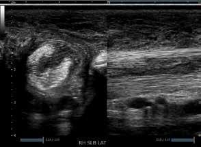
An ultrasound examination was performed on the right hind limb, and revealed diffuse cellulitis throughout the metatarsus and a large lesion within the lateral. At first glance, this could appear to be a core lesion that broke out the lateral margin of the branch, however, the character of the hyperechogenic material within the ligament was consistent with an abscess or hematoma. The nature of normal synovial fluid on ultrasound is typically black, or anechogenic. Cellular material appears more "sparkly" or hyperechogenic.


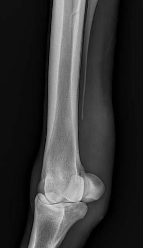
Ultrasonography cannot differentiate between white or red blood cells, hence the difficulty determining an abscess or hematoma. This pocket exited the branch and communicated with the surrounding subcutaneous tissue. Digital radiographs were obtained of the right hind fetlock and did not show bony involvement of the lateral sesamoid or third metatarsal bones.
The question arose whether to anesthetize the horse to get a sample. Being that it was a hind limb and a large degree of precision and accuracy are needed to place the needle within the lesion and minimize damage to healthy ligament, performing an aspirate standing seemed like an unnecessary risk. To help inform the decision, we ran blood work, and an elevated white blood count (12,300), elevated fibrinogen (600) and a serum amyloid A of 2,242 corroborated infection.
The filly was prepared for general anesthesia and a 14 g Mila catheter was placed in the right jugular vein and flushed with heparinized saline. No pre-operative antibiotics were given, as an aspirate and culture/sensitivity sample were to be taken in surgery.
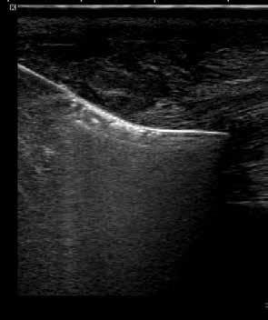
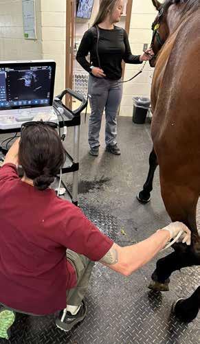
The horse was placed under general anesthesia in left lateral recumbency. The right hind leg was clipped from the coronet band to mid cannon bone and prepped using aseptic and sterile technique. I assisted the surgeon with the ultrasonography and covered the probe with a sterile rectal sleeve. A 20-g 1½" needles was directed into the intralesional abscess.


An aspirate of purulent fluid was obtained with the appearance of frank pus. White blood cells were too numerous to count so the sample was sent for culture and sensitivity. After the sample was taken, a mosquito hemostat was placed under ultrasound guidance to thread a ¼" Penrose drain as far proximally and distally as needed to ensure the abscess could not wall off. While under anesthesia a regional limb perfusion was performed by placing a tourniquet above the hock and inserting a catheter and extension set into the right saphenous vein. Amikacin (2 mL) was QS to 60 mL with sterile saline and left in for 20 minutes. After surgery a sterile half limb bandage was applied and the mare recovered without incident.
The following day, the cellulitis had improved and the filly was started on systemic antibiotics potassium penicillin and gentamicin. She was also scheduled to receive 1 g of phenylbutazone PO for 5 days. The regional limb perfusion was repeated under standing sedation. The culture grew heavy Staphylococcus aureus. There was no history of recent injections or lacerations prior to the episode of cellulitis, so the etiology of the infection is unknown.
The patient was released from the hospital 2 days after being admitted, 1/5 lame RH at the walk. Discharge instructions were to contain the filly to a box stall until the drain was removed 7 days post-surgery. Exercise could increase to hand walking only until she could have a follow up ultrasound in 30 days. After that, exercise could increase based on the healing of the suspensory branch.
Follow-up ultrasound showed that the abscess had resolved, and there was marked healing of the branch, though there was still a core lesion consisting of approximately 25% of the cross-sectional area of the ligament. The filly was cleared to continue stall rest and hand walking with a favorable prognosis, pending the continued healing of the ligament.
This case highlights the opportunities available for technicians to be trained to assist veterinarians in new and different areas of practice. With the specializations for veterinary technicians and increasing desire for veterinarians to let us take on more responsibility, with proper training and oversight the technician role could change quite a bit in the near future. MeV
Teaching Points
Ultrasonography is a common diagnostic tool for evaluating soft tissue and bony irregularity in veterinary practice. Compared with magnetic resonance imaging (MRI), it is less invasive and more cost effective. The role technicians play in acquiring ultrasound images lags far behind human medicine. With the declining number of veterinarians choosing to go into equine medicine, there is likely a need for technicians to step into more specialized and highly trained roles.
I was fortunate enough to be trained in ultrasonography after learning MRI, and the 2 modalities helped me to learn anatomy and recognize pathology.

Working closely with veterinarians, I have been able to perform the ultrasound exams for their interpretation, which saves valuable time during busy lameness days and emergency work ups.
There are a wide range of possibilities to extend the use of technicians and assistants in practice. It is already common practice for technicians to acquire radiographs, perform MRI, nuclear scintigraphy and computed tomography. In the absence of a radiologist on site or available during scans, there are many times that the knowledge and experience of a technician in imaging can be integral to finding the cause of lameness or disease in our patients.
About the Author
Emily Brooks began working as a veterinary technician in 2007 at Rood and Riddle Equine Hospital. There she developed an interest in diagnostic imaging and learned to run high field MRI, CT, and perform ultrasound and ultrasound-guided injections. She is now the Director of Imaging at Equine Athlete Veterinary Services in Simpsonville, Ky., and has added the modality of PET scan to her repertoire. Additionally, she works for Samsung, training on their ultrasounds. Emily also competes her offthe-track Thoroughbreds in eventing and dressage out of her farm in Lexington, Ky.


