5 minute read
Infectious Diseases
Navigating the Diagnostic Challenges ofEPM
By Nicola Pusterla, DVM, PhD, DACVIM
Equine protozoal myeloencephalitis (EPM) continues to be one of the most challenging maladies practitioners face, particularly when it comes to identification and diagnosis. The temptation to “treat and see” without a complete diagnostic picture can be tough to resist. However, with so many diseases causing clinical signs similar to EPM, it’s an urge practitioners must avoid. If a horse has a disease other than EPM, not only have we wasted money on unnecessary treatment, but also time that could have been better used pursuing the true cause of the horse’s problem.
Our diagnostic approaches to EPM have come a long way, as has our understanding of the causes. Reliable quantitative testing coupled with practical hands-on examination principles can help practitioners gain confidence in their EPM diagnosis.
What defines an EPM suspect case?
The best initial course of action is a combination of reviewing the horse’s health history and performing a thorough physical and neurologic exam. Any region within the central nervous system (CNS) can become parasitized, and the clinical signs may vary depending on which part of the nervous system is affected. Asymmetry is a telltale marker of a suspected EPM case. It is a progressive, multifocal disease, often with muscle atrophy. These signs along with ataxia and dysmetria are the most common clinical signs to watch for in EPM cases.
Let’s walk through a very basic ‘if-then’ scenario to gain a clearer picture of a proper diagnostic workup in a suspect case.
CRITICAL CASE QUESTION: After you’ve conducted a thorough physical and neurological exam, where does EPM sit on your list of differentials?
Upon physical and neurologic evaluation:
1. Does the horse have a history of EPM or has EPM been diagnosed in the resident population? a. Yes b. No
2. Is the horse exhibiting asymmetrical weakness and focal muscle atrophy? a. Yes b. No
3. What is the immune status of the horse?
a. High-stress, performance, travel, weaning, etc.
b. Presence of metabolic, endocrine and/or other chronic infectious diseases
c. Age-related immunosenescence d. Normal
HINT: If asymmetric gait and focal muscle atrophy are present, EPM should be considered a top differential.
If the horse does not appear to have any neurological deficits, rather a musculoskeletal condition such as lameness, or clinical signs point to a neurological disorder other than EPM, there is no need to test for EPM. If, however, you answered yes to the questions above and the horse’s immune status may be compromised, antibody testing is recommended to further differentiate EPM from other neurological diseases.
Antibody testing – a cautionary tale
The question I often get asked, “What is the right biological sample to use? Blood or cerebrospinal fluid (CSF)?”
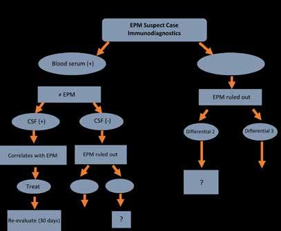
Evidence of intrathecal antibody production is the most accurate way to support an EPM diagnosis. However, blood is a good screening tool. If serum comes back negative, likely there is no infection or recent infection. EPM can generally be ruled out and you can proceed down your list of differentials. (Figure 1) A rare exception would be a very acute onset prior to antibody production, in which case a retest in 10 to 14 days is warranted.
A positive serum test result, however, presents a “gray zone” because it doesn’t necessarily equate to an EPM diagnosis. Knowing there is a high seroprevalence to the causative organisms of EPM in healthy U.S. horses, now what?
Blood serum (-) SERUM POSITIVE? PROCEED WITH CAUTION: 78% of healthy U.S. horses are seropositive to Sarcocystis neurona and 34% to Neospora hughesi. So, while you can find a seropositive horse in almost every pasture, a positive result doesn’t mean that horse has EPM.
To more definitively rule out (or in) EPM, a CSF tap is recommended. If the CSF sample is negative, EPM is ruled out and the practitioner should proceed to the next differential diagnosis. If the CSF sample is positive, consider it a pretty strong case for an EPM diagnosis. (Figure 1)
Be aware, a positive CSF result can happen for reasons other than antibody production within the CNS. For one, blood contamination. If there is a high blood titer to S. neurona, for example, this could give a positive result from blood-derived antibody and not production within the CNS.
Therefore, current best practice consensus is to collect spinal fluid and a blood sample and compare the antibody titers in each to determine if there is evidence of a CNS infection. This is done by evaluating the ratio of antibody titer in serum divided by antibody titer in CSF.

If EPM rises to the top of your list of differentials after a thorough physical and neurological exam, antibody testing should be used to further differentiate EPM from other neurological diseases. Intrathecal antibody production is the most accurate way to support an EPM diagnosis, while blood is useful for screening.
Treating the horse with an antiprotozoal drug for 2 weeks and then reassessing may be a practical approach if you feel strongly this is an EPM case and CSF sampling is not accepted. However, this is ONLY recommended if you can physically reassess the horse in 2 weeks to evaluate progress. At that time, critically evaluate the improvement by repeating the same thorough physical and neurologic work up. A significant improvement must be seen to continue with unfinished treatment (I like to see at least a 25% improvement). If the horse responds to treatment, you have further evidence to support an EPM diagnosis. If the horse does not respond, it’s back to the drawing board. This is a critical communication point with the owner to help them understand next steps and avoid potential frustration of treating for EPM with no improvement. (See sidebar on client communication tips.)
Tricky disease. Tricky diagnosis.
EPM got you scratching your head? Take heart. Each clinical presentation is different, and the best way to outwit this disease mimicker is with a solid dose of due diligence. There are no shortcuts when it comes to doing a proper EPM diagnostic work up. Look for hallmarks of clinical disease in your physical exam and don’t shy away from the need for immunodiagnostics to get to the root cause.
The good news: If EPM is diagnosed, there are safe and effective EPM treatment options. Come back for the third in our four-part series as we dive into the latest treatment advancements.
For more information:
James KE, Smith WA, Conrad PA, et al. Seroprevalences of anti-Sarcocystis neurona and anti-Neospora Hughesi antibodies among healthy equids in the United States. JAVMA. 2017;250( 11) :1291-1301 https://doi.org/10.2460/javma.250.11.1291
Reed SM, et al. Equine protozoal myeloencephalitis: An updated consensus statement with a focus on parasite biology, diagnosis, treatment and prevention. J Vet Intern Med. 2016;30:491–502. https://onlinelibrary.wiley.com/doi/10.1111/jvim.13834
NAHMS. Equine Protozoal Myeloencephalitis (EPM) in the U.S. In: USDA:APHIS:VS, ed. Centers for Epidemiology and Animal Health. Fort Collins, Colorado: NAHMS; 2001:1–46. https://www.aphis.usda.gov/aphis/ourfocus/animalhealth/monitoring-andsurveillance/nahms/nahms_equine_studies

