10 minute read
TECHINICIAN UPDATE


By Andrea Whittle, BS, LVT A 2-year old Standardbred gelding in central Kentucky presented recumbent to Rood and Riddle Equine Hospital in November 2018 with a detailed history from the referring veterinarian and a significant list of differential diagnoses.
Diagnostics, appropriate treatment and intensive nursing care over several weeks led to a very successful outcome for this gelding and his owners.
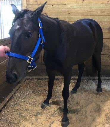
This gelding had been recently purchased by his owners in Ohio. He was actively in training at the time of purchase and was an exciting prospect for his owners. Shortly after purchase, they had him castrated in the field; it was stated on his admission notes that a tetanus toxoid injection was not given at the time of castration. The castration was performed 2 weeks prior to admission. The referring veterinarian gave a detailed history of the 24 hours prior to admission. The gelding was presented to him at the track with swelling in the scrotal area and weakness in the pelvic limbs. This progressed to the horse laying down and being unable to pull his hind legs up to rise without assistance. At this point he was able to dog sit and appeared strong through the thoracic limbs. He was given acepromazine to safely load, moved onto a thick tarpaulin and loaded into a trailer for referral. Upon presentation to Rood and Riddle, he was in right lateral recumbency (RLR) and unable to sit sternal with or without assistance. He was pulled onto the Large Animal Glide and moved to a well-bedded, padded stall with a hoist. He was aware and responsive to noise and contact. His weight was estimated at 1,000 lb. A physical examination was completed with unremarkable findings; temperature 99°F, heart rate 72 beats per minute (bpm), respiration rate 24 breaths per minute, slightly bright pink mucous membranes and a capillary refill time of 2 seconds. Upon palpation of his skeletal muscle his larger muscles groups were firm to touch but did not appear to elicit a pain response. All 4 limbs were difficult to flex and returned to a stiff extended position. Other than the lateral aspect of the right carpus his legs were free of abrasions. The scrotal swelling was approximately the size of a grapefruit, soft and cool to touch. An ophthalmic examination revealed a significant amount of debris in the right eye from the stall and trailer; the eye was rinsed with eyewash and lubricated with artificial tears ointment until his recumbency could be changed and the eye could be examined more Down But Not Out—Recumbency in a Standardbred Gelding
Images courtesy of Andrea Whittle, BS, LVT Fig 1. Ultrasound image of scrotal swelling
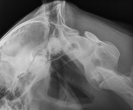
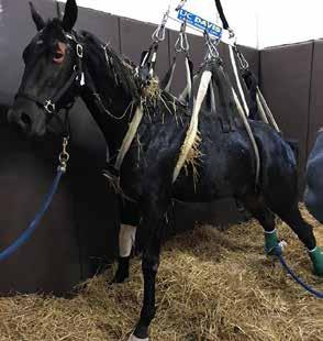
Fig 3. Standing in UCD lift
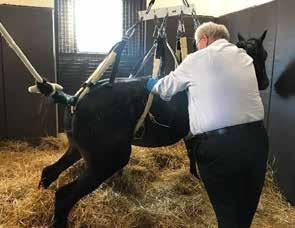
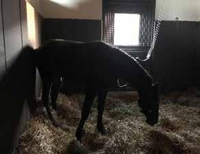
Fig 4. Standing without support
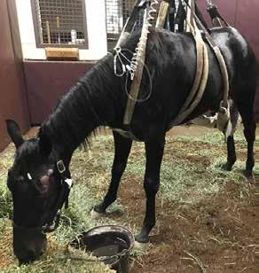
Fig 5. Knuckled and stiff in hind legs
thoroughly. No third eyelid prolapse was noted. At this point we were limiting any movement of the neck as a history of trauma had not been ruled out.
A 14 g 5.25" long-term Mila catheter was placed in the left jugular vein (LJV) after the vein was clipped and aseptically prepared. A complete blood count and chemistry panel were submitted. The pertinent results were white blood cell count (WBC) 12,400 x 10³, packed cell volume (PCV) 41.7%, serum amyloid A (SAA) 814 ug/mL, creatine kinase (CK) 64260 U/L and aspartate aminotransferase (AST) 1604 U/L; his serum potassium was a little low at 3.2 mmol/L. A 30" extension set was secured to the needle-free adaptor (microclave) of the catheter extension set to allow safe catheter access in either recumbency. The microclave was left in place as a safeguard in case the 30" set becomes disconnected when the catheter is on the recumbent side.
16 Issue 3/2020 | ModernEquineVet.com Fig 6. Situated in stall to eat
An ultrasound (US) examination of the scrotal swelling was completed; the swelling appeared uniformly edematous with no abscess formation.
Differential diagnoses were discussed with the owner after the PE, ultrasound and blood work results were completed. This list included the following: • Myositis • Tetanus • Botulism (suspect incisional) • EPM • Trauma and damage to the cervical spine After this discussion the decision was made to rule out or immediately counter several of the differentials. Radiographs of the cervical spine were taken stall side using a NEXT Equine DR system. Lateral views of the skull, poll and cervical spine revealed no significant abnormalities.
A lumbosacral cerebrospinal fluid (CSF) tap was completed through a clipped and aseptically prepared area; a 12 cc lidocaine block was administered via a 1.5" 20 g needle and an 18 g 6" BD spinal needle was used to obtain the sample. The CSF was clear and sent to Equine Diagnostic Solutions (EDS) for testing for Sarcocystis neurona and Neospora hughesi. Results were returned later that day and were negative for both organisms.
A 250 mL bag of Botulism Antitoxin was thawed and administered through a blood/plasma IV set. The available antitoxin was specific to Type B. The plasma ran without complication or reaction.
After these procedures were completed, the gelding was rolled dorsally to left lateral recumbency (LLR) with the use of shackles and ropes on each limb as well as head and neck support. In the process of doing this he was also rolled onto the 'down' straps of the UCD Large Animal Lift for ease of placement. A thorough examination of the right eye was completed; more eye wash was used and there was a small area of fluorescein stain uptake centrally on the cornea. Neobacimyx (QID) and atropine (SID) ophthalmic ointment treatment were started.
Continuing treatments were started with the administration of potassium penicillin (22,000 IU/kg IV QID), gentamicin (6.6 mg/kg IV SID) and flunixin (1.1 mg/ kg IV BID). The gelding was given a dimethyl sulfoxide (DMSO) bolus of 500 mL in 5 L of lactated ringers solution (LRS). He was started on a continuous rate fluid infusion of LRS with 20 mEq of KCl solution/liter after the DMSO bolus.
He was allowed to rest quietly in LLR with the barn lights off until the DMSO bolus was complete. Once this was disconnected the UCD Lift was fully fitted and attached to the hoist bar. For safety purposes the barn aisle and doorway were cleared of possible trip hazards and only minimal personnel were around the stall front; each person was allocated a clear job and a leader was identified. The UCD lift was used to assist with rolling the gelding to a sternal position. This was accomplished and he was offered a small pan of water to assess his ability to swallow—he passed the swallowing test. The first attempt to lift him to his feet was successful; he was stiff through all 4 limbs to lift but once he was vertical next to a padded wall for support, he was able to take his own weight and hold his head in a normal position. The pressure and support of the UCD lift was gradually released with no change to his ability to stand. He urinated once he was up; it was dark brown.
Once up, he was offered a bucket of water and a loosely packed hay net of grass hay. He was also started on Vitamin E (Elevate 4,000 iu PO SID) and methocarbamol powder (50 mg/kg PO BID). The UCD Lift was Fig 7. Grazing
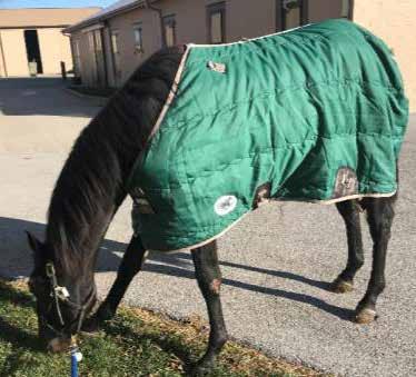
Fig 8. Loaded to leave RREH
removed after about 45 minutes; the gelding was able to walk around the stall. He was stiff through his hocks and dragged some straw with each step.
He stayed up for about 3 hours before laying down and resting quietly. He was allowed to rest overnight with his recumbency changed once. This was repeated the next day when he stayed up for 7 hours, allowed to rest and then stood again with the UCD for 16 hours. The next attempt to lift him in the UCD Lift was less successful. The stiffness in the pelvic limbs was drastically increased and he would hold them in a much exaggerated, hyperextended, stance on tiptoe.
Unfortunately the stall that he was housed in did not have sufficient height on the I-beam and hoist to be able
to pull his hind legs forward and help him place them, soles down, in the stall. After some troubleshooting and measuring, we realized that the second padded hoist stall in the barn had the I-beam placed one concrete block higher, giving us an additional 8 inches.
He was induced with xylazine, butorphanol and ketamine, maintained on a GKX CRI, placed on the glide and moved into the second stall. After sufficient time to wake up from sedation, he was lifted again with success. The biggest change to his mobility was an inability to support himself out of the sling, inability to walk and frequently knuckling on both hind fetlocks. Lightweight, padded, shipping wraps were placed around the hind fetlocks to protect them when he was knuckling.
Full blood work was submitted again with the following pertinent results: AST 5597 U/L, CK 214309 U/L, and SAA >3,000 µg/mL.
With myositis as the primary diagnosis at this point, he was placed onto a butorphanol CRI (0.01mg/kg/hr in LRS) for additional pain management and started on dantrolene (4 mg/kg PO TID). His attitude and appetite responded to the increased pain management, and he began to relax in the UCD lift; it was only in contact with him on the sternum and in front of the point of shoulder as he learned to lean forward for support. A tapering dose of IV dexamethasone was started at 30 mg IV.
He was very responsive to grooming, well-behaved enough to have his left knee cleaned and wrapped and only mildly evasive for his ophthalmic treatments. His owners elected not to undergo a muscle biopsy and subsequent testing and put that money toward continuing his care in the hospital for as long as needed as he was responding to his care and treatments.
Once he had learned to use the lift well, we started to work on his mobility to encourage movement and stretching within his bounds of comfort. His butorphanol CRI was discontinued at this time. His appetite was a good motivator and reward to use with him. His straw bedding was banked up to the sides of the stall to prevent him from bundling it up between his legs and a layer of peat moss was used for urine absorbance and traction. His hay was placed on a straw bale several feet in front of him, just out of his reach; his hind legs were initially lifted and manually placed forward for him to encourage steps forward. This was repeated every 2–3 hours, and he would slowly move around the stall. Occasionally he would knuckle both hind fetlocks and require help to regain his correct stance.
His next stage of physical therapy was to encourage locking and loading of the hind fetlocks. To do this, the UCD lift was raised slightly to support a small amount of hind-end weight and he was gently rocked about 6 inches from side-to-side to load and unload each fetlock. He was always compliant to these sessions; we did this 6 times a day for about 10 minutes at a time.
Since he was eating consistently now, his IV fluids were reduced in rate over 3 days then discontinued. After 6 days his SAA was rechecked at 220 µg/mL, his muscle enzymes decreased to CK 1545 U/L and SGOT / AST 7467 U/L.
His right eye wasn't responding as well as hoped to its current treatment; he started guarding it, and there was increased tearing and swelling. A thorough ophthalmic examination was performed again with proparacaine solution. The corneal ulcer had increased, and his medication regime was changed to gentocin (BID), terramycin (BID), miconazole (BID) and atropine (BID) ophthalmic ointments.
As his mobility increased and the frequency of the fetlock knuckling decreased, the UCD lift was taken off. His hay pile was still moved around the stall periodically Fig 9. At home on layup
After returning to his owners, the gelding was rested for >6 months before being gradually introduced back to work.