9 minute read
TECH UPDATE


Traumatic Facial Wound in a Miniature Donkey
By Megan Born, BT, AAS, LVT; Katherine MacGillivray, VMD, DACVIM; and Russell Freeland Jr, DVM, DACVS
A 32-kg male intact miniature 4-month-old donkey presented with extensive traumatic bite wounds over the left lateral mandible and maxilla after being attacked by a dog.
A large area of skin and muscles were no longer present after both the injury and necessary debridement. A portion of the masseter muscle, zygomaticus muscle, buccinator muscle and others were no longer intact. The nerves in this area that may have been affected by the trauma included the trigeminal nerve and the facial nerve.
The traumatized area was cleaned, lavaged, debrided and the patient was hospitalized for continued management of the wound and supportive care. Chest radiographs and ultrasound examinations of both the chest and abdomen were performed, and there was no evidence of trauma to other organ systems.
Bloodwork abnormalities showed:
• a lactate of 4.1 mmol/L, (ref. range 0.9 mmol/L);
• hematocrit of 33.60% (ref range 34-47%);
• WBC 17.9 K/uL (ref range 5.0-12 k/uL), (Neutrophils and platelets were high, hemoglobin, RBCs, and lymphocytes were low in the CBC);
• glucose 132 mg/dL (ref range 76-119 mg/dL);
• creatinine 0.7 mg/dL (ref range 0.8-2.0 mg/dL);
• total protein 5.0 g/dL(ref range 5.7-7.4 g/dL);
• albumin 2.8 g/dL (ref range 2.9-4.1g/dL);
• GGT 50 U/L (ref range 3-38U/L);
• AST 362 U/L (ref range 196-360 U/L);
• LDH 584 U/L (ref range 112-408 U/L); and
• CK 3235 U/L (ref range 120-320 U/L).
The low hematocrit and platelet, hemoglobin and RBC values were likely in response to blood loss from the traumatic injury. The initial increased white blood cell count, with increased neutrophils and decreased lymphocytes was suspected to be due to a “stress leukogram” after the traumatic attack. Hemoconcentration was likely the cause of the increased liver values. Creatine kinase was increased due to the muscle trauma.
A bacterial culture was obtained from the wound, which yielded heavy growth of both gram-positive and -negative bacteria of mixed antimicrobial sensitivity that aided in selection of antibiotic therapy.
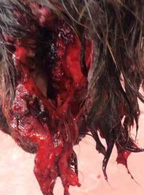
Initial wound showing hanging tissue

Wound after debridement
Images courtesy of Megan Born
Given the extensive damage and type of injury, wound management was focused on supporting healing by second intention.
Upon admission to the medicine facility, the colt’s heart rate was 86 beats per minute, temperature was 101.1° F, and the respiratory rate was 20 breaths per minute. The heart rate stayed in the range of 80-100 bpm over the course of the first day, and the respiratory rate decreased to 12 breaths per minute shortly after admission.
Silver sulfadiazine (SSD) was placed over the lesion.
A tetanus vaccine and 125 mg of SoluMedrol were administered at presentation. Amikacin and potassium penicillin were initiated for broad-spectrum antimicrobial activity and scheduled for serial IV administration. Flunixin Meglumine was also added to the treatment regimen.
A 16-gauge antimicrobial over the wire IV catheter was placed.
Crystalloid fluids of Normosol R with calcium, amino acids and dextrose were infused with a total of 4 L of fluids being given over the first 24 hours. The fluids provided circulatory support and replaced fluid losses from the large open wound. Dextrose and amino acids provided caloric and protein replacement as the donkey was not able to ingest feed material at this time.

Wound after granulation tissue started growing. SSD over large portion of exposed mandible

Out on a walk with stockinette and packing in place over wound
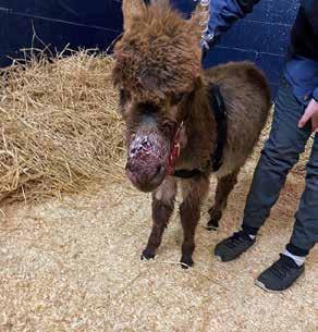
Showing edema over muzzle

Company in the stall!
A tracheostomy tube was placed several hours after admission due to the presence of increased respiratory effort, nasal edema and reduced airflow through the nasal passages. Prior to the tracheostomy air appeared to be passing predominantly through a hole in the dorsal aspect of the right nasal passage.
Small amounts of water were offered.
The second day, the wound was lavaged with betadine and saline. Manuka honey and SSD were placed over the wound edges and the exposed portion of the mandible. The open area was packed with moist gauze, and a stockinette was placed over the cranium to hold the packing in place.
In addition to the fluids, partial parenteral nutrition was initiated with lipids for continued caloric support.
Butorphanol was added to the analgesic plan and scheduled for IV administration every 6 hours.
Small amounts of grass and hay were offered. A large plush stuffed horse was placed in the stall with the donkey for environmental enrichment, and the donkey stayed close to the plush horse over the next couple days.
Triglycerides were monitored throughout the donkey’s hospital stay to assess for hyperlipidemia and hyperlipemia, which are common in stressed or ill donkeys. Unaddressed hyperlipemia in donkeys can further complicate recovery from the initial clinical condition. This is due in part to fat deposition in the liver and secondary liver dysfunction, which can lead to other more serious clinical conditions that affect patient prognosis.
On day 2 of hospitalization, the triglycerides were 105 mg/dL (ref range 13-55mg/dL). To deter additional fat mobilization, heparin sodium and corn syrup were initiated to increase fat uptake by peripheral tissues and decrease risk of hyperlipidemia and hyperlipemia.
Over the next few days, the wound was managed by periodic lavages, debridement and bandage changes. Debridement required sedation. Osteostixis was performed with a 2.5 drill bit in the lateral exposed mandible to help provide a blood flow to the area and promote granulation tissue formation.
The exposed bone was kept moist with Manuka honey and SSD.

Healthy granulation tissue filling in defect

Wound epithelialization
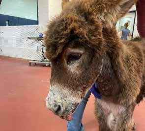
Final photo showing wound resolution and facial paralysis
The donkey was able to eat some grass and hay and a senior feed slurry, although some of the feed material would become lodged in the bandage due to the large cheek area deficit.
Scheduled walks during which the colt was allowed to walk on his own within a large confined indoor area were performed to help the patient’s activity and wellbeing.
PPN and fluids were weaned slowly as the donkey was able to ingest more of his nutrient requirement. At discharge, the donkey weighed 28 kg compared with 32 kg on arrival.
The tracheostomy tube was left in place at discharge, and recheck appointments were scheduled for further wound care and assessment.
The donkey went home and watched the Olympics with his family during recovery after day 5 of hospitalization.
The wound granulated very well with second intention healing. A releasing incision was necessary mid-way through the healing process to preserve mobility, because the face was healing with a tight band of scar tissue that was restricting movement.
The extensive damage of the initial wound caused nerve damage with a sequela of facial paralysis requiring a permanent tracheostomy.
The final picture shows a completely epithelialized site with hair growth filled in. This picture is from 12 weeks after the injury. The age of the animal likely helped with the quick and efficient healing as well as the presence of a large blood supply to the cranium.
This wound would probably not have been a candidate for grafts due to the motion of the jaw and the extensive area involved. MeV
Teaching Points
The successful practice of veterinary medicine involves treating the animal as a whole considering their behavioral and instinctual characteristics that contribute to their well-being. In addition to treating the primary wounds and secondary disease processes, this overall well-being of the animal is cared for with enrichment strategies. Environmental enrichment is defined as an improvement in the biological functioning of captive animals resulting from modifications to their environment.
Environmental enrichment for equine species in the hospital setting includes social companionship, the ability to find and eat forage, turnout or walking, and sensory stimulation, which can be provided by items such as jolly balls.
Caring for the animals well-being through environmental enrichment is particularly important for donkeys as they can become easily stressed in a hospital setting, which can lead to a negative energy balance and delay healing.
Donkeys prefer to be in a herd or with a bonded partner and providing this in a hospital can improve the overall wellbeing of the individual. This young donkey did not have a pasture mate, however; so the plush horse provided some initial company and hospital staff spent extra time with the donkey over the course of hospitalization.
Scientific studies have also shown that environmental enrichment accelerates wound healing. The wound on this donkey was extensive necessitating there be no impedance to growth by concurrent illness brought on by inadequate environmental enrichment. A quick return to the donkey's home environment with at home care and periodic veterinary support further increased this young donkey’s morale improving the wound’s biological support system.
About the Authors
Megan Born, MS, LVT, has worked in the Internal Medicine and Critical Care Unit at Hagyard Equine Medical Institute since 2012. She is also a part time instructor for veterinary technicians at the University of Missouri Online. She enjoys showing reining horses and attends Clays Mill Baptist Church.
Kathy C. MacGillivray,VMD, DACVIM, attended the University of Pennsylvania and obtained her veterinary degree in 1998. Following her internship at Peterson and Smith in Ocala Florida from June of 1998 through June of 1999, Dr. MacGillivray returned to New Bolton Center for her internal medicine residency. After her residency, Dr. MacGillivray worked as an emergency clinician at New Bolton Center and for some local private ambulatory practices. Dr. MacGillivray joined the Hagyard Equine Medical Institute McGee Medicine Center in Lexington, Kentucky as an Internal Medicine Associate in 2003.
Dr. Freeland grew up in agriculture in southwest Louisiana. He attended Louisiana State University earning a bachelor’s degree in wildlife ecology and a Doctor of Veterinary Medicine. Trained as a surgeon, Dr. Freeland has a special interest in orthopedics and lameness in the performance horse. He is a board-certified large animal surgeon by the American College of Veterinary Surgeons. He is a member of the American Association of Equine Practitioners, American Association of Bovine Practitioners, and the American Veterinary Medical Association. Outside of practice, Dr. Freeland enjoys the outdoors, spending most of his free time hunting, fishing, flying and diving.