HONORARY EDITOR Dr Cristiane da Mata BDS MFD (RCSI) Dip TLHE MPH Phd FFD RCSI
journaleditor@irishdentalassoc.ie
DEPUTY EDITOR Dr David McReynolds BA BDentSC MFDS RCSEd DChDent (Pros) FFD RCSI
EDITORIAL BOARD Dr Meriem Abbas BDS (NUI) MFDS RCSEd PGDip TLHE Una Farrell Dip Dental Hygiene
Dr Geraldine McDermott BA BDentSc MFDS (RCSI) PGradDip ConSed (TCD) MSc Healthcare Leadership (RCSI)
Dr Clair Nolan BDS (NUI) MSc (Endo) U. Lond
Dr Adedeji Daniel Obikoya BChD MFDS (RCSI) MSc
Dr Judith Phelan BDS (NUI) MDS (NUI) MSc (U Lond) MRD (RCS Eng and Glas)
Dr Patrick Quinn BCL BDS LLM MDPH
Dr Catherine Vaughan BDS (NUI)
IDA PRESIDENT Dr Rory Boyd
IDA CHIEF EXECUTIVE Fintan Hourihan CO-ORDINATOR Liz Dodd
The Journal of the Irish Dental Association is the official publication of the Irish Dental Association. The opinions expressed in the Journal are, however, those of the authors and cannot be construed as reflecting the Association’s views. The editor reserves the right to edit all copy submitted to the Journal. Publication of an advertisement or news item does not necessarily imply that the IDA agrees with or supports the claims therein. For advice to authors, please see: https://jida.scholasticahq.com/for-authors
Published on behalf of the IDA by Think Media, 537 NCR, Dublin 1 T: +353 1 856 1166 www.thinkmedia.ie
Audit issue January-December 2023: 3,867 circulation average per issue. Registered dentists in the Republic of Ireland and Northern Ireland.
34 PEER-REVIEWED Oral leukoplakia: an update for dental practitioners B. Maloney, S. Galvin, C.M. Healy
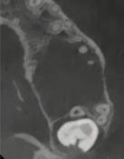
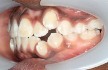
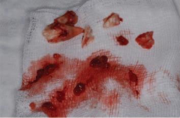
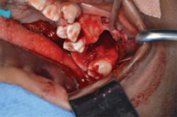
41 PEER-REVIEWED An inflammatory odontogenic cyst (unusual case): case report A.A. Alfurhud, S. Harrison, M. Alshammari
Navigating the orthodontics journey
Irish Dental Association Unit 2 Leopardstown Office Park, Sandyford, Dublin 18. Tel: +353 1 295 0072 Fax: +353 1 295 0092 www.dentist.ie Follow us on Facebook (Irish Dental Association) and X (formerly Twitter) (@IrishDentists).
Dr Cristiane da Mata Honorary Editor
Let’s talk about change
An older edition of this Journal offers a glimpse into past issues for dentistry, many of which are still relevant today.
The first edition of the year always brings out my most reflective side – it’s a time not only to plan for the future but also to contemplate the passage of time and its influence on both our personal and professional achievements and losses, and to learn from them. For our profession, 2025 brings with it a sense of hope and optimism, particularly with the opportunity to position dentistry at the forefront of political discussions. At the same time, the new year invites us to consider the enduring challenges that continue to shape our field.
A glimpse of the past
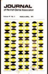
For those interested in history, like myself, I recently came across an older edition of the Journal of the Irish Dental Association from 1973 – a thoughtful present from a recently retired colleague. In it, an article addressed dental manpower needs in the Republic of Ireland, a topic that remains strikingly relevant in 2025.
For this edition, we are delighted to include an additional peer-reviewed article along with our usual scientific content. This is possible due to the increase in submissions to the Journal, and the continued high quality of the articles that we receive.
Then, as now, the uneven distribution of dental professionals between urban and rural areas was a pressing issue. However, the challenges of that era focused on the significant emigration of dentists from Ireland. Concerns were raised that the country was producing fewer dentists than it was losing, leading to fears of an impending shortage that could jeopardise adequate care for the population in the future. Today, while Ireland has witnessed changes in workforce movement and training capacities, the issues of workforce distribution and maintaining an adequate supply of dental professionals persist. We now have a shortage of dentists, as was predicted back then.
Remarkable progress
Another fascinating article in the same edition delves into ‘The prosthetic treatment of the elderly’, a topic that resonates deeply with my own area of interest. The title itself reflects the prevailing perspective of the time: treatment of older patients was primarily focused on complete dentures. While the authors briefly acknowledged situations where the patient had some remaining teeth, this was considered a secondary scenario, with their advice tailored more towards the patient’s age than their specific dental condition. The manuscript’s primary focus was on the fabrication and fitting of full dentures, which wouldn’t nowadays reflect the needs of a growing population of dentate seniors. Clinicians now face the challenges of treating older adults presenting with an array of chronic conditions affecting their oral health status and their ability to maintain oral health. In addition, the current range of restorative and preventive options available, including implant-supported prosthetics and comprehensive geriatric dental care, would mean that other topics would probably gain priority for publication. This glimpse into the past underscores the remarkable progress made in the oral health of the population, reflecting broader changes in the expectations and capabilities of modern dentistry.
Vital platform
Reflecting on the 1973 edition, it is striking to see how the Journal itself has transformed over the years. Once a smaller publication, it has grown into a more robust resource that reflects the evolving needs of our profession. For this edition, we are delighted to include an additional peer-reviewed article along with our usual scientific content. This is possible due to the increase in submissions to the Journal, both from Ireland and further afield, and the continued high quality of the articles that we receive. As we undergo a review of the Journal’s current format, further changes and improvements are on the horizon, ensuring that it continues to serve as a vital platform for knowledge sharing and professional dialogue. I renew my invitation to all dental professionals, researchers, and academics to contribute to our efforts by submitting your latest research, insights, and clinical experiences.
Dr Rory Boyd IDA President
A dental service we deserve
As the new Government starts its work, the IDA also has much work to do. We were delighted to see so many of the priorities we have raised on behalf of members over the years appearing in the new Programme for Government, but as ever, the devil will be in the detail.
Many of the commitments in the Programme, from updating the Dentists Act to improving paediatric dental services, are included in Smile agus Sláinte, the national oral health policy. The implementation plan for that policy is due to be published shortly, and we look forward to collaborating constructively with the Department to ensure the best possible outcome for patients and the profession.
There is much to be praised in the oral health policy, but much of the detail, from how new services might work and who will provide them, to what a new DTSS contract might look like, remains to be confirmed, and all elements of the plan will require consultation and engagement with the profession. Rest assured that we will make our concerns clear, and will work hard to create the dental service that patients, and the profession, deserve.
The commitment to amend the Dentists Act to enable the recognition of more dental specialties is timely, and the IDA will ensure that dentists’ voices are at the table in developing these roles. We have begun the processes to establish a committee within the Association to represent specialists, academics and those in limited practice. We hope that this committee will be a voice for that group within the IDA and beyond, helping us to hold Government to account to ensure that specialists are recognised in an appropriate manner.
Finally, I would like to congratulate Dr Bridget Harrington-Barry on her nomination by Council as IDA President-Elect. Bridget is a longstanding and well-respected member of the Association, and I know she will be a fantastic representative for our members.
Fintan
IDA
CEO
Hourihan
Opportunity for progress
The appointment of a new Government is an opportunity for the Association to forge a new relationship with key stakeholders in advancing our objectives as representatives of the dental profession. Our first task was to offer congratulations to our new Health Minister, Jennifer Carroll MacNeill TD, whom we hope to meet as soon as possible.
The newly published Programme for Government includes some very specific commitments in regard to oral health, and the first significant initiative will be the publication of the implementation plan for the national oral health policy, Smile agus Sláinte. This should identify the State’s ambitions over the next three years.
At its first meeting of the year, the GP Committee decided to prepare a new report on the future of general practice. The board has also decided to begin a consultative process with dentists in limited practice as we pursue recognition of new dental specialties. We have also resumed negotiations with senior HSE management, and have made progress in securing significant commitments to reverse the decline in numbers of dentists and dental team members.
The Association is the representative body for dentists in Ireland, and our role will be to shape and influence change to improve access to dental care, including proposed new laws and regulations, and addressing our staffing problems. We aim to be a trusted adviser and critical friend to the new Minister, whom we are certain will appreciate that reform can only succeed with the active support of the Association and its members.
For the Association to succeed on your behalf, we need your support and ideas. We need to ensure that we have energetic and solution-focused representatives that we can prepare to engage in arduous meetings with politicians and officials.
In return, we will listen to our members. We are ready to engage with the State if it is prepared to build a new relationship of trust and partnership with the profession, respects independent practice, and supports public service. It has never been more important to be a member of your Association.
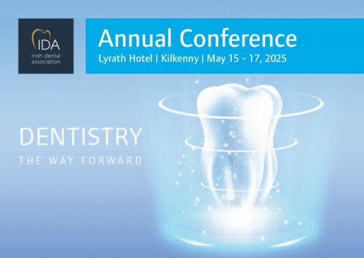
Annual Conference 2025
Bookings are now open for Annual Conference 2025, which takes place from May 15-17 at Lyrath Estate Kilkenny.
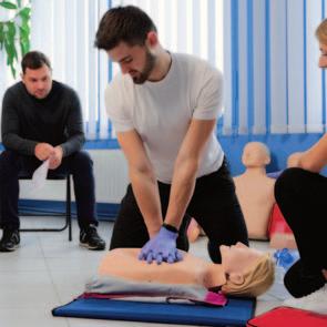
This year delegates have the choice of no less than eight pre-Conference courses on Thursday, on endodontics, composites, facial aesthetics, sleep apnoea, polypharmacy, a dental implants workshop, and prosthodontics. A dedicated full-day programme on facial aesthetics is a new addition to the Friday programme, along with leading experts and presenters on topics such as trauma, paediatric dentistry, dentistry for the older patient, oral surgery, medical emergencies, and much much more.
For full details, go to www.dentist.ie or our social media platforms.
Education/CPD spring-summer programme
The IDA is delighted to bring you our calendar of events for February to May 2025. We have loads on, and with a mixture of hands-on courses, basic life support/immediate life support (BLS/ILS), webinars and annual scientific meetings (ASMs), there’s plenty to keep your CPD up to date over the next few months.
February
February 21
February 21
Hands-on digital photography with Dr Minesh Patel –Hilton Hotel, Charlemont, Dublin 2, 9.00am-5.00pm
Dental immediate life support for sedation teams –Radisson Blu Dublin Airport, 9.00am-5.00pm
February 22 Basic life support and medical emergencies –Radisson Blu Dublin Airport, 10.00am-4.00pm
February 25 North Eastern Regional Meeting –Fairways Hotel, Dundalk, 7.00pm
February 28
South Eastern ASM –Faithlegg Hotel, Waterford, 9.00am-5.00pm
February 28 Western Regional Meeting –The Galmont, Galway, 7.00pm
March
March 7
Eastern Region ASM –Hilton Hotel, Charlemont, Dublin 2, 9.00am-5.00pm
March 26 Webinar, 8.00pm
March 28
Dental immediate life support for sedation teams –Limerick Strand Hotel, 9.00am-5.00pm
March 29 Basic life support and medical emergencies –Limerick Strand Hotel, 10.00am-4.00pm
April
April 26
General practice meeting –venue to be confirmed, 10.00am-3.00pm
April 30 Webinar, 8.00pm
May
May 8
May 15-17
IDA Annual General Meeting –Hilton Hotel, Charlemont, Dublin 2, 6.00pm
IDA Annual Conference 2025 –Lyrath Estate, Kilkenny
May 28 Webinar, 8.00pm
To book these events, go to www.dentist.ie and click on ‘Book CPD’. For any queries, please contact IDA House.
Get behind the IDA’s social media campaigns
Our campaigns rely on communicating the issues, and mobilising public and political sentiment to influence the Government. This is where you come in! Like and share the IDA’s content, and create your own content to drive the conversation on behalf of your profession and make the Government take notice of oral health policy in Ireland.
Scan the QR code for some handy social media tips.
Check out the IDA’s extensive webinar library and bank your CPD points – exclusive for members
LearnUpon is the IDA’s learning management system that makes it easy for members to manage all of their CPD. Whether it is booking one of our upcoming hands-on courses, or national and regional events, watching one of the monthly webinars we provide, catching up on JIDA articles, or recording non-IDA CPD, LearnUpon is a one-stop shop for IDA members. We have a library of over 60 webinars available exclusively for our members to watch back at a time convenient for them. Topics covered include clinical presentations, practice management, employment law, and compliance.
Save the date
The South Eastern Region will hold its Annual Scientific Meeting (ASM) in Faithlegg Hotel, Co. Waterford, on Friday, February 28. A full day’s programme is scheduled along with trade show.
The Eastern Region ASM returns to the Hilton Hotel Charlemont on Friday, March 7. The ASM is a great way to catch up with colleagues old and new, and to see outstanding presenters offering verifiable CPD.
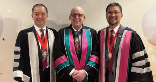
Congratulations to Prof. Christopher Lynch
Prof. Christopher Lynch has been awarded a prestigious Honorary Fellowship from the College of Dental Surgeons of Hong Kong. The citation for Prof. Lynch highlighted that his “contributions to the fields of restorative dentistry and dental education have been exemplary and groundbreaking”.
Prof. Christopher Lynch is Professor & Consultant in Restorative Dentistry at Cork University Dental School & Hospital/University College Cork.
The College of Dental Surgeons of Hong Kong highlighted that “throughout his illustrious career, Prof. Lynch has excelled in academia and demonstrated exceptional leadership qualities.
”As the Editor-in-Chief of the esteemed Journal of Dentistry since 2011, and as a Dean at the Faculty of Dentistry, Royal College of Surgeons in Ireland since 2023, he has showcased his commitment to advancing research and education in dentistry”.
Survey on sustainable dental care
Stephen Walsh, a PhD researcher at RCSI, is exploring the knowledge and attitudes of dentists in general practice in Ireland regarding delivering dental care in a greener and more sustainable way. Members in general practice (principal, associate or locum) are encouraged to spare 10 minutes to complete an anonymous survey to share your views and perspectives on delivering dental care more sustainably.
Scan the QR code to complete the survey.
From left: Prof. Ka Kit Gilberto Leung, President, Hong Kong Academy of Medicine; Prof. Christopher Lynch; and, Prof. Wai Keung Leung, College of Dental Surgeons of Hong Kong.
IDA Golf Society 2025 diary
Friday, April 4 Lyttle Cup – Co. Louth GC Baltray 10.30am **IDA members only**
Thursday, May 15 President’s Cup – Carlow GC 11.00am All delegates at the IDA Annual Conference can play. Only IDA members who are delegates at the Conference can win the President’s Cup.
Sunday, June 22 Cotter Cup – Luttrellstown 1.00pm Guests welcome
The deadline to buy a UK State Pension for anyone who has worked in the UK for three years is approaching. The temporary arrangement to top up your entitlement ends on April 5, 2025. However, it is strongly advised to start immediately, as setting up the account and making the application can take some time. An income equivalent to the full UK pension bought as an annuity at age 67 would cost ¤260,000 to ¤300,000 in today’s terms. The outlay that the majority have paid to secure this benefit is approximately ¤5,000.
For more information, scan the QR Code to read a Q&A by Colm Moore of Moore Wealth Management.
Have your say on the future of general practice
A vital National Meeting for Practice Owners and Associates is to take place in the Midlands Park Hotel, Portlaoise, on Saturday, April 26, from 10.00am to 3.00pm.
This meeting is open to both IDA members and non-members who wish to discuss their input, experience and vision for general dental practice in Ireland. Free for all!
Book through the IDA website or contact IDA House.
Mandatory BLS training every two years for dental teams
Did you know that all dental practitioners and dental team members must complete certified basic life support (BLS) training every two years?
Any practice that uses sedation should do the immediate life support (ILS) training.
The good news is that the IDA provides both training programmes for members to complete in one day. Courses will take place in Dublin and Limerick in February and March:
Dublin (Radisson Blu Dublin Airport): February 21 (ILS) and 22 (BLS)
Limerick (Strand Hotel): March 28 (ILS) and 29 (BLS)
Bespoke coaching for dentists
Lasting tooth preservation
Are you a dentist feeling overwhelmed by unmotivated teams, financial pressures, or inefficient systems that you know need to change, but don't have the time?
Lisa Grogan, an expert dental coach, states that she offers tailored solutions to help dental professionals across Ireland achieve remarkable growth and feel more in control of their business.
Lisa states that she has extensive experience and specialises in optimising dental practices by identifying opportunities within clinics, streamlining operations, and increasing profitability.
According to Lisa, her bespoke coaching programmes empower dentists to attract more patients, develop effective marketing strategies, and confidently promote high-value treatments.
Working with Lisa means having a dedicated partner who truly understands the unique challenges of running a dental business.
From creating efficient workflows to enhancing the patient journey, Lisa states that her guidance is designed to help you build a successful and reputable practice.
Lisa also runs in-person training programmes in a variety of areas to support dentists and their teams.
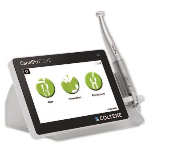
Coltene states that it has a high-quality solution for every aspect of endodontic treatment to support the clinician in preserving natural teeth. For example, the company notes its CanalPro Jeni endomotor, which has a digital assistance system for controlled root canal preparation. Coltene states that the guidance system automates the navigation through the root canal, guiding the mechanical and chemical preparation process at every stage. According to the company, the user can work from coronal to apical with light pressure – the motor independently calculates the course of movement. Coltene notes that speed and motion are continuously controlled using complex algorithms, based on intensity, torque and file stress.
Small but mighty
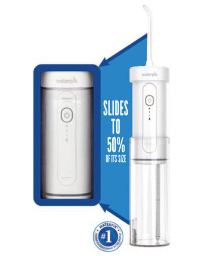
Waterpik states that dentists’ patients can save space in their bathrooms, while protecting their dentition, with a compact interdental cleaning solution – the Waterpik Cordless Slide water flosser.
According to the company, the device’s compact design means the power of Waterpik now fits in a device that collapses to just half of its full size. Waterpik states that for busy or small homes, it is a perfect adjunct for maintaining a highly effective oral hygiene regimen. According to the company, the device is 50% more effective than traditional dental floss for improving gum health, and can remove up to 99.9% of plaque from treated areas after a threesecond application. Waterpik states that the Cordless Slide is an effective oral health adjunct for patients with implants, crowns, bridges and veneers, and the orthodontic tip makes it fantastic for those with braces.
Holiday Cheer from Henry Schein
Henry Schein’s Holiday Cheer programme delivered gifts to children in hospital over the festive period. Company team members joined forces to bring joy to the children at Ronald McDonald House in Crumlin. Since 1999, this corporate flagship initiative has helped more than 20,000 underserved children, their families, and senior citizens in many countries to experience cheerful holidays, with Henry Schein donating clothing, comfort items, toys, gift cards, and more.
In Ireland, Henry Schein contributed to kids aged 0 to 16 receiving care at Children’s Health Ireland (CHI) in Crumlin. Team members’ contribution involved the donation of presents to 'The Magic Press’, an initiative by Ronald McDonald House aimed at bringing joy to the lives of sick children in the hospital.
This year, Schein employees demonstrated their generosity by providing more than 80 gifts, helping to create a festive and uplifting holiday experience for these young patients.
Joe Kenny, CEO of Ronald McDonald House Charities Ireland, said: "On behalf of Ronald McDonald House Charities Ireland, I would like to extend our heartfelt thanks for your very generous donation. Your kindness and generosity have brought immense joy and comfort to many children and families spending Christmas at Ronald McDonald House”.
RCSI sign with Schein
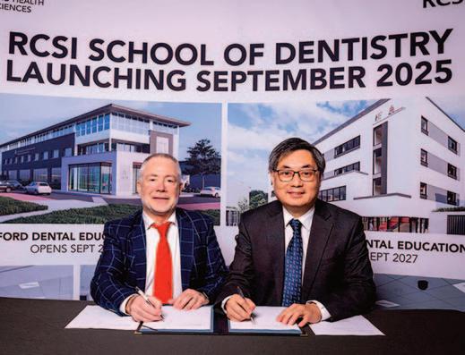
RCSI's School of Dentistry has entered into an agreement with Henry Schein to provide a wide range of equipment for students enrolled in its new Bachelor of Dental Surgery programme.
Through Henry Schein Ireland, the company will supply world-class dental equipment to RCSI’s Dental Education Centre in Sandyford, Dublin which is currently under construction, and will welcome its first students in September 2025.
The Centre will be equipped with an extensive range of dental equipment, such as dental chairs and state-of-the-art patient simulators, supplied and installed by Henry Schein Ireland, providing a protected environment where clinical skills can be acquired and developed.
It will also host a community-based clinical facility, which will include 12 dental chairs for treating patients. The Centre’s simulation unit will have 55 phantom heads, and a dental laboratory to support the dental students’ learning journey.
Henry Schein team members taking part in the company’s Holiday Cheer programme (from left): Paul Shortall; Jennifer McGrath; Darren Murphy; Siobhán Cleary (kneeling); Tracey Maher; and, John Archbold.
From left: Paddy Bolger, Managing Director of Henry Schein Ireland; and, Prof. Albert Leung, Head of the RCSI School of Dentistry.
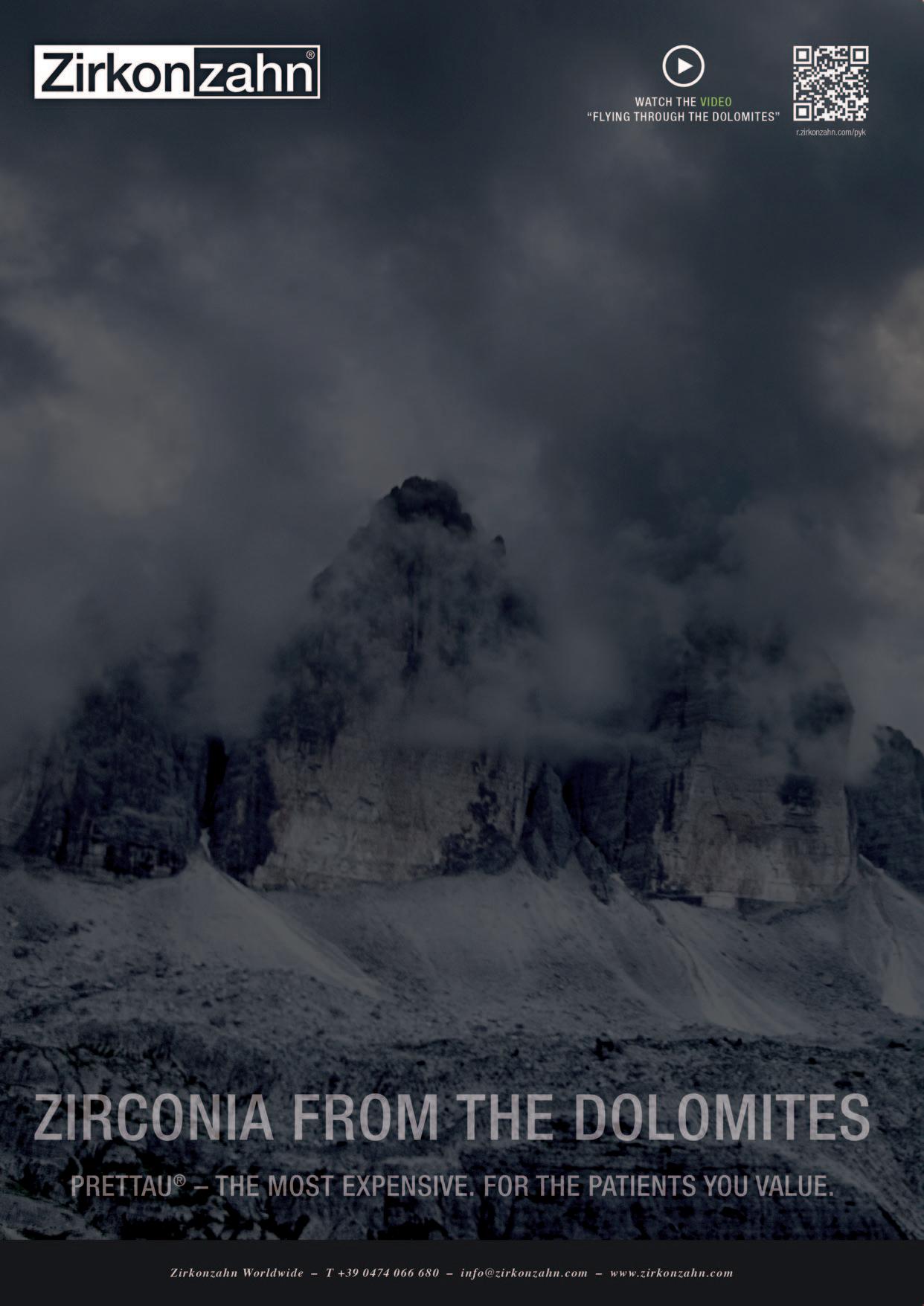
From the Alps to the world
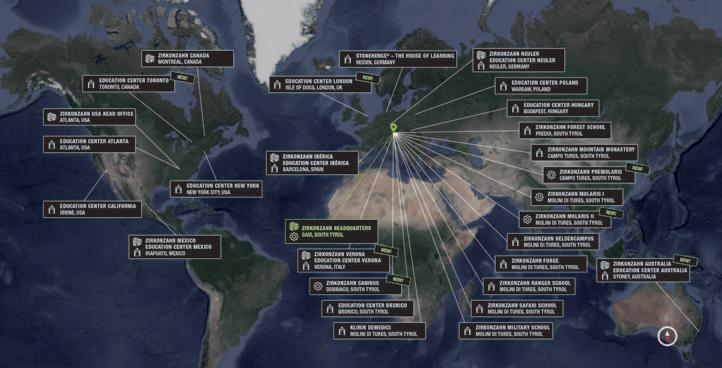
In the heart of the Italian Alps, founded by MDT Enrico Steger, the family-run company Zirkonzahn states that it has the core values of discipline, innovation, trust and responsibility, and has been providing innovative solutions for the dental sector since 2003. Under the motto ‘Everything from a single source’, all Zirkonzahn’s devices, tools, and materials are designed and produced in house, with full control over the entire production process to guarantee the highest quality standards.
According to the company, this approach means that all teams are united under one roof to ensure easy communication and quick knowledge transfer. As a result, Zirkonzahn states that customers can benefit from 360° support covering all technical, dental, and methodological assistance, with extremely fast response times.
Zirkonzahn is headquartered in its homeland of South Tyrol in Italy. Here it has its main education centres and dental laboratory, as well as four
The Ivoclar Group has announced that it will be in attendance at the International Dental Show (IDS) 2025 in Cologne, Germany. From March 25-29, Ivoclar will showcase what it states are innovative solutions and workflows at two exhibition stands during the global trade show. The company is especially looking forward to connecting directly with its customers and highlights a dynamic line-up of presentations and live events that will enhance the trade fair experience. According to Ivoclar, as the leading trade fair in the dental industry, IDS sets the stage and serves as a key platform for presentations and a place for everyone in the market to meet and discuss topics related to dentistry and dental technology. Ivoclar states that it has a long-standing and successful partnership with IDS in Cologne. Over the years, the company has often used the trade show to launch new innovations. In 2025, Ivoclar states that it will once again bring groundbreaking new products to IDS – innovations that will provide real value to users in their everyday work.
production sites, including the recently built Caninus factory: a new facility overlooking the Dolomite Mountains, specially conceived to expand the production of the company’s Prettau zirconia.
Ivoclar at IDS
Clinical dental photography: part II
The second of this two-part series will aim to cover framing in dental photography, the full clinical photographic series, and troubleshooting some common mishaps in order to capture reproducible clinical images of our dental patients.
Introduction
As with any form of clinical record, it would be highly beneficial for photographs of patients to be standardised and reproducible. This would allow for accurate monitoring of the dentition over time, forming a baseline pretreatment record (i.e., orthodontics, teeth whitening, aesthetic rehabilitations) as well as aiding comprehensive treatment planning.
The first part of this series covered the required equipment as well as camera settings used to capture reproducible intra-oral images. In this second part, we will explore framing of dental images, what constitutes a full clinical photographic series, and troubleshooting in cases of incorrect exposures – all of which should enable the user to take images that are standardised, consistent and reproducible.
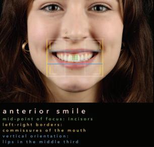
Framing
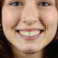
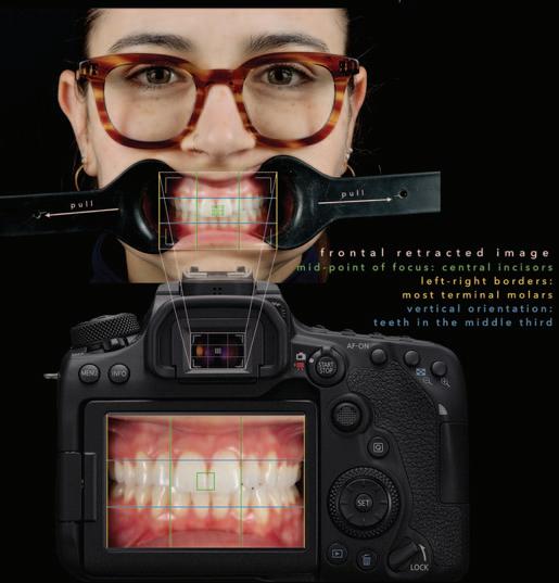
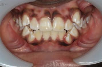
Framing your subject (i.e., the smile, full arch or individual teeth) is vital to ensure the point of focus and purpose of the image being captured (Figures 1 and 2).
This is done primarily by looking into the viewfinder (the eyepiece on the body of your camera) and adjusting the distance between you and the patient. Being too far away will result in capturing more than you require and reducing the flash exposure on the subject, leading to underexposure (i.e., a darker image), and potentially losing macro detail. On the other hand, being too close can lead to overexposure (i.e., an image that is too bright) due to too much light, not capturing sufficient information, and again losing macro detail (see troubleshooting).
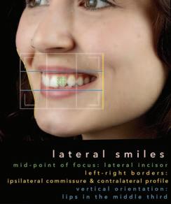
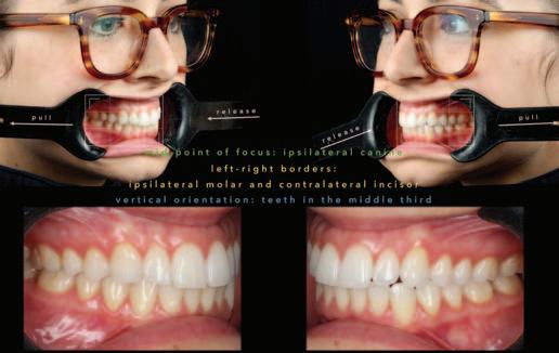
A systematic approach to taking these images is beneficial when starting out. With experience, these rules can be altered as required:
1. Determine the point of focus of the image (i.e., what structures do we need to capture – teeth, gingival margins, soft tissue lesions, etc.). This structure is placed in the middle of the frame.
2. Horizontally orientate the left and right borders of the frame accordingly to include the precise amount of detail you require for that image.
3. Vertically orientate the subject as close to the middle of the frame as possible.
The ‘rule of thirds’ is a guiding principle in photography. It helps the photographer to position their subject within lines in both the vertical and horizontal planes, dividing the frame into thirds. The focus of clinical images should be placed in, or as close as possible to, the middle third, thus ensuring well-composed images.
Dr Ambrish Roshan
BA BDentSc(Hons) DipPCD(RCSI)
Restorative and cosmetic dentist
FIGURE 1: Framing your subject is vital to ensure the point of focus and purpose of the image being captured.
Docklands Dental, 1 Forbes St, Sir John Rogerson’s Quay, Dublin 2
Corresponding author: Dr Ambrish Roshan E: ambrish@docklandsdental.ie
These lines are also beneficial to ensure standardised angulation of the image, i.e., along the occlusal and incisal planes, as well as the left-right orientation. From the horizontal, keeping the barrel of the lens in line with the maxillary occlusal plane ensures that the occlusal plane is not too ‘smiley’ or ‘frowny’ (see troubleshooting).
The camera should also be level with the incisal plane horizontally, keeping the dental midline as close to the vertical orientation as possible.
TIP: It is useful to orientate anatomical landmarks (commissures of the mouth, gingival margins, incisal edges, occlusal planes, etc.) within the viewfinder of your camera, as well as ensuring the angulation between your camera and these landmarks (Figures 1 and 2).
Full clinical photographic series
A full series of clinical photographs is extremely useful in the medicolegal documentation of a patient’s records. It certainly captures much more detail than any graphical electronic dental recording system. These photographs act as a record of the dental status of our patients, as well as the progression or stabilisation of disease over the duration of their care. It is also valuable to
be able to share these photographs with other dental professionals should a patient need to attend a new practice or clinic.
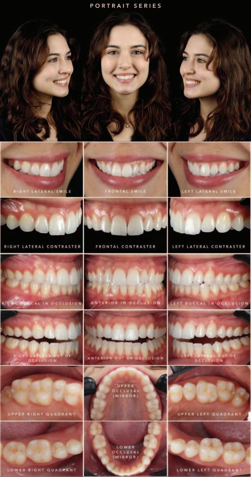
While there is no list of standard images required by any regulatory or indemnity body, the photographs shown in Figure 3 form a comprehensive set. The rationale for each view includes the following:
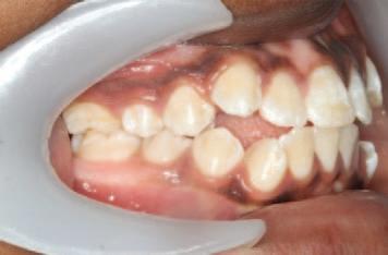
1. Smile line (frontal and lateral): lip line, dental and gingival display, aesthetics.
2. Retracted intraoral in occlusion (frontal and lateral): caries or restorations, gingival margins, occlusal relationship.
3. Intraoral out of occlusion (frontal and lateral): caries, cracks or restorations, incisal edge and cuspal integrity (or tooth substance loss).
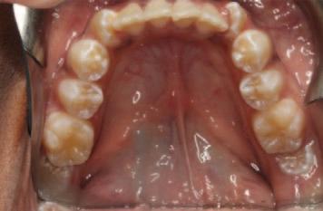
FIGURE 2: A systematic approach to taking clinical dental images is beneficial when starting out.
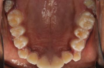
FIGURE 3: Example of a full clinical photographic series.
CLINICAL TIPS
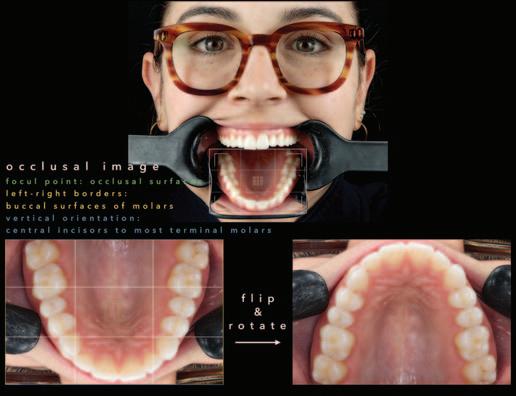
4. Occlusal surfaces: caries, cracks or restorations.
TIP: Get patients’ help to hold their own cheek retractors while your hold the occlusal mirror. Pre-heat the occlusal mirror in hot tap water, dry it with paper towel, and then take the occlusal shots – this helps to prevent fogging of the mirror.
Troubleshooting
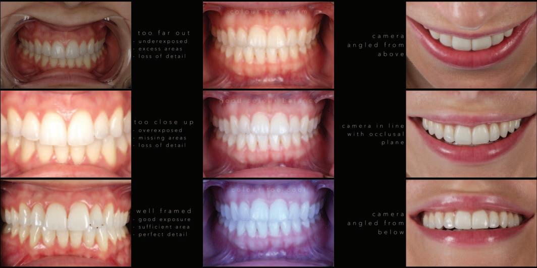
At times, we might find ourselves in a position where an image captured is inadequate ( Figure 4 ). It may be too dark (underexposed) or too bright (overexposed), too yellow (warm) or too blue (cool), out of focus, or perhaps framed incorrectly. In any event, it is useful to know how to rectify these in the hope of obtaining standardised and reproducible images. There are multiple ways in which these common errors can be corrected, but here are some guiding steps to consider:
1. Exposure discrepancy (i.e., image being too bright or too dark).
■ Check that all the settings are correct first (see part one of this series);
■ for direct images (i.e., no mirror use), once all camera settings are correct, adjust the flash exposure (increase if images are dark and vice versa); and,
■ for indirect images (i.e., using a mirror), once direct images are correctly exposed, increase the ‘ISO’, as the mirror tends to absorb some light.
2. Colour discrepancy (i.e., image looking too warm (yellow) or cool (blue)).
■ Check the white balance compensation on the camera body (custom 5,000K to 5,500K or ‘flash’);
■ if image is too cool, i.e., blue – increase white balance value; and,
■ if image is too warm, i.e., yellow – decrease white balance value.
3. Inadequate focus, i.e., centrals in focus but premolars back are out of focus.
■ Increase the f-stop to a minimum of f22 (higher if required) to reduce the size of the aperture and increase the depth of field.
4. Angulation of camera to the subject is incorrect (see Figure 4).
TIP: Practice your clinical photographic sequence and the camera settings with family or colleagues – practice makes perfect.
Conclusion
Dental photography is a fantastic tool in helping us to communicate with our patients, as well as dental colleagues. In addition, it can also make our careers more rewarding, helping to develop our skills by reflecting on our work and providing a higher level of care for our patients. Reflecting on patients’ whose dental diseases have been managed, mouths rehabilitated, and lives improved because of the oral care provided, gives us as dental practitioners satisfaction too, in the knowledge that we have contributed towards that. We cannot treat what we cannot see, and photography helps both patient and practitioner in the recognition and management of disease concurrently.
Camera angled from above Too far out – underexposed – excess areas – loss of detail
Too close up – overexposed – missing areas – loss of detail
Well framed – good exposure – sufficient area – perfect detail
in line with occlusal plane
Camera angled from below
FIGURE 4: Examples of correct and incorrect images.
Colour too warm
Camera
Colour too cool
Good colour balance
MEMBERS’
Dentistry prioritised in new Programme for Government
The new Programme for Government has quite a number of provisions in regard to dentistry and oral health, so we can all look forward to some busy times ahead as the Association represents the profession in numerous negotiations.
In fact, this is the first Programme for Government in recent times that has specific provisions in regard to oral health. Here are a few topline mentions of priorities as they relate to dentistry:
“This Government will:
■ recruit additional doctors, nurses, dentists, and health and social care professionals, and reduce reliance on contract and agency workers; and,
■ increase the number of healthcare college places in nursing, medicine, dentistry, pharmacy, and health and social care professions (including physiotherapy, occupational therapy, and speech and language therapy).”
Priorities
The document indicates some very specific priorities as regards oral health in its section on health:
Fintan Hourihan IDA CEO
“This Government is committed to making dental services more accessible for everyone. This Government will:
■ implement Smile agus Sláinte;
■ hire more public dentists;
■ agree a new Dental Treatment Service Scheme for medical card holders;
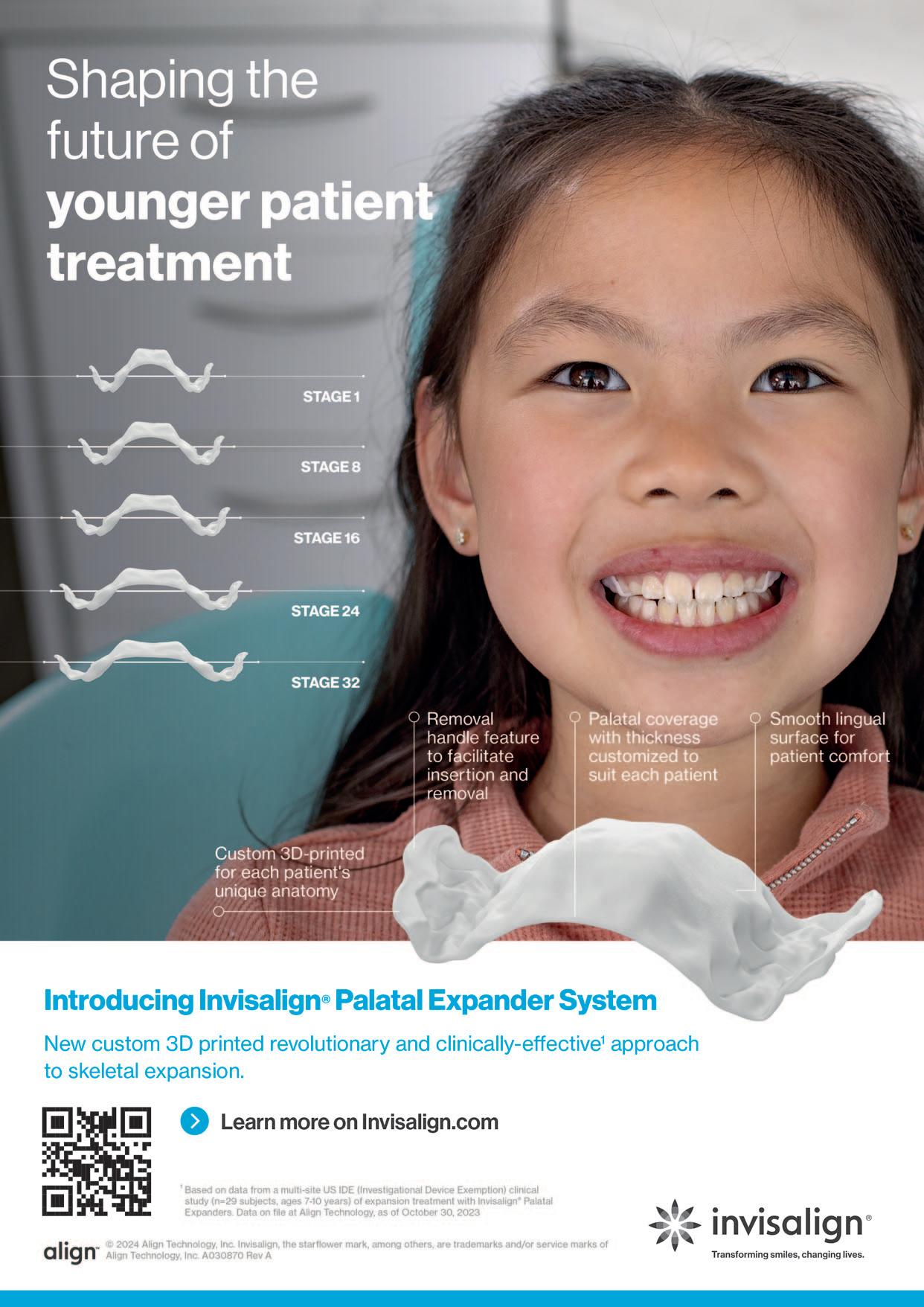
■ expand access to the orthodontic scheme for children and strengthen the School Dental Programme;
■ update the Dentists Act 1985; and,
■ recognise and regulate more dental specialties.”
It is encouraging to see many of the Association’s priorities reflected in the above. As always, these are early commitments from a new Government, and it will be interesting to follow how these points are addressed and progressed in the coming weeks/months and throughout the term of Government.
There is plenty of work ahead, but these undertakings also represent recognition of the powerful advocacy work undertaken by the Association in recent times!
Roisin Farrelly IDA Director of Communications & Advocacy
IDA seeks early meeting with new Minister for Health
Council to propose new President-Elect
Dr Bridget Harrington Barry will be nominated by Council as President-Elect of the Association at this year’s Annual General Meeting.
Bridget is currently working for the HSE as a senior dental surgeon (special needs) in Galway. She has been a longtime member of the Association and is currently a member of the HSE Dental Surgeons Committee. A keen tennis player, Bridget is a graduate of Cork Dental School and also graduated with business and legal degrees. She has served previously as a Board member with the Association, and has extensive experience in representing dentists and advocating for patients.
Dr Will Rymer will succeed Dr Rory Boyd as President of the Association at the 2025 AGM.
The Association has written to congratulate the newly appointed Minister for Health, Jennifer Carroll MacNeill TD (left), and sought an early meeting to discuss the dental reforms signalled in the new Programme for Government.
Jennifer Carroll MacNeill is a TD for Dún Laoghaire, and was first elected to Dáil Éireann in 2020. Before entering elected politics, she qualified as a solicitor and barrister, and holds a PhD in public policy. She most recently served as Minister of State for European Affairs and Defence, and was previously Minister of State in the Department of Finance. She has also served on a number of Oireachtas Committees, including the Public Accounts Committee, the Justice Committee, and the Committee on the Implementation of the Good Friday Agreement.
Submission to Covid-19 inquiry
The Association recently notified the then Taoiseach, Simon Harris TD, of our intention to make a submission to the recently established independent Covid-19 inquiry.
His office responded in writing to the IDA to say that our letter to the Taoiseach was being copied to the secretariat to the Evaluation Panel.
The secretariat is currently engaged in preliminary set-up work. The IDA is to be provided with follow-up information regarding consultation when that phase of the evaluation begins.
The inquiry will be chaired by Prof. Anne Scott, and will begin its work in the coming weeks. The evaluation will be undertaken by a multidisciplinary panel with relevant expertise.
The Government’s “evaluation” is to be entirely voluntary, will have no powers of compellability, and its secretariat will be drawn from the civil service.
Preparing for recognition of more dental specialties
The IDA board has decided to arrange a meeting of representatives from societies representing areas of limited practice in order to prepare a plan on how to achieve greater recognition of dental specialties.
The Association has welcomed the promise in the Programme for Government to recognise and regulate more dental specialties.
Both the Association and the Dental Council have lobbied for the recognition of up to nine additional specialties in addition to orthodontics and oral surgery.
Future of general dental practice
The GP Committee of the IDA is to prepare a report on the future of general dental practice in Ireland.
At a time of rapid change, the Association wishes to examine how it can help members to navigate their way to a rewarding and fulfilling career in general practice.
A survey of members will be complemented by a strategy planning exercise to be carried out by the GP Committee. The Committee proposes to share its findings and recommendations at a national meeting for practice owners and associates to take place on Saturday, April 26.
Public service dentists update
Sectoral bargaining – CPD allowance
The Association has submitted a claim for a CPD allowance worth 1% of salary for all serving HSE dental surgeons, including specialist orthodontists, as well as members employed in other publicly funded agencies. For our retired members, we are seeking a 1% increase to be applied to the value of their pension.
We have submitted these claims under the sectoral bargaining clause of the Public Service Agreement.
Dental staffing and restructuring
The IDA is engaged in a talks process with senior HSE management where we have highlighted the ongoing staffing crisis within the public dental service. In a comprehensive paper presented to the HSE last October, we highlighted the flaws in the Pay and Numbers policy, given the decline in staffing in the HSE dental service and the impact this is having on the service, on morale, and on eligible patients. In a recent meeting, the HSE committed to prioritising the filling of vacant dental posts and to streamlining the recruitment process. The HSE has also committed to additional posts. It is also encouraging that there is a commitment to hire more public dentists included in the Programme for Government.
With regard to the HSE restructuring process, the IDA strongly asserted that oral health must be given the same standing as the other clinical care programmes, and must feed in and report at the highest level.
Membership renewals 2025
IDA subscriptions for 2025 are now due.
A huge thanks to all members who have already paid your 2025 membership or set up a payment plan. This year will be a busy one, with many of the Association’s priorities reflected in the new Programme for Government. There is plenty of work ahead and we thank you for your continued support.
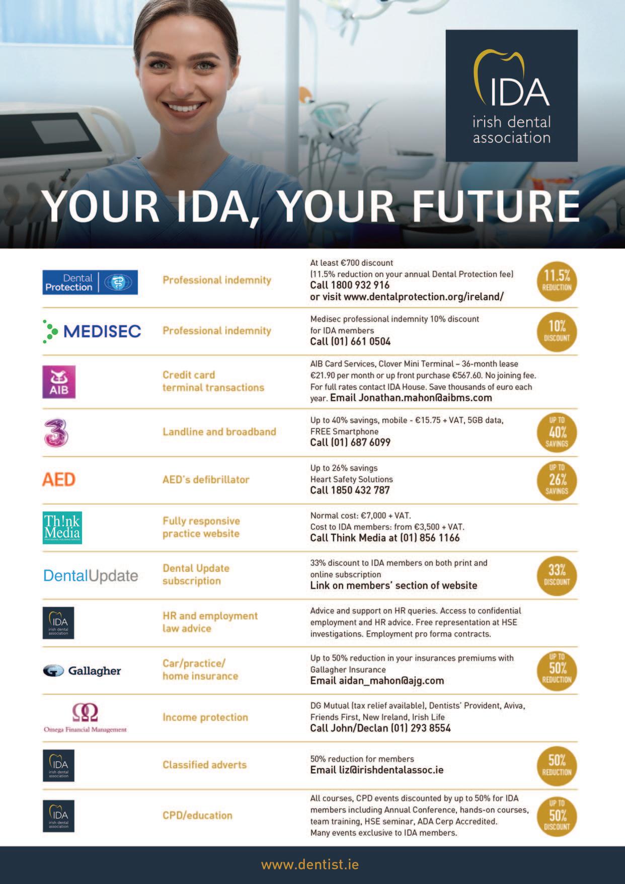
We have independently verified that membership of IDA saves members in private practice thousands of Euro per year. Savings include:
■ discounted indemnity;
■ discounted CPD;
■ discounted landline and broadband packages;
■ income protection;
■ insurance;
■ discounted access to Dental Update and Ortho Update; and,
■ discount at Press Up Group venues.
Renew today and make sure you are availing of all these savings.
Did you know you can pay in 12 monthly instalments, quarterly, biannually, or as a once-off annual payment by direct debit? If you usually pay by direct debit, then you don’t need to do anything. If you would like to pay by credit card, please contact Membership (Cindy) at cindy@irishdentalassoc.ie, or alternatively you can pay by bank transfer or cheque.
Severe weather policy
At this time of year, it is wise to review your practice policy on severe or inclement weather, or to implement one if you do not currently have a policy in place.
A practice policy on absence due to a bad or severe weather event should aim to address the situation where employees are unable to attend for work or the practice has to close or limit its normal working hours due to weatherrelated circumstances.
Legally, employers do not have to pay employees for days that they do not work. However, it is important to be aware of any custom and practice in the organisation, or a contractual clause, which may override this position.
Clarify options
Employers have various options to consider when deciding how to treat the periods of time when employees are unavailable for work due to inclement weather or other natural events. The company’s approach should ideally be clarified beforehand in a policy.
Where an employee has the capacity to carry out his or her work from home for the duration of the disruption, this should be agreed with his or her line manager. This will not be feasible for a number of roles where the employee’s presence is required. In this case, where an employee cannot attend and cannot carry out his or her normal duties, the options of annual leave or unpaid time off should be presented. Some employers may choose to pay employees as normal, particularly in cases where the practice is shut due to bad weather or where this is custom and practice.
Where employees arrive late or leave early due to limited public transport or worsening conditions, for example, flexibility should be provided. In such circumstances, employers may consider paid leave where the employees will work up the time missed at a later date. Alternatively, the option of unpaid leave or annual leave (broken into hours) may be considered. In the case of schools or
crèches closing, some staff may be forced to take leave at short notice. Where the employee is unable to make alternative arrangements, annual leave or unpaid leave could be considered on a case-by-case basis.
In circumstances where a practice is forced to close as a result of weather conditions, a lay-off situation or short-time working may arise. Employers who have reserved the right to place employees on lay-off or short-time working in their contracts of employment may utilise this clause.
Duty of care
At all times and in all decisions, safety considerations must be paramount. Employers have a duty of care for all employees under the Safety Health and Welfare at Work Act. A key consideration during a period of severe weather needs to be whether, in the circumstances, it is safe to ask employees to travel to work, or to undertake their work.
For further advice or discussion on the matter, please contact me in IDA House or email me at roisin@irishdentalassoc.ie
Roisin Farrelly
IDA Director of Communications & Advocacy
How can the IDA help protect you against costly HR compensation awards?
The Workplace Relations Commission (WRC) handed down two decisions within one week recently resulting in compensation awards of ¤20,000 and ¤40,000, respectively, against two dental practice owners.
In one case, a WRC adjudicator determined that a dental practice discriminated against a pregnant worker, having demonstrated a series of failings in its duty of care to a pregnant employee.
In a separate case, the WRC awarded over ¤40,000 against a dental practice in an unfair dismissals case.
The IDA is here to help practice owners avoid costly trips to the WRC in a number of ways:
■ Roisín Farrelly in our Advisory Service provides excellent advice on all HR issues to IDA members free of charge (some 200 queries were dealt with in 2024);
■ we have produced a comprehensive Dental Practice Handbook, which is essential to ensure that you have good policies and procedures in place in your practice to deal with HR issues;
■ we publish regular HR advice for IDA members exclusively in the Journal of the Irish Dental Association;
■ we arrange regular HR webinars exclusively for IDA members; and,
■ we can provide in-person presentations to IDA regional meetings, free and exclusively to IDA members.
Minimally invasive dentistry part 2: caries risk assessment
Learning outcomes
This article aims to assist the reader to:
■ understand the role of caries risk assessment as part of a minimally invasive approach to oral healthcare;
■ be aware of some of the available caries risk assessment tools that can be used in practice; and,
■ appreciate how caries risk status can be used to tailor oral healthcare for children and adults.
Introduction
Dental caries continues to be a major public health problem in Ireland and worldwide. Untreated dental caries in permanent teeth is the most common health condition in the world according to the Global Burden of Disease 2019 study.1 Dental team members are acutely aware of the negative impacts of untreated dental caries on the health and well-being of our patients, not to mention the wider societal and economic consequences.2 In Ireland each year, approximately 7,000 children are referred for dental extractions under general anaesthesia.3 Although perceived by many as a disease of childhood, the risk and consequences of caries continue into adulthood, and indeed may initiate a lifetime burden of care.
Dental caries is a multifactorial disease, which progresses when pathological factors outweigh preventive factors by tipping the ‘caries balance’ towards demineralisation and breakdown of the dental hard tissues (Figure 1). While restorative dental techniques for caries management and tooth retention have improved significantly over the years, it is clear that preventing dental caries is still preferable to cure.4,5 Successful management of dental caries requires both preventive measures and behaviour change, in addition to clinical intervention, preferably using a minimally invasive approach.
Caries risk is the likelihood of a patient developing new caries lesions in the near future. Assessment of a patient’s caries risk level can aid clinicians in predicting development of new caries lesions and allow for an individualised approach to caries management. This is in keeping with the concept of minimal intervention dentistry (MID), which deals with the causes of dental disease and not just the outcomes. The aim of MID is to maintain as much healthy tooth structure as possible and keep teeth functional for life.6
Dr Gavin Nugent
BDS MFDS MSc PGCert TLHE
Senior House Officer Paediatric Dentistry
Cork University Dental School & Hospital
Protective factors
Biological and environmental risk factors Disease indicators
No caries
Caries progresses
FIGURE 1: The ‘caries balance’ as depicted in the CAMBRA system.12
Caries risk assessment tools
Caries risk can be determined by means of a caries risk assessment (CRA). Several different CRA methods have been developed globally, including the Cariogram, the American Dental Association’s (ADA) CRA forms, Caries Management by Risk Assessment (CAMBRA), and the CariesCare Practice Guide systems.7-10 The Caries Risk Assessment Checklist (CRAC) has been developed to encourage a formal, risk-based approach to the management of caries in Irish school children.11 Common to all of these systems is the assimilation of information from the medical, behavioural, social and dental histories, and the clinical and radiographic assessment, to inform the CRA.
The dental team may choose any system that best suits local needs and preferences. For the purposes of illustration, we have chosen CAMBRA 123,12 as it has been relatively well evidenced, and caters for both adults and children. CAMBRA provides a CRA form for two age ranges, namely 0-5 years and six years to adult. The caries risk level is determined by the clinician as low, moderate, high or extreme after evaluating the protective factors, biological and environmental risk factors, and disease indicators. CAMBRA 123 results in a numerical score, which can be used by clinicians to guide decision-making. Both CAMBRA 123 forms and instructions are freely accessible online.12
Dr Siobhán Lucey
BDS MFDS MClinDent FDS RCSI
PGDip TLHE
Consultant/Senior Lecturer
Paediatric Dentistry
Cork University Dental School & Hospital
Corresponding author: Dr Gavin Nugent
Dr Rose Kingston
BDS(NUI) MDPH PGCert TLHE
Clinical Tutor
Paediatric Department
Cork University Dental School & Hospital
Paediatric Department, Cork University Dental School & Hospital, Wilton, Cork, Ireland E: gnugent@ucc.ie
Table 1: Risk-based caries management options (for children aged under six years).
Risk category Oral hygiene instruction
Low ■ Assisted/supervised
Topical fluoride (F-) Dietary advice
■ For children aged
Recall interval
Bitewing interval (from age three)
■ Reinforce healthy 12 months Not likely to be indicated. brushing by adult twice daily under two years, brush eating and tooth-
■ Spit, don’t rinse with soft brush and friendly drinks advice. water only as soon as
■ Provide toothfirst tooth appears. friendly weaning
■ For children aged and snacking advice two to five years, use a for infants and toddlers. small pea-sized amount
■ Support breastfeeding of toothpaste containing if applicable. at least 1,000ppm F-
■ Drink fluoridated twice daily. tap water if available.
Moderate ■ As above
■ Consider site-specific
■ As above
■ Consider professional
■ As above Six months
Depends on detection of proximal
■ Consider dietary caries: enamel caries = two- to interdental cleaning application of varnish analysis and three-year interval; dentinal caries containing 22,600ppm individualised advice. = one-year interval.
F- at recall visits from ■ Review diet at age one. recall visits.
■ Consider requesting sugar-free medications if applicable and/or take with meals if appropriate.
High As above
■ As above
■ As above Three months As above
■ For children aged ■ Dietary analysis and under two years, use a individualised advice. small amount of toothpaste (comparable to a grain of rice) containing at least 1,000ppm F- twice daily.
■ Professional application of varnish containing 22,600ppm F- two to four times a year from age one.
Caries risk assessment in practice
The caries risk status informs the development and implementation of a personalised caries management plan for each patient. Preventive measures, bitewing radiograph intervals and recall planning can be tailored for each patient, in accordance with national and international guidelines.11,13-15 Furthermore, restorative treatment decisions may also be influenced, e.g., interim highviscosity glass ionomer restoration for a high-caries-risk patient with multiple lesions, in contrast to a definitive composite restoration in a patient for whom caries risk can be more readily controlled. Tables 1 and 2 illustrate this tailored approach for different age groups, categorised by caries risk status. These tables represent sample protocols and it is acknowledged that variation will exist depending on local needs, preferred guidance and clinical experience. The personas in Figures 2-4 illustrate the practical application of CRA in general dental practice.
Discussion
CRA is integral to MID, which aims to maintain oral health and preserve tooth structure in the long term. Proactive identification of caries risk status, followed by tailored preventive advice, is also well aligned with the common risk factor approach, which is widely advocated within public health strategies to tackle the
rising prevalence of non-communicable diseases. Conditions such as type 2 diabetes, cardiovascular disease and obesity all share common risk factors with caries. Incorporating CRA into clinical practice also helps to ‘put the mouth back in the body’ by linking oral health to general health.
CRA is a continuous and dynamic process, and a patient’s caries risk can change over time. It is important for the dental team to review caries risk at regular intervals. CRA should be performed at least once every second year throughout life. Furthermore, increased attention should be paid to caries risk at certain stages, such as before the eruption of permanent molars, before orthodontic treatment, during pregnancy, and at the onset of chronic diseases such as diabetes.16
The re-orientation of health services towards prevention is essential for successful implementation of risk-based approaches, as advocated in Smile agus Sláinte, the National Oral Health Policy, and by the World Health Organization (WHO).17,18 State-funded dental schemes such as the Dental Treatment Services Scheme (DTSS) do not make provision for preventive measures; rather, they focus on the treatment of disease. Evidence indicates that risk-based programmes result in both reduced costs and improved outcomes for individuals and policymakers.19 Implementation of CRA can also help to focus resources where they are most needed. In Sweden, geo-mapping of caries risk in children has been used to allocate public resources for preventive care.20
Risk category
Low
■ Brush twice daily
CLINICAL FEATURE
Table 2: Risk-based caries management options (for ages six and over).
Brush with toothpaste
■ Reinforce healthy Consider fissure 12-24 months Two years3
■ Two minutes’ duration containing at least eating practices and sealants on a
■ No rinsing after 1,000ppm F- twice daily. tooth-friendly tooth-by-tooth
■ Assisted/supervised drinks advice. basis. as appropriate for
■ Drink fluoridated tap younger children water if available.
Moderate ■ As above
■ Brush with toothpaste
■ Consider site-specific containing 1,450ppm F-
■ As above Consider sealing Six to 12 months 12-18
■ Consider dietary pits and fissures months interdental cleaning twice a day. analysis and of first and
■ Consider professional individualised advice. second application of fluoride ■ Review diet at permanent varnish containing recall visits. molars. 22,600ppm F- at
■ Consider requesting appropriate recall visits. sugar-free medications if applicable and/or take with meals if appropriate.
High ■ As above
■ At a minimum, ■ As above Consider sealing Three to six Six to 12
■ Alcohol-free fluoridated toothpaste advice ■ Dietary analysis and pits and fissures months months mouthwash as above. individualised advice. of all permanent
■ Consider prescribing teeth toothpaste containing 2,800ppm F- for children aged ≥10 years or 5,000ppm F- for people aged ≥16 years.
■ Professional application of varnish containing 22,600ppm Ftwo to four times a year.
■ Consider mouthwash containing 225ppm F- at a different time to brushing for ages seven and over.
Extreme ■ As above
■ Prescribe toothpaste As above As above Three months Six months
■ Avoid sodium lauryl containing 2,800ppm Fsulfate (SLS) toothpaste for children aged ≥10
■ Saliva substitutes years or 5,000ppm F- for people aged ≥16 years.
■ Otherwise, as above
Martha is a 26-year-old mature student who has just commenced a nursing degree programme. She has moved into campus accommodation in the city. She is enjoying her course and goes to the library every day to study and work on her assignments. She also works in the evening and at weekends in a cinema.
Martha’s general health is important to her. She attends her family dentist once a year for a check-up and has never required a filling. She brushes her teeth twice a day with fluoridated toothpaste. However, since returning to university her habits have
changed and she sometimes forgets to brush at night. She has also started to treat herself more regularly with a packet of sweets while she is studying and working.
Actions:
Communicate risk to Martha.
Reinforce advice to brush twice daily, including at night, acknowledging the current barriers. Provide advice for healthy, nourishing meals and snacks. Aim to discontinue association of sweets with studying.
Obtain bitewing radiographs if not taken within last 12 months.
Apply topical fluoride varnish to at-risk sites; consider sealants on a tooth-by-tooth basis.
Set recall interval at six months.
FIGURE 2: Persona 1 – Martha.
Persona 1 – Martha
Tim is a 67-year-old farmer who lives two miles from his local village in the countryside. He continues to farm with his adult son and is a keen sports fan. He also enjoys spending time with his family, including three grandchildren living nearby. Tim has enjoyed good heath throughout his life. Last year, his GP prescribed an antihypertensive following a routine medical check-up.
Tim’s oral health has never been a major concern for him. He gives his teeth a quick brush every morning and most evenings with toothpaste. Since starting his medication, he has noticed his mouth feeling dry. He has started to suck hard sweets as he works on the farm. He has also noticed food tending to get stuck between two of his back
Personae 3 and 4: Daniel and Kayla
Daniel and Kayla are seven-year-old twins. They live with their parents in an estate in a large town. They are both outgoing and busy children. They enjoy school and activities with their friends. Daniel has mild autism. He has a special needs assistant in his classroom who helps him with his reading and language activities.
Daniel and Kayla have had uneventful visits to their family dentist once a year since infancy. Their teeth are brushed twice daily by their father. It is more challenging for Daniel as he does not always cope well with the flavour of the toothpaste. Recently, Daniel has found it more difficult due to sensitivity. Their dentist advised that Daniel has molar incisor hypomineralisation (MIH) and there was enamel breakdown on his newly erupted lower first permanent molars. Kayla’s teeth appeared normal. Bitewing radiographs showed that Kayla’s teeth were intact, but there were uncavitated lesions evident on Daniel’s primary molars.
teeth, with occasional sensitivity. He attended his dentist for a check-up. His dentist confirmed that his mouth was dry, and radiographs showed that he had two cavities, which required fillings.
F toothpaste at least once a day
Frequent snacking
Factor Score
Fluoridated water -1
F toothpaste at least once a day -1
F toothpaste twice daily or more -1
Normal salivary function -1
New cavities +3
New non-cavitated lesions in enamel +3
Total 6; High*
*Even though the numerical score is 2, Daniel has a high risk of developing caries when considering the overall caries balance. This is due to the presence of MIH and the sensory challenges experienced in this case due to autism.
Actions:
Communicate risk to Daniel’s parents. Provide information regarding MIH. Advise continuing to brush Daniel’s teeth twice daily. Suggest unflavoured F toothpaste, e.g., Oranurse. Consider diet diary to provide tailored dietary advice. Plan and agree acclimatisation and preparation for Daniel’s future visits.
Actions:
Communicate risk to Tim. Consider liaising with GP with Tim’s consent to enquire if an alternative antihypertensive may be prescribed. Reinforce advice to brush twice daily, including at night. Advise Tim regarding risk of frequent sweet consumption.
Suggest sips of water throughout the day. Consider saliva substitution products.
Apply topical fluoride varnish to at-risk sites; consider prescriptions of toothpaste with increased fluoride concentration, e.g., Duraphat 5000.
Complete restorations and set recall interval at three months.
FIGURE 3: Persona 2 – Tim.
Apply topical fluoride varnish to at-risk sites; fissure seal uncavitated permanent molars. Consider glass ionomer sealant if isolation compromised or hypomineralised enamel present.
Stabilise molars with breakdown, e.g., using highviscosity glass ionomer cement, and set recall interval at three months. Further planning required regarding definitive management of first permanent molars.
Persona 4: Kayla
Factor Score
Fluoridated water -1
F toothpaste at least once a day -1
F toothpaste twice daily or more -1
Normal salivary function -1
Total -4; Low
Actions:
Communicate risk status to Kayla’s parents.
Reinforce advice to brush twice daily, and advice regarding tooth-friendly drinks and snacks.
Consider risk–benefit analysis of topical fluoride varnish and fissure sealants with Kayla’s parents. Agree recall interval. May be up to 12 months given low caries risk.
Persona 2 – Tim
Persona 3: Daniel
FIGURE 4: Personae 3 and 4 – Daniel and Kayla.
In summary, implementation of CRA in clinical practice can benefit patients by facilitating a shift towards a health outcomes model of dental care. CRA is aligned with MID and a common risk factor approach, encouraging the integration of oral health with general health.
References
1. Global Burden of Disease (GBD). Disease and Injury Incidence and Prevalence Collaborators. Global, regional, and national incidence, prevalence, and years lived with disability for 354 diseases and injuries for 195 countries and territories, 1990-2017: a systematic analysis for the Global Burden of Disease Study 2017 [published correction appears in Lancet. 2019;393(10190):e44]. Lancet. 2018;392(10159):1789-1858.
2. Casamassimo PS, Thikkurissy S, Edelstein BL, Maiorini E. Beyond the dmft: the human and economic cost of early childhood caries. J Am Dent Assoc. 2009;140(6):650-657.
3. Health Service Executive. Parliamentary Questions Response. 2019(a). PQ 41522/19 link. https://www.hse.ie/eng/about/personalpq/pq/2019-pq-responses/october2019/pq-41522-19-bernard-j-durkan.pdf
4. Fraihat N, Madae’en S, Bencze Z, Herczeg A, Varga O. Clinical effectiveness and costeffectiveness of oral-health promotion in dental caries prevention among children: systematic review and meta-analysis. Int J Environ Res Public Health. 2019;16(15):2668.
5. FDI World Dental Federation. FDI policy statement on preventing oral diseases: adopted by the General Assembly: September 2016, Poznan, Poland. Int Dent J. 2017;67(1):10-11.
6. FDI World Dental Federation. FDI policy statement on minimal intervention dentistry (MID) for managing dental caries: adopted by the General Assembly: September 2016, Poznan, Poland. Int Dent J. 2017;67(1):6-7.
7. Bratthall D, Hänsel Petersson G. Cariogram – a multifactorial risk assessment model for a multifactorial disease. Community Dent Oral Epidemiol. 2005;33(4):256-264.
8. American Dental Association (ADA). Caries Risk Assessment Forms. https://www.ada.org/resources/ada-library/oral-health-topics/caries-riskassessment-and-management#:~:text=ADA%20Caries%20Risk%20Assessment%20for ms,and%20older%20than%206%20years. Accessed October 21, 2023.
9. Young DA, Featherstone JD, Roth JR, et al. Caries management by risk assessment: implementation guidelines. J Calif Dent Assoc. 2007;35(11):799-805.
10. Martignon S, Pitts NB, Goffin G, et al. CariesCare practice guide: consensus on evidence into practice [published correction appears in Br Dent J. 2019;227(11):988]. Br Dent J. 2019;227(5):353-362.
11. Irish Oral Health Services Guideline Initiative. Strategies to prevent dental caries in children and adolescents: guidance on identifying high caries risk children and developing preventive strategies for high caries risk children in Ireland (full guideline). 2009. https://www.dentalhealth.ie/assets/files/pdf/full_strategies_finaleb.pdf
12. Featherstone JDB, Crystal YO, Alston P, et al. Evidence-based caries management for all ages – practical guidelines. Front Oral Health. 2021;2:657518.
13. Kühnisch J, Anttonen V, Duggal MS, et al. Best clinical practice guidance for prescribing dental radiographs in children and adolescents: an EAPD policy document. Eur Arch Paediatr Dent. 2020;21(4):375-386.
14. Faculty of General Dental Practice (UK). Selection Criteria for Dental Radiograph. 3rd ed. London, UK: 2018.
15. National Collaborating Centre for Acute Care (UK). Dental Recall: Recall Interval Between Routine Dental Examinations. October 2004. NICE Clinical Guidelines, No. 19.
16. Twetman S, Fontana M, Featherstone JD. Risk assessment – can we achieve consensus? Community Dent Oral Epidemiol. 2013;41(1):e64-e70.
17. Department of Health Ireland. Smile agus Sláinte: National Oral Health Policy. Department of Health, April 3, 2019.
18. World Health Organization (WHO). Achieving better oral health as part of the universal health coverage and non-communicable disease agendas towards 2030. Report by the Director-General (EB148/8) 148th session of the Executive Board. Provisional Agenda Item 6. December 23, 2020. https://apps.who.int/gb/ebwha/pdf_files/EB148/B148_8-en.pdf. Accessed February 27, 2024.
19. Davidson T, Blomma C, Bågesund M, et al. Cost-effectiveness of caries preventive interventions – a systematic review. Acta Odontol Scand. 2021;79(4):309-320.
20. Stromberg U, Magnusson K, Holmen A, Twetman S. Geo-mapping of caries risk in children and adolescents – a novel approach for allocation of preventive care. BMC Oral Health. 2011;11:26.
Submitted by Dr Shane O’Dowling Keane BDS NUI.
Questions
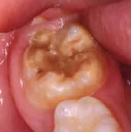
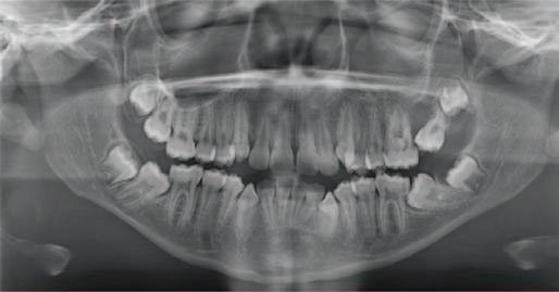
1. What condition is affecting the first permanent molar in Figure 1?
2. What is the estimated global prevalence of the condition according to the European Academy of Paediatric Dentistry (EAPD) Best Clinical Practice Guidelines?
3. List the main aetiological factors that have been linked to this condition.
4. What signs and symptoms of the condition influence clinical management of affected teeth?
Answers on page 45
FIGURE 1: What condition is affecting this first permanent molar?
Oral leukoplakia: an update for dental practitioners
Précis: This narrative review presents an evidence-based overview of oral leukoplakia, discussing its diagnosis and treatment, and the challenges involved in its management.
Abstract
Statement of the problem: Oral leukoplakia is a common mucosal pathology frequently encountered in general dental practice, which belongs to a group of conditions known as oral potentially malignant disorders. This inferred risk of progression to oral squamous cell carcinoma (OSCC) warrants an understanding of the aetiology of this condition, its clinical presentation, and how patients diagnosed with oral leukoplakia are managed in both general and specialist care practices.
Purpose of the review: To update the dental practitioner on the current understanding concerning the diagnosis and management of oral leukoplakia.
Methods: A search strategy was conducted in the MEDLINE, Ovid and Embase databases, and the Cochrane Library. No time limit was applied. The search results were limited to those in the English language.
Discussion: The aetiology for oral white patches can range considerably from innocuous frictional keratosis to OSCC. A thorough history and clinical examination should precede referrals to secondary care, with the elimination of risk factors a priority. In cases where white patches are suspicious, or remain despite managing known risk factors, prompt referral to a specialist centre is warranted. Despite the extent of research in this field, controversy remains in oral leukoplakia management and there is currently no agreed international consensus. Therefore, management is primarily governed by local contemporaneous guidelines, and is based on the most reliable predictor of malignant transformation: the grade of dysplasia. Despite various treatments, oral leukoplakia may still undergo transformation to malignancy.
Conclusions: General dental practitioners (GDPs) are the healthcare practitioners best placed to detect oral leukoplakia on a daily basis, given the volume of patients encountered from various backgrounds. An understanding of the causes and presentation of oral leukoplakia will allow GDPs to recognise this entity in practice, and facilitate further management and treatment in a bid to prevent transformation.
Journal of the Irish Dental Association February/March 2025;71(1):34-40
Introduction
Oral white patches are frequently seen in general dental practice and represent a wide spectrum of possible conditions, with various aetiologies and prognoses, ranging from benign, reactive mucosal disease to oral squamous cell carcinoma
Dr Brian Maloney BA BDentSc PCD Non-Consultant Hospital
Doctor
Dublin Dental University
Hospital
Trinity College Dublin
(OSCC). Oral leukoplakia is a common cause of such white patches. Oral leukoplakia is defined as a white patch of questionable risk having excluded all (other) potential causes that carries no increased risk of carcinoma.1 Oral leukoplakia is the most common oral potentially malignant disorder (OPMD)
Dr Sheila Galvin
BDentSc MFD MB BAO BCh MRCPI FFD
RCSI (OM) FRCSEng (OM)
Assistant Professor/Consultant in Oral Medicine
Division of Oral and Maxillofacial Surgery, Oral Medicine, and Oral Pathology
Dublin Dental University Hospital
Trinity College Dublin
Prof. Claire M. Healy
BDentSc MB BCh BAO FDS RCS Eng (OM), FFD RCSI, PhD
Professor/Consultant in Oral Medicine
Division of Oral and Maxillofacial Surgery, Oral Medicine, and Oral Pathology
Dublin Dental University Hospital
Trinity College Dublin
Corresponding author: Dr Brian Maloney BA BDentSc PCD E: Brian.Maloney@dental.tcd.ie
Table 1: Oral potentially malignant disorders (adapted from Warnakulasuriya et al. , 2021).1
Disorder Clinical description Source
Leukoplakia “A predominantly white plaque of questionable risk having excluded (other) known diseases or WHO Collaborating Centre, 2007 disorders that carry no increased risk for cancer.”
Erythroplakia “A predominantly fiery red patch that cannot be characterised clinically or pathologically as any other WHO Collaborating Centre, 2007 definable disease.”
Proliferative verrucous “Progressive, persistent, and irreversible disorder characterised by the presence of multiple WHO Collaborating Centre, 2020 leukoplakia (PVL) leukoplakias that frequently become warty.”
Oral lichen planus “A chronic inflammatory disorder of unknown aetiology with characteristic relapses and remissions, displaying white reticular lesions, accompanied or not by atrophic, erosive and ulcerative and/or WHO Collaborating Centre, 2020 plaque-type areas. Lesions are frequently bilaterally symmetrical. Desquamative gingivitis may be a feature.”
Oral lichenoid lesion “Oral lesions with lichenoid features but lacking the typical clinical or histopathological appearances of WHO Collaborating Centre, 2020 oral lichen planus, i.e., may show asymmetry or are reactions to dental restorations or are drug-induced.”
Oral lupus “An autoimmune connective tissue disease, which may affect the lip and oral cavity, where it presents WHO Collaborating Centre, 2020 erythematosus as an erythematous area surrounded by whitish striae, frequently with a ‘target’ configuration.”
Dyskeratosis congenita “A rare cancer-prone inherited bone marrow failure syndrome caused by aberrant telomere biology. It is characterised clinically by the presence of the diagnostic triad of dysplastic nails, lacy reticular skin Ballew and Savage, 2013 pigmentation and oral leukoplakia.”
Oral submucous fibrosis “A chronic, insidious disease that affects the oral mucosa, initially resulting in loss of fibroelasticity of World Workshop on Oral Medicine the lamina propria and, as the disease advances, results in fibrosis of the lamina propria and the V (Kerr et al., 2011) submucosa of the oral cavity along with epithelial atrophy.”
Actinitc keratosis
“A disorder that results from sun damage and affects exposed areas of the lip, most commonly the vermillion WHO Collaborating Centre, 2020 /cheilitis border of the lower lip, with a variable presentation of atrophic and erosive areas and white plaques.”
Palatal lesions in “White and/or red patches affecting the hard palate in reverse smokers, frequently stained with nicotine.” WHO Collaborating Centre, 2020 reverse smokers
Oral graft vs host “Clinical and histopathological presentations similar to oral lichen planus in a patient developing an WHO Collaborating Centre, 2020 disease autoimmune, multi-organ complication after allogenic hematopoietic cell transplantation.”
(Table 1). The term OPMD refers to any oral mucosal abnormality that is associated with a statistically increased risk of developing oral cancer.1 Given the risk of malignant transformation associated with leukoplakia, it is imperative that general dental practitioners (GDPs) recognise oral leukoplakia and understand the appropriate management of this condition.
Aetiology
The development of oral leukoplakia appears to be multifactorial in nature. However, the definitive cause is unclear. Smoking has been identified as the predominant risk factor, with oral leukoplakia six times more common in smokers.2 Alcohol is recognised as an independent risk factor for oral leukoplakia.3 However, its aetiological role is less clear in oral leukoplakia than in OSCC. Oral leukoplakia also arises in non-smokers and non-alcohol drinkers, suggesting a potential genetic predisposition.4 Betel quid is a significant aetiological factor in Southeast Asia and is responsible for the increased prevalence of oral leukoplakia in this region.
Epidemiology
While reported rates of oral leukoplakia vary among different geographic regions and demographical groups, a recent systematic review and meta-analysis reported a pooled prevalence of 4.11% globally.5 Oral leukoplakia is more commonly seen in men and is increasingly common with age.
Clinical presentation
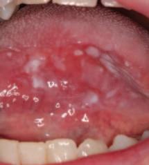
Oral leukoplakia can affect any part of the oral mucosa, either as solitary or multiple white patches. The sites most commonly affected include the lateral and ventral tongue, buccal mucosa, and floor of the mouth, the latter site being frequently affected in populations with a high prevalence of smoking.6
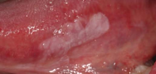
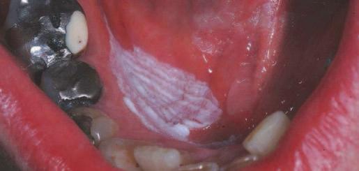
Oral leukoplakia may be subclassified into homogeneous and non-homogeneous forms. Homogenous oral leukoplakia (Figure 1) is characterised by a predominantly flat, uniform, often well-demarcated white patch, with a consistent surface topography, and it usually lacks symptoms.1 When homogenous oral leukoplakia is found on the floor of the mouth, it can have a distinctive ebbing tide appearance (Figure 2).
The non-homogeneous form is any white patch that deviates from the above.
FIGURE 1: Homogenous oral leukoplakia on the lateral border of the tongue.
FIGURE 2: Oral leukoplakia involving the floor of the mouth with characteristic ebbing tide appearance.
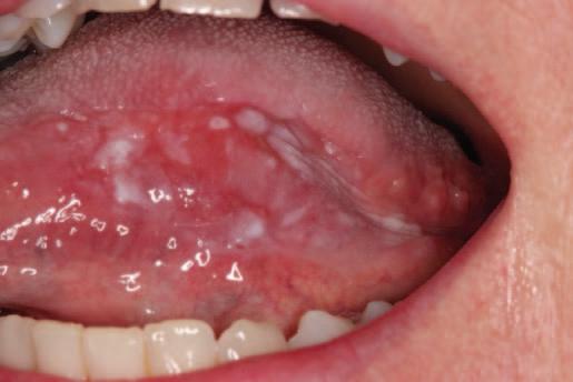
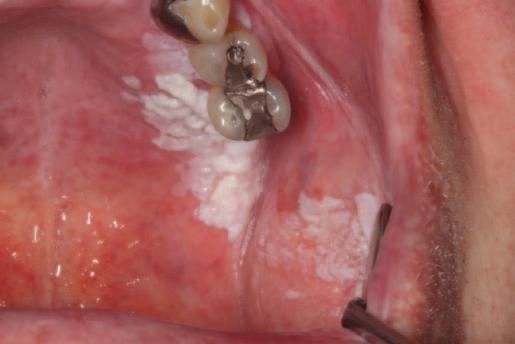
Non-homogenous oral leukoplakia should be regarded with significant suspicion as it carries a higher risk of malignant transformation than homogenous oral leukoplakia.7 There are several diverse clinical presentations including erythroleukoplakia (Figure 3), which is defined as a mixed white and red patch, but retaining a predominantly white colour. Non-homogenous oral leukoplakia may show focal superficial ulceration and the margins can be more diffuse. Nonhomogenous oral leukoplakia with red or ulcerated areas can be symptomatic. Finally, proliferative verrucous leukoplakia (PVL) (Figure 4) is a rare form of oral leukoplakia, characterised by an exophytic, wrinkled, corrugated surface. PVL commonly affects the gingivae, is often multifocal, and is most commonly found in elderly females.
Diagnostic procedures
History and clinical assessment
White patches suggest the classic differential diagnostic spectrum, ranging from benign hyperkeratosis to frank carcinoma. Therefore, the first stage in managing a white patch is a comprehensive history and examination to rule out other more common causes (Table 2). Assessment should begin with a history of the patch itself, including its onset and evolution, as well as any associated symptoms, including any pain or bleeding from the site. Patients should be asked about any skin, genital, scalp, or nail involvement, and about joint or muscle pain, which could indicate lichen planus or lupus as possible causes.
An up-to-date medical and social history is essential to identify risk factors for oral leukoplakia. This should include questions relating to a family history of white patches or oral cancer, genetic conditions, and immunosuppression. The medication list should be reviewed to identify if the patient is taking any drugs that can elicit lichenoid reactions. Patients should be asked about past or current smoking habits and, if current smokers, they should be asked details about previous quit attempts, including number of attempts, duration of quit periods, smoking cessation aids used, and triggers for resumption of tobacco. Similarly, alcohol consumption should be quantified, and the use of betel nut queried, as appropriate.
A thorough intra- and extraoral examination is necessary. All cervical lymph nodes should be palpated in case carcinoma is already present and has spread to involve regional lymph nodes. The size and site of the patch should be inspected and noted, as well as its homogeneity. Palpation to assess texture is important, as induration (hardness on palpation) or tethering to tissue planes
Developmental
■ Leukoedema
■ Fordyce granules
■ Darier’s disease
Inflammatory
■ Lichen planus
■ Lichenoid reaction
Trauma
■ Frictional keratosis
■ Mucosal burns
■ Scarring
Infectious
■ Acute pseudomembranous candidiasis
■ Chronic mucocutaneous candidiasis
■ Chronic hyperplastic candidiasis
■ Oral hairy leukoplakia (EBV)
■ Squamous cell papilloma (HPV)
■ Syphilitic leukoplakia on the dorsum of the tongue
Neoplastic
■ Oral squamous cell carcinoma (OSCC)
Idiopathic
■ Leukoplakia
■ Proliferative verrucous leukoplakia
Iatrogenic
■ Graft versus host disease
Tobacco-induced
■ Tobacco-related leukoplakia
■ Stomatitis nicotina (smoker’s palate)
FIGURE 3: An extensive, non-homogenous leukoplakia showing intermixed red and white areas involving the left lateral border of the tongue. This appearance is also termed erythroleukoplakia.
FIGURE 4: Proliferative verrucous leukoplakia (PVL) involving the hard palate and alveolar ridge.
Table 2: Differential diagnosis of oral white patches.
Table 3: World Health Organisation (WHO) criteria for epithelial dysplasia (2017).
Architectural changes
■ Irregular epithelial stratification
■ Loss of polarity of basal cells
■ Drop-shaped rete pegs
■ Increased number of mitotic figures
■ Abnormal superficial mitosis
■ Premature keratinisation in single cells
■ Keratin pearls within rete pegs
■ Loss of epithelial cohesion
Cytological changes
■ Abnormal variation in nuclear size (anisonucleosis)
■ Abnormal variation in nuclear shape (nuclear pleomorphism)
■ Abnormal variation in cell size (anisocytosis)
■ Abnormal variation in cell shape (cellular pleomorphism)
■ Increased number and size of nucleoli
■ Hyperchromasia
are early signs of carcinoma, requiring urgent referral. An assessment of the entire oral mucosa and dentition is necessary to identify any other white or red patches, and to identify any possible sources of trauma that could be inducing frictional keratosis. Finally, photographic documentation is essential for monitoring changes in the patch and to aid in triaging patients referred to specialist care.
The clinical findings along with the history must be interpreted by the practitioner to decide the next stage of management. This will most likely involve referral to secondary care. A detailed referral letter using institutional referral proformas, if available, and including clinical photographs of the oral leukoplakia, should be sent to facilitate appropriate triage at the specialist centre. If there is an obvious source of trauma from adjacent teeth or a denture, this should be addressed first, and the area reviewed two to three weeks later. Referral to secondary care should proceed if there is no improvement in the white patch. If there is no obvious cause, then a referral should be made immediately. Of note, white patches on edentulous alveolar ridges are generally due to friction when chewing and can be monitored in general dental practice, only requiring referral if atypical in appearance.
GDPs should also address potential risk factors and encourage smoking cessation and alcohol reduction, if applicable, directing patients to available support services, e.g., HSE Quitline. In this regard, the HSE provides online training to all healthcare professionals in Ireland (Making Every Contact Count; MECC), which provides tools for brief interventions in health promotion.8
Upon receipt of a referral for an oral leukoplakia in a specialist unit, the patient will be assessed and will likely proceed to biopsy. Histological examination is important, firstly to exclude other conditions that can present as a white patch (e.g., lichen planus, chronic hyperplastic candidiasis), and secondly to determine the presence and degree of epithelial dysplasia. Usually, an incisional biopsy is carried out from the most clinically suspicious area of the patch, which will usually correlate with the most severe histological findings. If the oral leukoplakia is small, however (e.g., <5mm), an excisional biopsy may be performed.
Table 4: Red flag signs and symptoms that warrant concern according to NICE guidelines.
■ Non-healing ulcer >2 weeks, yellow with red rolled borders
■ Exophytic growth (lump)
■ Non-homogenous leukoplakia
■ Erythroplakia
■ Induration
■ Tethering of tissue planes
■ Tooth mobility (in the absence of periodontal disease elsewhere)
■ Failure of healing of socket following extraction
■ Pathological fracture
■ Cervical lymphadenopathy
On histopathological examination of oral leukoplakia, hyperkeratinisation is always present, which is responsible for the white appearance of the patch. However, epithelial atrophy and hyperplasia may also be evident. The pathologist will look for the presence of oral epithelial dysplasia (OED), which is a disturbance in the differentiation of the epithelium. Several classification systems have been proposed over the last two decades in an attempt to standardise the reporting of OED. A three-tier grading system proposed by the World Health Organisation (WHO) is used to grade OED as mild, moderate, or severe (Table 3). The most recent iteration of this system has some notable changes, including an absence of the terms ‘squamous hyperplasia’ and ‘carcinoma in situ’, the latter term now being used synonymously with severe OED. Occasionally, an early OSCC is identified on biopsy and then the patient will proceed to definitive cancer management.
Malignant transformation of leukoplakia
Oral leukoplakia is an OPMD and, as such, a common question from patients following a diagnosis of leukoplakia is the risk of transformation into OSCC. The likelihood of malignant transformation cannot be reliably predicted and is patient specific. The pooled proportion of malignant transformation of leukoplakia is estimated at 9.8%.9 While no single factor can reliably determine the risk of carcinoma, several have been highlighted as important predictive factors:
Dysplasia grade
The degree of dysplasia is regarded as the most important determinant for progression to invasive carcinoma,10 with the risk of malignant transformation increasing with increasing degrees of OED.9 Warnakulasuriya et al. (2011) reported 10-year malignant transformation rates (MTRs) of 4.8% and 26.7% for mild dysplasia and severe dysplasia, respectively.11
Non-homogeneity
The presence of redness or nodularity in an oral leukoplakia is associated with a greater risk of developing carcinoma9 and these are red flags for GDPs to be aware of, as they require urgent referral (Table 4). PVL in particular has a significantly increased 10-year MTR of 49.5% compared to homogenous oral leukoplakia.12
Site
Floor of the mouth, ventral and lateral tongue, and soft palate oral leukoplakias are at increased risk of malignant transformation compared with those on the buccal mucosa.9
Size
Malignancy
Referred and managed by OMFS
No potential risk factors to address
Provisional diagnosis of OLK (based on clinical findings and exclusion of other causes)
Referral to oral medicine/oral surgery/oral maxillofacial surgery (OMFS) unit
Confirmation of other pathology
Revise diagnosis and treat accordingly
Elimination of risk factors, including tobacco (observation 2-4 weeks)
No response following risk factor cessation
No/mild dysplasia
Confirmation of OLK
Management in oral medicine/oral surgery/OMFS
Conservative management:
■ manage risk factors
■ re-biopsy (in cases of clinical deterioration)
Routine monitoring for up to five years in specialist centre in conjunction with primary care, followed by discharge to primary care where appropriate
Oral leukoplakias greater than 200mm2 are at greater risk of developing carcinoma.9
Female sex
While oral leukoplakia is more common in males, oral leukoplakias in females are more likely to undergo malignant transformation.6,9
Age
The incidence of malignant transformation increases with age.9
Smoking status
Paradoxically, some studies have found that oral leukoplakias in non-smokers are at increased risk of malignant transformation.13
Regression
Smoking-induced leukoplakia:
■ surveillance ■ maintenance of risk factor cessation
Moderate/severe dysplasia
Management in oral epithelial dysplasia clinic
Treatment as per no/mild dysplasia, plus surgical excision/ablation (where feasible)
Close, long-term surveillance at regular 3-6 monthly intervals in conjunction with primary care
16
Management of leukoplakia
General considerations
Currently, no consensus exists on the management of oral leukoplakia, and the management algorithm is dictated by a combination of patient, clinical, and histological factors, through a shared decision-making process. The British Association of Head and Neck Oncologists (BAHNO) Head and Neck Cancer Multidisciplinary Management Guidelines offer recommendations for the management of oral leukoplakia.14 This protocol emphasises the use of histological assessment and cessation of risk factors, as well as surgical excision where permissible. A simplified algorithm can be found in Figure 5
The primary objective for treatment is the prevention of transformation into OSCC, considering the poor prognosis of OSCC and the considerable morbidity associated with its management. While there is an elevated risk of OSCC developing from oral leukoplakia, many leukoplakias do not progress to
FIGURE 5: Management algorithm for oral leukoplakia (adapted from the Liverpool Management Algorithm for the management of OED)
Biopsy
carcinoma. Unfortunately, it is not possible to reliably predict which patches will undergo malignant transformation and which will not. Therefore, management must be based on individual risk assessment. Given that the degree of OED is at present the most reliable predictor of malignant transformation risk, this is the primary consideration in risk stratifying oral leukoplakias and patients.
Low-risk oral leukoplakias
Oral leukoplakias that demonstrate no/mild OED can often be managed conservatively. This involves addressing known risk factors for malignant transformation, i.e., alcohol and smoking. If a patient has other risk factors, however, the risk may need to be reassessed. Photographs at each review are essential to monitor the evolution of the oral leukoplakia. Surgical removal of areas may be advocated if the dimensions are small.15
High-risk oral leukoplakias
Risk factors must be addressed in patients with oral leukoplakia showing moderate/severe OED, and excision is generally advocated if feasible. Several methods are available. Laser ablation and cryosurgery are not advised, however, as they have the significant disadvantage of causing tissue destruction, so there is no specimen available for histopathological examination. An important factor that must be considered when deciding if surgery is the best option is whether the morbidity likely to arise from the surgery is justified.16 A part or the entirety of the area can be removed, a decision that is often dictated by the size and site of the oral leukoplakia, and the functional status and wishes of the patient. Multifocal oral leukoplakia poses a particular challenge in this regard. A significant advantage of surgical removal of the area includes the benefit of providing the entire specimen for histological analysis, with one study identifying foci of OSCC in 7% of excised oral leukoplakias.17 While surgical excision is the treatment of choice in the presence of moderate or severe OED, 10-35% of cases demonstrate recurrence post excision, particularly if there is dysplasia present at the excision margins or persistence of risk factors.18
While some medical treatments, such as the use of agents either topically or systemically (e.g., anti-inflammatory agents) have been shown to result in oral leukoplakia improvement or resolution, adverse effects and relapse after discontinuation of treatment are exceedingly common.19
Surveillance and follow-up
Patients must be informed that progression of oral leukoplakia to malignancy can occur and that recurrence of oral leukoplakia following excision is possible. Patients should be taught how to perform oral self-checks regularly and to report any changes associated with the oral leukoplakia. Long-term follow-up is advocated, which may even be lifelong.20 GDPs therefore play an important role in the ongoing surveillance of these patients. There are no strict guidelines on the frequency of follow-up for patients with oral leukoplakia, with periodicity most often dictated by the degree of OED on biopsy. Although there is little evidence to indicate that follow-up of patients with OED has any influence on preventing the development of cancer, regular review facilitates early detection of OSCC in the event of malignant transformation, which is associated with less destructive surgical treatment and a significantly improved long-term prognosis. Given that it may take up to ten years for oral carcinoma to develop from oral leukoplakia, long-term follow-up is considered best practice, at three- to 12-month intervals, depending on the degree of OED and other risk factors.14
Repeat biopsy/biopsies should be carried out in the event of a clinical change in the oral leukoplakia, e.g., development of ulceration, or induration, which could indicate the development of an OSCC. There is no evidence for serial monitoring biopsies in the absence of clinical change.
Challenges with leukoplakia
Oral leukoplakia rarely presents with any symptoms, which can lead to delayed diagnosis. While clinical examination of the oral mucosa has a high sensitivity for the detection of oral leukoplakia (93%), its specificity can be as low as 31%.21
Meticulous screening of all patients for oral leukoplakia and recognising the need to refer to secondary care are essential to allow for early diagnosis, addressing of modifiable risk factors, and further specialist management.
The cessation of tobacco habits and alcohol consumption can result in the resolution of oral leukoplakia and therefore the risk of oral cancer. It has also been shown to reduce the risk of recurrence post excision and the risk of postoperative infection.22 Difficulties with adherence to cessation programmes are a concern for the recurrence or progression of oral leukoplakias, with up to 30% of patients failing to sustain smoking cessation long term.23
Patients with oral leukoplakia are also at increased risk of developing OSCC in other parts of the oral cavity and upper aerodigestive tract due to field change. Field change refers to the concept that cells within the same region have been exposed to the same environmental and genetic influences as the mucosa where the oral leukoplakia has arisen, and so have an increased susceptibility to dysplasia/OSCC. The full extent of the field, however, cannot be assessed histologically,24 and it is not feasible to remove what is clinically normal mucosa throughout the region.
Despite our understanding of the predictive factors associated with the malignant transformation of oral leukoplakia, based on our current knowledge there are no objective measures to determine an individual’s likelihood of transformation to OSCC. Additional challenges arise with the management of multifocal oral leukoplakia, i.e., PVL. Its extensive involvement and persistent spread render it difficult to completely excise. There is also a high rate of recurrence and transformation to OSCC, as mentioned previously, warranting frequent reviews and repeat biopsy procedures in the event of clinical change.
Conclusion
Oral leukoplakia, a common OPMD that is usually asymptomatic, is likely to be encountered in general dental practice. Its clinical importance is derived from its association with the development of OSCC, a disease with high morbidity and mortality. The main strategy in the management of oral leukoplakia is early diagnosis, the addressing of modifiable risk factors, regular surveillance, and surgical management, as dictated by the clinical situation. GDPs are the healthcare practitioners best placed to detect oral leukoplakia, given the volume of patients from various backgrounds encountered daily. An understanding of the aetiology and presentation of oral leukoplakia will place GDPs in a better position to detect this entity, address risk factors, refer appropriately, and participate in its long-term surveillance.
References
1. Warnakulasuriya S, Kujan O, Aguirre-Urizar JM, et al. Oral potentially malignant disorders: a consensus report from an international seminar on nomenclature and classification, convened by the WHO Collaborating Centre for Oral Cancer. Oral Dis 2021;27(8):1862-1880.
2. Freitas MD, Blanco-Carrión A, Gándara-Vila P, Antúnez-López J, García-García A, Gándara Rey JM. Clinicopathologic aspects of oral leukoplakia in smokers and nonsmokers. Oral Surg Oral Med Oral Pathol Oral Radiol Endod. 2006;102(2):199-203.
3. Maserejian NN, Joshipura KJ, Rosner BA, Giovannucci E, Zavras AI. Prospective study of alcohol consumption and risk of oral premalignant lesions in men. Cancer Epidemiol Biomarkers Prev. 2006;15(4):774-781.
4. Khan Z, Khan S, Christianson L, Rehman S, Ekwunife O, Samkange-Zeeb F. Smokeless tobacco and oral potentially malignant disorders in South Asia: a systematic review and meta-analysis. Nicotine Tob Res. 2017;20(1):12-21.
5. Mello FW, Miguel AFP, Dutra KL, et al. Prevalence of oral potentially malignant disorders: a systematic review and meta-analysis. J Oral Pathol Med. 2018;47(7):633-640.
6. Warnakulasuriya S, Ariyawardana A. Malignant transformation of oral leukoplakia: a systematic review of observational studies. J Oral Pathol Med. 2016;45(3):155-166.
7. Warnakulasuriya S, Johnson NW, van der Waal I. Nomenclature and classification of potentially malignant disorders of the oral mucosa. J Oral Pathol Med 2007;36(10):575-580.
8. O’Brien M, Scott A. Making every contact count: a health and behaviour change framework and implementation for health professionals in the Irish health service. Health and Wellbeing Division. Health Service Executive (HSE), 2016. https://www.hse.ie/eng/about/who/healthwellbeing/making-every-contactcount/making-every-contact-count-framework.pdf
9. Aguirre-Urizar JM, Lafuente-Ibáñez de Mendoza I, Warnakulasuriya S. Malignant transformation of oral leukoplakia: systematic review and meta-analysis of the last 5 years. Oral Dis. 2021;27(8):1881-1895.
10. van der Waal I. Oral potentially malignant disorders: is malignant transformation predictable and preventable? Med Oral Patol Oral Cir Bucal. 2014;19(4):e386-e390.
11. Warnakulasuriya S, Kovacevic T, Madden P, et al. Factors predicting malignant transformation in oral potentially malignant disorders among patients accrued over a 10-year period in South East England. J Oral Pathol Med. 2011;40(9):677-683.
12. Iocca O, Sollecito TP, Alawi F, et al. Potentially malignant disorders of the oral cavity and oral dysplasia: a systematic review and meta-analysis of malignant transformation rate by subtype. Head Neck. 2020;42(3):539-555.
13. Paglioni MP, Khurram SA, Ruiz BII, et al. Clinical predictors of malignant transformation and recurrence in oral potentially malignant disorders: a systematic review and metaanalysis. Oral Surg Oral Med Oral Pathol Oral Radiol. 2022;134(5):573-587.
CPD questions
To claim CPD points, go to the MEMBERS’ SECTION of www.dentist.ie and answer the following questions:
1. Which feature of oral leukoplakia is regarded as the most important determinant of progression to malignancy?
l A. Site
l B. Size
l C. Degree of dysplasia
l D. Gender
l E. Smoking status
14. Kanatas AN, Fisher SE, Lowe D, Ong TK, Mitchell DA, Rogers SN. The configuration of clinics and the use of biopsy and photography in oral premalignancy: a survey of consultants of the British Association of Oral and Maxillofacial Surgeons. Br J Oral Maxillofac Surg. 2011;49(2):99-105.
15. Diz P, Gorsky M, Johnson NW, et al. Oral leukoplakia and erythroplakia; a protocol for diagnosis and management. 2011.
16. Field EA, McCarthy CE, Ho MW, et al. The management of oral epithelial dysplasia: The Liverpool algorithm. Oral Oncol. 2015;51(10):883-887.
17. Holmstrup P, Vedtofte P, Reibel J, Stoltze K. Oral premalignant lesions: is a biopsy reliable? J Oral Pathol Med. 2007;36(5):262-266.
18. Gilvetti C, Soneji C, Bisase B, Barrett AW. Recurrence and malignant transformation rates of high grade oral epithelial dysplasia over a 10 year follow up period and the influence of surgical intervention, size of excision biopsy and marginal clearance in a UK regional maxillofacial surgery unit. Oral Oncol. 2021;121:105462.
19. Lodi G, Franchini R, Warnakulasuriya S, et al. Interventions for treating oral leukoplakia to prevent oral cancer. Cochrane Database Syst Rev. 2016;7(7):Cd001829.
20. van der Waal I. Historical perspective and nomenclature of potentially malignant or potentially premalignant oral epithelial lesions with emphasis on leukoplakia – some suggestions for modifications. Oral Surg Oral Med Oral Pathol Oral Radiol 2018;125(6):577-581.
21. Watabe Y, Nomura T, Onda T, et al. Malignant transformation of oral leukoplakia with a focus on low-grade dysplasia. Journal of Oral and Maxillofacial Surgery, Medicine, and Pathology. 2016;28(1):26-29.
22. Vladimirov BS, Schiodt M. The effect of quitting smoking on the risk of unfavorable events after surgical treatment of oral potentially malignant lesions. Int J Oral Maxillofac Surg. 2009;38(11):1188-1193.
23. Burris JL, Studts JL, DeRosa AP, Ostroff JS. Systematic review of tobacco use after lung or head/neck cancer diagnosis: results and recommendations for future research. Cancer Epidemiol Biomarkers Prev. 2015;24(10):1450-1461.
24. Gomes CC, Fonseca-Silva T, Galvão CF, Friedman E, De Marco L, Gomez RS. Interand intra-lesional molecular heterogeneity of oral leukoplakia. Oral Oncol 2015;51(2):178-181.
2. What is the average rate of malignant transformation of severe oral dysplasia?
l A. 1%
l B. 5%
l C. 15%
l D. 26%
l E. 50%
3. Which of the following conditions is not an OPMD?
l A. Oral leukoplakia
l B. Oral lupus
l C. Oral lichenoid reaction
l D. Leukoedema
l E. Erythroplakia
An inflammatory odontogenic cyst (unusual case): case report
Précis: Inflammatory odontogenic cysts may develop from the presence of dental anomalies such as enamel pearls or cervical enamel extension.
Abstract
Different types of inflammatory lesions, such as odontogenic cysts, can affect the oral and maxillofacial regions. The conventional aetiology of inflammatory periapical lesions is triggered by dental caries or traumatic injury to the teeth, leading to the death of the dental pulp and subsequent bacterial infiltration of the periapical area. In rare cases, inflammatory odontogenic cysts may originate from non-inflammatory causes, such as the presence of enamel pearls located within the furcation of teeth with multiple roots. This case report illustrates a case of an asymptomatic inflammatory cyst associated with the upper left first molar in a young female patient. The cyst developed without any obvious clinical explanation and appeared in a typical sound tooth without evidence of pathology-related conditions. The radiographic findings demonstrated a notably aggressive nature not atypical for inflammatory cysts. The cyst expansion resulted in significant displacement of the adjacent upper left second molar, causing it to deviate from its original position within the dental arch. Additionally, the inferior border of the maxillary sinus was displaced in a superior direction. The diagnosis of non-specific inflammatory cyst was confirmed via histopathological examination. In this particular case, it is important to acknowledge that no singular factor is attributed to the cyst development. However, it is worth noting that the tooth in question is associated with enamel pearls, which have been discussed in the field of oral and maxillofacial pathology as a relatively uncommon cause of inflammatory cyst development.
Journal of the Irish Dental Association February/March 2025;71(1):41-45
Introduction
According to the WHO classification (4th edition, 2017), odontogenic cysts of inflammatory origin are classified into radicular cysts including residual cysts or inflammatory collateral cysts. The latter is also subclassified into paradental cysts and mandibular buccal bifurcation cysts.1
In a retrospective study conducted to determine the range of diagnosed odontogenic cysts with age, researchers analysed 55,446 specimens to determine the prevalence of diagnosed odontogenic cysts. Out of the total specimens, 7,121 (12.8%) were identified as having odontogenic cysts.2 The predominant diagnostic category was the radicular cyst, constituting 52.3% of all odontogenic
Ahmed Ata Alfurhud
BDS MMedSci Oral Pathology
DClinDent Oral Surgery
Oral Surgery, Institute of Dentistry, Faculty of Medicine and Dentistry,
Queen Mary University of London, UK
Institute of Dentistry, Al Jouf University, KSA
cysts (3,724 cases). Adults exhibited a higher prevalence of radicular cysts (3,359 cases), comprising 52.6% of the overall cases, compared to paediatric patients (241 cases) at 43.6%. Residual cysts made up 8% of the total cases (573), with 555 cases found in adults, indicating a higher occurrence in adults than in paediatric patients. A total of 402 paradental cysts were observed, with a twofold higher incidence in adults (5.9%) compared to children (2.7%).2 Buccal bifurcation cysts have a higher prevalence among paediatric populations, primarily manifesting in the buccal region with the eruption of the first molar.3 Radicular cysts are chronic inflammatory lesions developed pathologically by inflammation to proliferate the epithelial rests of Malassez, and they are
Sheelah Harrison
BDS PhD
Consultant Oral Surgery
Institute of Dentistry, Faculty of Medicine and Dentistry
Queen Mary University of London UK
Corresponding author: Ahmed Ata Alfurhud E: ha21916@qmul.ac.uhk
Mohammed Alshammari
BDS DClinDent Oral Surgery
Oral Surgery, Institute of Dentistry, Faculty of Medicine and Dentistry
Queen Mary University of London UK
Prince Sattam Bin Abdulaziz University, KSA.
associated with non-vital teeth. Radiographically, the cyst is characterised by an unilocular, well-defined corticated periapical radiolucency associated with the apex of non-vital teeth.4,5
Histologically, the central lumen is supported by a fibrous connective tissue wall and lined with non-keratinised stratified squamous epithelium. Inflammatory cell infiltrates ranging from mild to heavy are frequently observed, with fluid or cellular debris filling the lumen.4
The term ‘inflammatory collateral cyst’ refers to cysts that develop on the buccal
surface of the roots of partially or recently erupted teeth due to peri-coronal tissue inflammation.1 Paradental cysts are associated with partially erupted mandibular wisdom teeth with a history of pericoronitis, whereas mandibular buccal bifurcation cysts develop on the lower first or second molars.1
This report presents a detailed analysis of a very uncommon occurrence of an inflammatory odontogenic cyst that probably originated from an enamel pearl located in the furcation of the upper left first molar (UL6) in a young patient.
Case report
A 14-year-old girl was referred to the Orthodontic Department at Royal London Dental Hospital (RLDH) with crowding in her permanent canines, which required orthodontic treatment. The patient reported to the Department of Oral and Maxillofacial Surgery at RLDH with a chief complaint of a large radiolucent lesion attached to the UL6. On extra-oral examination, nothing abnormal was discovered and the intra-oral examination of UL6 revealed that the tooth was sound with no evidence of pathology, but demonstrated non-vitality with application of Endo Frost Spray. Pre-operative clinical photographs are shown in Figure 1
Radiographic examination
An orthopantomogram (OPG) (Figure 2) showed an enamel pearl measuring up to 5.7mm by 5mm lying within the furcation of UL6 with a large, well-defined corticated radiolucency lesion measuring up to 45mm by 43mm attached to the apices of the UL6. The radiolucency overlapped the left maxillary sinus and extended superiorly. The upper left second molar (UL7) was mesioangular, abutted to the distal aspect of the UL6, and was severely displaced due to the cyst development.
Cone-beam computed tomography (CBCT) showed a large, well-defined corticated radiolucency extending beyond the superior edge of this volume (Figure 3B). The radiolucency filled the left maxillary sinus and displaced the sinus floor superiorly. The radiolucency was centred on UL6, with an enamel pearl lying within its furcation, as shown in Figure 3D. There was a dehiscence of the alveolar crest between the crowns of UL6 and UL7 associated with slight buccopalatal expansion, as shown in Figure 3C. The UL7 was mesioangular, and abutted the distal aspect of UL6 at the approximate level of the cemento-enamel junction (CEJ), as shown in Figures 3A and 3B
FIGURE 1: Pre-operative clinical photographs.
FIGURE 3A-D: Cone-beam computed tomography of the left maxilla.
FIGURE 2: Full peri-operative orthopantomogram.
Frontal view.
Mandibular arch.
Lateral view (right).
Maxillary arch.
Lateral view (left).
A. Buccal view.
D. Coronal view.
B. Sagittal view. C. Axial view.
FIGURE 4: Postoperative clinical photographs
Differential diagnosis
The clinical and radiographic findings were suggestive of a differential diagnosis as follows: (i) radicular cyst or large periapical granuloma; (ii) collateral inflammatory cyst; or, (iii) odontogenic keratocyst. The first stage of the treatment plan was surgical extraction of UL6 with cyst enucleation and histopathological examination, to confirm the diagnosis and monitor the eruption of UL7 and the bone levels around it. Ultimately, after reviewing the benefits and risks of these procedures, the patient and her mother consented to have all surgical procedures performed in a single appointment under general anaesthesia.
Management and intervention
One of the challenges was preserving the UL7. During the procedure, the UL6 extraction was difficult due to the existence of the enamel pearl engaged in the buccal bone, which necessitated surgical extraction of UL6 to preserve UL7. The surgery started with the surgical extraction of UL6 with a three-sided flap and the UL6 was sectioned into many pieces so that it could be extracted without applying additional force on the adjacent UL7, (Figure 4). The cyst was enucleated with special care for the maxillary sinus. Subsequently, UL6 and the cyst pieces were sent for histopathology examination to confirm the diagnosis. Despite the presence of a substantial bone defect at the surgical site, UL7 was eventually saved in its position.
Macroscopic and microscopic findings
Hematoxylin and eosin (H&E) staining demonstrated multiple, brown-coloured soft tissue pieces. Microscopic examination showed fibrous and granulationwalled cyst fragments lined in part by non-keratinised stratified squamous epithelium with a dense, patchy, mixed inflammatory infiltrate. The histological examination confirmed that the lesion was an inflammatory odontogenic cyst with non-specific features.
Discussion
The process of definitively diagnosing oral lesions may be challenging due to the presence of some lesions that closely resemble others. Therefore, the correlation between clinical and radiographic findings and microscopic features is vital to confirm the diagnosis and formulate a treatment plan for such lesions. The typical pathogenesis of inflammatory periapical lesions is initiated by caries or dental trauma, which results in pulp necrosis and bacterial invasion of the periapical region. This may progress into an acute or chronic dentoalveolar abscess or the formation of a periapical granuloma or radicular cyst.6 Paradental cysts are often seen in cases when mandibular wisdom teeth have only partially erupted and had an underlying diagnosis of pericoronitis. In these instances, the
inflammatory response triggers the proliferation of the reduced enamel epithelium or the sulcular or junctional epithelium, leading to the formation of the cyst lining.
In the literature review, a number of terminologies are used to describe the same lesion, including paradental cyst, infected buccal cyst, and buccal bifurcation cyst. From a histological perspective, these entities exhibit histopathological characteristics similar to radicular cysts, but they are identified as pericoronal in terms of their morphological and radiographic features.7
Extremely rarely, cervical enamel extension, also known as enamel pearl, can develop along the surface of dental roots, particularly molars, and may be one of the rare causes of inflammatory cysts.8,9 Enamel projection into the furcation of molars is one of the uncommon causes of this type of cyst, but it is of note that the source of the epithelium in such cases could be derived from the apically directed epithelial attachment and post-eruptive expansion of the reduced enamel epithelium during the tooth eruption.5 Therefore, this must be associated with a source of infection to proliferate the lining epithelium, which contributes to the formation of the cyst. Scientific evidence has shown that bacterial plaque is the main component responsible for the development and advancement of periodontal disease. This is particularly true when combined with anatomical factors like ectopic enamel, which contribute to advanced localised periodontal destruction,10 and are associated with a predisposition to attachment loss.11
The enamel pearl can be considered a risk factor that contributes to the development of inflammatory cysts rather than being the direct cause. This is because the presence of an enamel pearl increases the likelihood of periodontal disease progressing at the furcation of teeth with multiple roots within the course of the epithelium, which derives from the apically directed epithelial attachment and post-eruptive expansion of the reduced enamel epithelium. There is a lack of literature on this issue; no high-quality study has discussed the risk of inflammatory cyst development for such reasons in as our case, and no case report has described a cyst that develops from enamel pearls. Moreover, there is confusion regarding the terminologies for collateral inflammatory cysts and what has been discovered is only related to paradental cysts raised in partially erupted third molars or buccal bifurcation cysts raised by periodontal diseases. In future dental examinations, it may be reasonable to consider the presence of enamel pearls as a risk factor for the development of inflammatory cysts. Furthermore, it may be concluded from the present case report that regular monitoring may be necessary for such instances to detect any early cystic-like changes and prevent the need for invasive jaw surgery.
Conclusion
Inflammatory odontogenic cysts are less prevalent in paediatric populations compared to adults. They are often present with clinical indicators including
carious dentition, retained roots, failed root canal therapy, or a previous history of dental trauma resulting in necrotic pulp infiltration into the periapical area. Nevertheless, the occurrence of cysts in sound teeth is rare. The identification of enamel pearls located at the furcation of multi-rooted teeth may indicate a potential aetiological factor in the formation of inflammatory cysts, as discussed in the context of oral and maxillofacial pathology, which is considered to be rather infrequent. It is important to note that no case report like the present case has been identified, and the underlying disease process lacks a definitive aetiology for discussion.
Acknowledgement
Our greatest appreciation goes to our colleagues, who provided unconditional support during the preparation of this report. We would like to acknowledge the Orthodontic Department at RLDH and staff nurses for their assistance.
References
1. EI-Naggar AK, Chan JKCG, Randis JR, Takata T, Slootweg P.J. WHO Classification of Head and Neck Tumours (4th ed.). IACR: Lyon, 2017.
2. Jones AV, Craig GT, Franklin CD. Range and demographics of odontogenic cysts diagnosed in a UK population over a 30-year period. J Oral Pathol Med 2006;35(8):500-507.
3. Martin L, Speight PM. Odontogenic cysts. Diagnostic Histopathology. 2015;21(9):359369.
4. Arce K, Streff CS, Ettinger KS. Pediatric odontogenic cysts of the jaws. Oral Maxillofac Surg Clin North Am. 2016;28(1):21-30.
6. Pimenta Carvalho S, Estrela C, Franco EV. Clinical differential diagnosis between nonodontogenic and endodontic radiolucent lesions in periapical location: a critical review. Iran Endod J. 2021;16(3):150-157.
7. Silva TA, Batista AC, Camarini ET, Lara VS, Consolaro A. Paradental cyst mimicking a radicular cyst on the adjacent tooth: case report and review of terminology. J Endod 2003;29(1):73-76.
8. Cawson RA, Odell EW. Cawson’s Essentials of Oral Pathology and Oral Medicine (9th ed.). Elsevier Health Sciences, 2017.
9. Neville BW, Damm DD, Allen CM, Chi AC. Oral & Maxillofacial Pathology (4th ed.). WB Saunders, Elsevier, Missouri, 2017.
10. Askenas BG, Fry HR, Davis JW. Cervical enamel projection with gingival fenestration in a maxillary central incisor: report of a case. Quintessence Int. 1992;23(2):103-107.
11. Hou GL, Tsai CC. Cervical enamel projection and intermediate bifurcational ridge correlated with molar furcation involvements. J Periodontol. 1997;68(7):687-693.
CPD questions
To claim CPD points, go to the MEMBERS’ SECTION of www.dentist.ie and answer the following questions:
1. The most prevalent form of odontogenic cyst is:
l A. Radicular cyst
l B. Paradental cyst
l C. Residual cyst
l D. Dentigerous cyst
2. The common aetiologies of inflammatory odontogenic cysts are:
l A. Dental caries
l B. Dental trauma
l C. Failed endodontic treatment
l D. All of the above
3. One of the uncommon aetiologies of inflammatory odontogenic cysts are:
l A. Dental caries
l B. Enamel pearls
l C. Dental trauma
l D. Failed endodontic treatment
Is zirconia surface etching a viable alternative to airborne particle abrasion? A systematic review and meta-analysis of in vitro studies
D’Alessandro C, Josic U, Mazzitelli C, et al.
Objectives: This systematic review aimed to determine the effectiveness of various etching surface treatments on zirconia bond strength with the following research question: “Can zirconia etching serve as a viable alternative to airborne particle abrasion (APA) for achieving reliable bonding?”.
Data: In vitro studies comparing APA, performed with either conventional or silicacoated aluminium oxide (Al2O3) particles, with various etching protocols in terms of bonding performance, were included. The risk of bias of the included studies was assessed using the QUIN’s tool for in vitro studies. Meta-analyses were performed using RevMan; random-effects models were applied, and heterogeneity was tested using the I2 index. The significance level was set at p<0.05.
Sources: A comprehensive literature search was conducted across electronic databases, including Clarivate Analytics’ Web of Science, Cochrane Library, EMBASE, PubMed, Scopus and ProQuest.
Study selection: Fifty-four relevant articles were included in this systematic review. According to the QUIN’s tool, seven studies were rated as “high risk of bias”, 46 studies were rated as “medium risk”, and one study was rated as “low risk”. Nineteen studies were used for meta-analyses. Mostly, APA demonstrated significantly higher bond strength compared to various etching protocols (p<0.05). However, no statistical difference was found between APA and high concentrations (40-48%) of hydrofluoric acid (HF) in terms of immediate- and medium-term bond strength to resin composite (p>0.05). On enamel, an experimental hot etching solution performed significantly better than APA in short-term follow-up (p<0.05). A novel multi-acid solution exhibited significantly higher immediate shear bond strength to resin cement than APA (p<0.05).
Variable heterogeneity, ranging from low to high, was observed.
Conclusions: APA remains the surface treatment with the strongest evidence in the literature and it is usually more efficacious than zirconia etching. However, highly concentrated HF and an experimental hot etching solution have demonstrated similar or significantly higher bond strength values over time compared to APA, depending on the adhesive substrate. A recently introduced multi-acid solution (Zircos-E) needs to be further explored, especially with regard to long-term bond durability.
Clinical significance: This systematic review provides a comprehensive analysis of the existing in vitro evidence on the potential of zirconia etching and the bond
Quiz answers
Questions on page 33
1. Molar incisor hypomineralisation (MIH).
2. Recent systematic reviews and meta-analyses have estimated global prevalence at 12.9% and 14.2%.
3. Main aetiological factors:
• maternal illness during pregnancy;
• maternal smoking;
• maternal stress;
• use of certain medications during pregnancy;
• gestational diabetes and hypertension;
• prematurity;
• low birth weight;
NEW DENTAL SCIENCE
durability of resin-based materials after artificial ageing. Selecting appropriate surface treatment protocols is crucial for achieving optimal clinical outcomes.
J Dent. Published online October 5, 2024.
Effect of cervical margin relocation on marginal adaptation and microleakage of indirect ceramic restorations
Adel M, Hamdy A, Sabet A, Ebeid K.
Purpose: This study evaluated the effect of cervical margin relocation (CMR) with two different materials and contamination with haemostatic agents on the margin adaptation and microleakage of ceramic restorations.
Materials and methods: Mesial-occlusal-distal cavities were prepared in 60 human first molars and distributed to three groups (n=20) according to the margin relocation procedure. The groups were: group F – flowable composite applied in two 2mm increments; group B – bulk-fill flowable composite applied as a bulk increment of 4mm thickness; and, group C (control) – no CMR was done. Each group was subdivided into two subgroups (subgroup N – no haemostatic agent applied, and subgroup H –haemostatic agent was applied). In all groups, ceramic inlays were prepared and cemented. The samples were subjected to thermocycling (10,000 cycles). The adaptation of the cervical margin was evaluated with scanning electron microscopy (200×). Samples were then assessed for microleakage analysis with the dye penetration method. Marginal adaptation data were normally distributed and analysed using twoway ANOVA followed by Tukey’s post hoc test. Ordinal microleakage score data were analysed using cumulative link models followed by the analysis of deviance using Wald chi-square tests.
Results: Both CMR and contamination with a haemostatic agent had significant effects on the margin adaptation of the cervical margin. Group C showed the highest adaptation with no significant difference from group F. The lowest adaptation was revealed in group B with a significant difference from group C. Subgroup N (in all groups) showed a statistically higher adaptation than subgroup H. Regarding microleakage assessment, CMR had no significant effect, but haemostatic agent application showed a significantly higher microleakage score for all groups.
Conclusions: Both the CMR procedure and contamination with AlCl3 haemostatic agent had a negative effect on marginal adaptation. For microleakage assessment, only contamination with haemostatic agent negatively affected the microleakage, with no effect on the margin relocation procedure.
J Prosthodont. 2024;33(4):374-381.
• perinatal hypoxia (oxygen deprivation at birth);
• complications during delivery, including Caesarean section;
• environmental pollutants and exposure to toxins;
• nutritional deficiencies, including vitamin D deficiency; and,
• family history of MIH.
4. Signs and symptoms that influence clinical management:
• severity of the defect;
• hypersensitivity;
• aesthetic concerns;
• post-eruptive breakdown;
• caries status and extent;
• pulpal status; and,
• age and co-operative ability of the patient.
Navigating the orthodontics journey
When it comes to orthodontics, there are a number of issues to take into account to ensure a satisfactory experience for dentist and patient.
Since Covid times, with people working from home and spending much more time looking back at themselves on screens in virtual meetings, there has been a significant uptick in demand for all forms of cosmetic dentistry, including orthodontics for cosmetic reasons – the ‘Zoom boom’. Any rise in demand unfortunately carries with it a rise in the number of patients who are dissatisfied with their care. At Dental Protection, we see this especially when reviewing more elective or cosmetically driven cases where patients perceive that their expectations are not being met. We know that most orthodontic cases involve a time span that can be viewed as a patient journey, from initial visit/referral to finish/review. Along that road, Dental Protection regularly sees defined ‘pinch points’ that can lay the ground for a patient complaint. This article will go through a potential orthodontic case from start to finish, highlighting some of the common themes and issues that arise. When referring to the patient, it is assumed that this includes the parent/guardian/responsible person if the patient does not have capacity, such as a minor.
Initial presentation – taking a thorough history and having a good understanding of the patient’s presenting concerns and expectations as the starting point. This should involve taking a history of previous treatments – for example an adult seeking clear aligner treatment due to relapse of previous orthodontic treatment may signal potential compliance discussions to be had, prior to any further course of care.
Communication – effective and clear communication is paramount from the start. Building a trusting relationship starts with something as simple as open disclosure about the practitioner’s qualifications – regrettably we sometimes see that general practitioners can be the subject of a complaint where the patient reports that they were under the impression the dentist was a specialist.
Assessment – the quality and quantity of information gathered should be sufficient to evidence an assessment that is to standard, including consideration of a lateral ceph and analysis of this. Pre-treatment records such as models/scans and good-quality clinical photos are prudent practice. Are there any other dental considerations that may be impacted by treatment – for example periodontal considerations, existing recession, or previous trauma? If so, this should be carefully assessed and documented, and will serve as a baseline reference at the start of care. If there are sufficient concerns, it may be in the patient’s best interest to undergo treatment or even see a specialist to ensure dental fitness prior to orthodontic treatment.
Diagnosis – we often see cases where there is no diagnosis noted, and it is difficult to justify relatively complex treatment without a diagnosis.
Options – following diagnosis, are all reasonable options presented (including benefits/risks/limitations/costs of each) appropriate to the case? Is the option of no
treatment included? Are clear aligners going to achieve the outcome the patient expects? Is it an extraction versus non-extraction case?
Consent – a two-way conversation ensuring that the patient has a good understanding of treatment options/limitations. The obligation is on the practitioner to ensure that the patient also understands their responsibilities (compliance, oral hygiene, care of appliances, attendance, etc.). As orthodontic treatment is often elective, expensive and relatively complex, it is prudent to affirm any conversations with a written consent form that reiterates mutual understanding (signed prior to treatment) and/or a treatment letter setting out the diagnosis and proposed care.
Financial consent – what is the cost, including any known additional costs such as retainers? Who is responsible for payments? Treating minors can create an additional level of consent if a child lives with one parent, and another parent is paying for the treatment. To avoid dispute, there needs to be full clarity about who is paying for what and who is entitled to discuss the child’s care, give consent on their behalf (or withdraw it) and access their records, prior to the commencement of treatment.
During treatment – discussion of any concerns or unanticipated problems, for example a tooth that is not moving as anticipated or standards of oral hygiene that may put teeth at risk. Practitioners should work with patients to address these concerns wherever possible, in an open and transparent manner. If a tooth is not moving or is going dark, it may be in the patient’s interest to have another opinion, such as from an endodontist. This may be a difficult conversation, especially if there are additional costs. If there are difficulties finding a mutually agreeable resolution, there may be a need to reconsider the merits of proceeding. For example, if a patient is not compliant with oral hygiene measures and is at risk of causing permanent damage, it may be in the patient’s best interests for the treatment to pause or cease.
Finish – at end of the planned course of care, was there a satisfactory outcome/an outcome within the limitations discussed and agreed to at the start of treatment? This links back to options chosen and mutual understanding of limitations (consent) at the start. Dispute about this is very common in surgical cases, where patients may have chosen a non-surgical compromised approach only to be unhappy with the outcome at the end of treatment – everyone needs to be on the same page from the start.
Burnout – we sometimes see patients/families who are tired and burned out with treatment and want to finish/deband early – are they fully aware of any compromises to the outcome agreed at the start? For example, if there is still misalignment or bite discrepancies, is everyone aware and accepting of compromises before a deband?
Dr
Colm Harney Dentolegal Consultant at Dental Protection
Consent at end of treatment – this should be a reiteration of conversations had at the start, such as the need for retainers, ongoing compliance, any further potential costs (for example in case of lost or broken retainers), and spelling out what could happen in case of non-compliance (such as relapse and future need for partial/complete course of treatment again at the patient’s expense).
In summary, if we consider orthodontic care as a journey that practitioner and patient travel on together, it is best practice to ensure that everyone is on the same page on what the journey looks like, including the end point or destination, before the journey starts. Once travelling, any unexpected roadblocks or diversions should be flagged and discussed. At the journey’s end, the practitioner needs to ensure that the patient is ‘dropped off safely’ at their destination, and any ongoing mutual obligations are discussed and understood.
SITUATIONS VACANT
Associates
Associate dentist required full time, Tralee, Co. Kerry. Busy, modern practice. Computerised, OPG/CBCT scanner. Strong established books. Provate/PRSI/VHI. Two years+ experience. Full IDC registration. Replacing a departing colleague. Friendly, experienced team in place. Contact fiacloir@gmail.com
Greater Dublin: Experienced, full-time associate for a very busy expanding practice. Supportive, progressive environment. Excellently equipped, superb support staff. Endo/oral surgery experience helpful. Good long-term prospects. Own transport important. Email niall@innovativedental.com
Associate required for busy Meath practice. New clinic with excellent support staff, OPG, digital X-rays, intraoral scanners. IDC registration required. Full-time preferred, but for the right candidate can be flexible days/hours. Contact anfiacloirdeirdrejob@gmail.com
Experienced full-time associate required for busy mixed practice in south Dublin. Work visa assistance will be provided if required. Contact hrdental22@gmail.com
Dental associate position available in fully private practice in Dublin. Flexible days/hours. Excellent remuneration. State-of-the-art equipment. Specialists and hygienist on site. Friendly and experienced support staff. CBCT scanner, 3Shape digital scanner on site. Contact northdublindentalassociate@gmail.com
Dental associate position available three days/week, fully private practice in Sandymount, Dublin 4. March 2025 start. Fully computerised, digital OPG, X-rays
Classified advertisements are accepted via the IDA website –www.dentist.ie – only, and must be pre-paid. The deadline for receipt of advertisements for inclusion in the next edition is Friday, March 7, 2025. Classified ads placed in the Journal are also published on www.dentist.ie for 12 weeks.
Please note that all prices are inclusive of VAT.
Advert size Members Non-members up to 25 words ¤135.30 ¤270.60 26 to 40 words ¤161.70 ¤330.65
The maximum number of words for classified ads is 40.
If the advert exceeds 40 words, then please contact: Think Media, The Malthouse, 537 North Circular Road, Dublin 1. Tel: 01-856 1166 Fax: 01-856 1169 Email: paul@thinkmedia.ie
Please note that all classified adverts MUST come under one of the following headings:
4 Situations wanted
4 Practices for sale/to let
4 Equipment for sale/to let
4 Situations vacant
4 Practices wanted
Classified adverts must not be of a commercial nature. Commercial adverts can be arranged by contacting Paul O’Grady at Think Media.
and intra-oral scanners. Hygienist, friendly and experienced support staff. IDC registered, two years’ post-qualification experience. Contact dr.elizabeth.melvin@gmail.com
Kilkenny: Dental associate required one to two days for modern, private practice. Friendly, supportive team. Fully computerised, OPG, hygienist. Excellent remuneration for the right candidate. Contact practicemanager2024@gmail.com
General associate required full-time. Westport, Co. Mayo. Four-surgery restorative/implant practice. Computerised, CBCT/IOS scanners. Busy book, good remuneration. Private/small GMS commitment. Experience preferable but not essential. Full IDC registration. Replacing a departing colleague. Friendly, experienced team. Contact shane@mayodentalclinic.com
Dental associate position available in north Dublin, 15-minute drive from city centre. Fully private practice, large multidisciplinary team with specialists and hygienists on site. CBCT scanner, 3Shape digital scanner. Flexible days available. Experienced and friendly support staff. Contact dublindentistposition@gmail.com
Experienced dental associate required Newbridge Town. Full-time position in exceptional clinic. Full book guaranteed. Be part of a great multidisciplinary team with visiting specialists. Excellent backroom support. Cerec, in-house laboratory, digital scanner, CBCT/OPG. Please send CV to bpm.gmedical@gmail.com
South east: associate required to replace departing colleague. Full/part-time. Busy general practice. April 1. IDC registration essential. CV to info@rogersdental.ie
Experienced dental associate required. Part-time position building to full in growing clinic, Merrion Square Dental, Dublin. Cerec, microscope, biomimetic, digital scanner. Please send CV to info@merrionsquaredental.ie
Dental associate required two to four days/week in private clinic in Co. Louth. Join our excellent team with supportive clinical and clerical staff. General dentistry, cosmetic, orthodontic and facial aesthetic treatments provided. Three minutes from M1 motorway. Current IDC registration and CV required. Contact info@dunleerdental.ie
Dental associate position in a state-of-the-art, very busy, fully private dental practice. Flexibility on days. Excellent remuneration. 3Shape digital scanner and CBCT facilities available. Experienced and friendly support team. Multidisciplinary specialists and hygienist present. Mentorship available. Contact skerriesprivatedentalpractice@gmail.com
Part/full-time associate dentist required for specialist/general modern and friendly private practice. We offer great remuneration with flexible working hours and a very supportive team. Forward your CV to info@naasdental.ie
Experienced associate required in busy, modern practice, 30 minutes from M50. Excellent earning potential, great patients and support staff. Private/PRSI patients. Flexible start for the right candidate. CV and cover letter to aidan@kinnegaddental.ie
Associate required for a well-established, busy Cork City practice. Excellent earning potential, great patients, and friendly and experienced support staff. Fully private/PRSI. Eight minutes from Cork City centre. Part-time position growing to full-time. Contact info@cantydental.ie
Experienced associate required in busy, modern, state-of-the-art practice in Galway. Excellent and friendly support staff. Private and PRSI patients. Please forward on your CV to Laura at office@renmoredental.ie
Dentists
Dentist required in private practice in Athlone. Part time, one to three days a week. May progress to full-time. Send CVs to reception@mearesdental.ie
Exciting opportunity for experienced, motivated dentist to join a dynamic team in Co. Kerry. State-of-the-art equipment. Multi-award-winning practice.
Seeks associate for one to three days. Specialist interest desirable. Fifteen minutes from airport. Winner of employer of the year 2024. Contact creansusan@yahoo.co.uk
Swords Dental seeks an experienced dentist for a part-time role at our busy practice with excellent support staff. Large volume of high-end restorative and endodontic treatment and high remuneration for the right candidate. Contact colinpatricklynam@hotmail.com
Owl Dental is seeking a general dentist to join our thriving practice in Wexford Town. Competitive compensation, supportive team, and growth opportunities. Enjoy a fulfilling career in a modern facility, with a scanner and a very friendly patient base. Contact owldentaljobs@gmail.com
We are looking for a caring dentist to join our ever-expanding team at Shackleton Clinic Newbridge and Celbridge in Co. Kildare. Fully digital, great remuneration and a great patient base. Full- or part-time position will be considered. Contact shackletonclinic@gmail.com
We are seeking a skilled and compassionate dentist to join our team at Galway Dental Group. The ideal candidate will have a passion for providing excellent patient care and the ability to work collaboratively in a team-oriented practice. Contact ethna@galwaydentalgroup.ie
Experienced dentist required to join our team at Dental Suite Carlow + Rathdrum. Fully digital, great remuneration and patient base. Full/part-time position. Contact info@dentalsuite.ie
Dentist required two days a week in busy practice in Monaghan. We have a fantastic team in a modern practice. Maternity leave post starting February 2025. Contact info@lakesidedental.ie
Eden Medical seeks a passionate dentist for facial aesthetic medicine. Part-time role available in Carlow. Join our team to blend dental expertise with cutting-edge cosmetic treatments. Enhance smiles and confidence in patient-focused environment. Contact careers@eden-medical.ie
Eden Medical seeks a passionate Dentist for Facial Aesthetics in Dublin. Part-time role. Blend dental expertise with cutting-edge cosmetic treatments. Enhance confidence in a patient-focused environment with a high-demand schedule. Competitive salary. Contact: careers@eden-medical.ie
Due to exciting growth plans for 2025, we will be recruiting general dentists for various locations across Ireland. Please get in touch to register your interest to join an exciting and experienced team of dentists with a strong support team. Contact jobs@shieldsdentalclinic.ie
Dental Care Ireland Ennis – flexible part- or full-time options, strong patient books within our established modern practice are on offer. IDC registration is essential and must be eligible to work in Ireland. Friendly and experienced clinical team in place. Contact careers@dentalcareireland.ie
General dentist required part-time in our very modern surgery in Naas, Co. Kildare. Fully digital, CBCT, 3Shape intra-oral scanner. Contact hungariandentist@gmail.com Kiwi Dental is looking for a superstar to replace a departing colleague who is leaving us for foreign lands. Experience preferred. Come join our family. February start, fulltime. Contact caroline@kiwidental.ie
Cube Dental, Ennis, Co. Clare, requires experienced dentist to replace departing clinician. Full book, very busy, excellent remuneration. Full support for postgraduate training. Brand new equipment, cloud-based software, AI note taking assistance and Pearl AI. Immediate start. Overseas applicants welcome. Contact Jobs@cubedental.ie
We are looking for a motivated team player dentist to join our team at Smiles Creation Raheny, Wednesday, Thursday, and potential Saturdays. Contact soumandent@gmail.com
Specialist/limited practice
Orthodontist wanted for state-of-the-art, fully computerised practice with intraoral scanner and CBCT – 75 patients on waiting list. Multidisciplinary clinic. Maxillofacial surgeon. City centre location, near five secondary schools. Main referral centre for tech company. Contact billywaters@thenationalimplantcentre.ie
Orthodontist required. South Dublin, Blackrock and north Dublin practices available. Fully equipped. Multiple chairs. Fully digital. Private. iTero. Highly trained, experienced support staff. Very flexible working days/hours. High demand. Weekly remuneration. Contact alex@whitesmiledental.ie
Orthodontist required. Rathdrum and Donnybrook. Fully equipped. Multiple chairs. Fully digital. Private. iTero. Highly experienced support staff. Very flexible working days/hours. High demand. Contact info@dentalsuite.ie
Part-time periodontist required with flexible days/hours within our busy, fully private practice. Established patient books, excellent remuneration. Experienced and friendly support staff. Multidisciplinary team with specialists on site. CBCT scanner and 3Shape digital scanner present. Contact dentalassociatepositiondublin@gmail.com
Hygienists
Dental hygienist required for very busy state-of-the-art private practice in Tralee, Co. Kerry. Full books guaranteed. Two days per week. Fully supportive staff. Very good rate. Immediate start. Contact creansusan@yahoo.co.uk
Dental hygienist required Tuesdays in Midleton, Co. Cork. Excellent facilities and support. Large, busy practice, lovely patients and team. Please email for more information. Contact carmel@corabbeydentalclinic.ie
Dental hygienist required in Malahide, Co. Dublin. Excellent facilities and support. Well established practice, lovely patients and team. Please email cv to: cirociao4@gmail.com
Dental Care Ireland Ennis – due to growth, hygiene hours, part- or full-time flexible options, strong patient book within our established practice. Friendly and experienced clinical team in place. IDC registration essential, indemnity insurance and eligible to work in Ireland. Contact: careers@dentalcareireland.ie
Hygienist position available to replace our departing colleague who is returning to her home country. Flexible shifts. Fully private, very busy, established dental practice. Experienced and friendly supportive team. Excellent remuneration. Free parking. Multidisciplinary team with specialists. Contact northdublindentalassociate@gmail.com
Hygienist position in Sligo. Full-time position, busy, employee status. Every surgery has an intra-oral camera, screen on the chair, movies on the ceiling. To discuss please contact info@clearyfitzgeralddentalpractice.ie, or 071-914 3927. Dental hygienist required for part-time work for award-winning, established practice in Westmeath. Great patients and excellent support. Please send CV to aidan@kinnegaddental.ie
Full-time dental hygienist positions available to join our existing experienced team. Busy, modern practice with full book. Excellent and friendly support staff. Cavitron and intra-oral camera in each surgery. Contact office@renmoredental.ie
Experienced dental nurse required to join lovely team in centre of Malahide. Co. Dublin. Reply with CV to cirociao4@gmail.com
Experienced dental nursing position available with our multidisciplinary team in north Dublin. Fully private practice located a five-minute walk from the Dart station and 15-minute drive from city centre. Flexible working hours and excellent remuneration. CPD provided. Promotion opportunities. Contact northdublindentalassociate@gmail.com
Rare opportunity for dental nurse/treatment co-ordinator to join our awardwinning team in Co. Meath. Knowledge of dental software, Exact, would be desirable but not essential. Candidate must be friendly and patient focused. Start February 2025. Contact meathdentists@gmail.com
PRACTICES WANTED
Room wanted to let for intermittent use. All locations considered. Daily rate and access to OPG essential. Call 085-163 3156 to discuss.
PRACTICES FOR SALE/TO LET
Galway City: For sale/lease. Three surgeries, room to expand if needed. Longestablished, private, PRSI, plus medical cards. Principal retiring. Excellent equipment plus OPG. Good parking. Contact 086-225 4708 (evenings), or email des.kelly3@gmail.com
Room to let in south Dublin, Blackrock dental practice. Specialist or limited practice dentist only. New chair. Fully equipped. Access to OPG/X-rays. Free parking. Bright and modern. Use of reception and waiting area/WC. ¤2,000 per month. All building costs covered. Contact alex@whitesmiledental.ie
South east. Strong, private, three-surgery practice, ample room for expansion. Leasehold/freehold. Flexible options. Modern equipment, computerised, digitalised. Separate decontamination room. Long-established hygienist service. Loyal patient base, High new patient numbers, principals available for transition. Significant growth potential. Contact niall@innovativedental.com
As a member of the Irish Dental Association you can use this logo on your website and other practice material. Contact rosalba@irishdentalassoc.ie for details.
MEMBER 2025
A global view
The IDA’s International Affairs Committee represents the Association on the European and global stages.
The International Affairs Committee ensures that Irish dentists are represented at European and global levels, contributing to policy development and lobbying on issues that matter to the profession.
This involves membership of and representation at two international organisations – the Council of European Dentists (CED) and FDI World Dental Federation. Committee members attend meetings, and sit on working groups to ensure that Irish voices are heard. Dr Robin Foyle (left), a GDP based in Wexford, is the Committee Chair, reporting to Council on its behalf. He is also a Board member of CED, having just begun his second three-year term: “Membership of the Board means I’m involved in the day-to-day running of the CED. We meet in person four times a year, with additional online meetings as appropriate. IDA representatives also attend the CED’s two plenary meetings each year, in May and November, and sit on a number of CED Working Groups. The CED’s work is about political representation in Europe, lobbying on EU legislation and directives that will affect dentists across the EU, to give the profession’s perspective”.
The CED has five Working Groups (WGs): Education and Professional Qualifications; eHealth; Patient Safety, Infection Control and Waste Management; Oral Health; and, Dental Materials and Medical Devices. Robin is a member of the latter group, which is currently working to address issues arising from the Medical Devices Directive: “By 2028 everything will have to recertified, from the fabric of dentists’ chairs, to the materials we use. The WG is working to try and negotiate grandfathering for existing materials and devices that have a clear track record of safety and reliability”.
Dr Kieran O’Connor is the IDA’s representative at the FDI, which represents more than one million dentists worldwide, working to raise awareness about the importance of good oral health and its vital role in securing overall health and well-being.
Robin says that membership of these organisations has a real value for Irish dentists: “Most regulation in dentistry comes from the EU and there has been a huge increase in the last 20 years. We play our part in lobbying for sensible regulation, not overregulation, so as not to overburden dentists”.
He says they “don’t always win”, citing the decision to completely phase out amalgam, which the CED argued against, but says they are hoping for success on the medical devices issue: “We have had good access at EU level, and some good meetings”.
Membership can have a direct influence on issues of specific importance to Irish dentists, such as when Revenue proposed charging VAT on fees paid to associates: “The International Affairs Committee surveyed CED colleagues on arrangements in their countries and the IDA presented this information to Revenue. That made a difference to the final decision not to impose VAT”.
Issues like these show the value of representation, whether national or international:
“Some of these issues might be under the radar for IDA members, but if CED wasn’t there, and if it wasn’t lobbying, things could be very different”.
Meet the members
Dr Michaela Dalton
Michaela is a general dental surgeon in the HSE Dublin South region. She joined the Committee as she felt that its work is very relevant to the profession and to the IDA. “I felt it was something to which I could contribute meaningfully. We work to ensure that the IDA’s voice is heard in relation to any European or international laws or agreements that will directly affect members, e.g., Minamata or the European Medical Devices Directive. It is vital that we stay ahead of the curve, to understand legislation that may be in development and to communicate its impact to the profession and consequently to the patients for whom we care before those laws are introduced or agreements signed. The IDA’s involvement in the groups that represent the profession at European and international level is crucial to ensure that our members’ voices are heard in these discussions.”
Dr Kieran O’Connor
Kieran is a general dental practitioner in Youghal, Co. Cork, and a past President of the IDA. He is a member of the CED’s Oral Health, and Patient Safety, Infection Control and Waste Management Working Groups, and is the National Liaison Officer to the FDI. “My previous experience in various IDA roles showed me the importance of being outward looking, and I feel exchanging information with international colleagues is not just helpful, it is essential. I coordinated feedback from Irish dentists to the eight FDI Policy Statements adopted at the 2024 meeting and our feedback altered the tone and content of the adopted statements. Sharing knowledge and experience with international colleagues means that the successes and failures of oral healthcare policies and delivery systems, education programmes, advocacy, etc., across the globe can be brought back to the IDA to help us navigate current and future issues and developments.”