4 minute read
FDC2022 Speaker Preview: Erosive Tooth Wear
erosive tooth wear:
Etiology, Diagnosis, Risk Factors and Management
By Alex J. Delgado, DDS, M.S.
Dental erosion is the irreversible loss of tooth structure by a chemical process that does not involve bacteria.1 Diagnosis at an early stage may decrease the risk of wear reaching a pathological state, in which the wear requires restorative intervention.2
Enamel and root dentin begin to decalcify at pH values of 5.2-5.5 and 6.7. There are two types of dental erosive causes, intrinsic and extrinsic. The intrinsic type results from gastric acids entering the oral cavity, most often due to gastroesophageal reflux disease (GERD) or eating disorders. The extrinsic type is mainly due to the ingestion of acidic diet. A thorough medical history is essential to identify the causative factors, and treatment should never be initiated until the factors at play are under control.
Intrinsic Causes
The intrinsic causes are mainly from gastric acids, which can reach a pH value of less than 1. The prevalence of GERD in the adult U.S. population has been found to be 6-10%.3 Clinical symptoms of GERD are heartburn, non-cardiac chest pain, chronic cough and hoarseness. Dental erosion is the primary oral clinical manifestation of patients who suffer from this condition.3 Systematic reviews have reported the prevalence of dental erosion among GERD-positive patients to be in the range of 32.5-48%.5
Eating disorders, such as anorexia nervosa (AN) and/or bulimia also may cause dental erosion. Excessive vomiting, in addition to eroding the teeth, leads to dehydration and low levels of salivary flow.4
Extrinsic Causes
Extrinsic erosion is due to acidic dietary habits. Several beverages such as citrus juices, carbonated drinks, teas, wine and designer drinks have been shown to have erosive potential. The erosive potential of beverages is not only related to their pH value; beverage composition and the titratable acidity can be even more important. The consumption of these drinks has increased tremendously in the last decade.
Clinical Signs
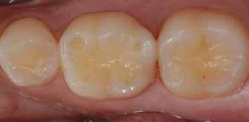
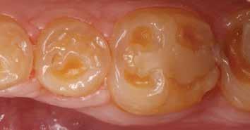
The three main clinical signs of erosion are cupping of cusps, restorations standing proud or alone, and the absence of anatomical features (Figs. 1-3). It is essential to determine the location of these clinical signs to understand the cause.
Prevention and Management of Dental Erosion
When a patient is diagnosed with dental erosion, one needs to identify whether it is intrinsic, extrinsic or both. No matter what the eating disorder the patient is suffering from, open communication between the patient and dentist must be established, using a nonjudgmental approach.4 A multidisciplinary approach is crucial to treat these patients, and the role of the dentist is to assess the case and make the appropriate referral to a psychologist, nutritionist and physician. Keep in mind that dentists often are the first health provider to identify these situations.
In case of extrinsic erosion, a diet analysis should be made, including at least two weekdays and a weekend. The patient needs to be educated about the nature of the condition and encouraged to change his/her dietary habits to a less erosive pattern. Acidic consumption should be reduced with emphasis on reducing the frequency of acidic attacks. Acidic beverages should be substituted with water, or milk ideally, or the mouth at least rinsed with water, fluoride or bicarbonate after acidic drinks.
If the posterior teeth are affected by erosive tooth wear, the vertical occlusal relationship can be raised with either direct
composite resin or bonded onlays on the affected teeth, thus gaining space to restore the anterior dentition adhesively. Evidence is supporting the use of bonded ceramic onlays, which defy traditional retention and resistance form, a very favorable treatment modality for eroded teeth. Long-term clinical evidence on this treatment modality is still being established, but short- to medium-term studies and reports have shown very good results. Numerous case reports have been published where cases of erosive tooth wear have been successfully treated using bonded ceramic onlays, palatal and facial veneers, and direct composite.6
For more severe cases, a full-coverage restoration may be the most reasonable treatment to offer. Still, our goal should always be to delay such invasive treatment for as long as possible, and whenever possible detect this condition early and employ preventive measures and minimally invasive treatment modalities to protect what is remaining.

Dr. Delgado is a clinical associate professor and director of dental continuing education at the University of Florida College of Dentistry. He will be speaking at the 2022 Florida Dental Convention and presenting two courses. On Saturday, June 25, “Conservative Approaches in the Aesthetic Zone” will be at 9 a.m. and “Erosive Tooth Wear: Etiology, Diagnosis, Risk Factors and Management” will be at 2 p.m.
References
1. Pindborg J. Pathology of Dental Hard Tissues. Copenhagen: Munksgaard; 1970.
2. Amaechi BT, Higham SM, Edgar WM. Influence of abrasion in clinical manifestation of human dental erosion. J Oral Rehabil 2003;30(4):407-13.
3. Barron RP, Carmichael RP, Marcon MA, Sandor GK. Dental erosion in gastroesophageal reflux disease. J Can Dent Assoc 2003;69(2):84-9.
4. Hellstrom I. Oral complications in anorexia nervosa. Scand J Dent Res 1977;85(1):71-86. 5. Milosevic A. Gastro-oesophageal reflux and dental erosion. Evid Based Dent 2008;9(2):54.
6. Dietschi D, Argente A. A comprehensive and conservative approach for the restoration of abrasion and erosion. Part I: concepts and clinical rationale for early intervention using adhesive techniques. Eur J Esthet Dent 2011;6(1):20-33.

Fig. 1: Cupping

Fig. 2: Restorations standing alone

Fig. 3: Loss of anatomical features, such a cusp ridges, occlusal anatomy.


