16 minute read
Decoding DNA by Next Generation Sequencing
Features
Nathan Pitt, University of Cambridge
Advertisement
Decoding DNA by Next Generation Sequencing Shankar Balasubramanian (1994)
I returned to Cambridge in early 1994, having spent a couple of years in a lab in the USA. When I started my independent laboratory in the Department of Chemistry at Lensfield Road, I set out to explore a number of different problems, each at the interface of chemistry and biology. Looking back, I might interpret this phase as either being pioneering and exploratory, or one where I didn’t really know what I wanted to pursue. Being in Cambridge it was perhaps inevitable that I would soon converge on the study of DNA, which I have stuck with ever since. My current research explores the nature of alternative folded DNA secondary structures and the natural chemical modification of DNA. Here, I will focus on some early work that led to a method for reading the DNA code very quickly, which is now widely used.
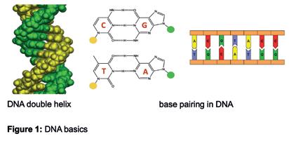
DNA DNA is a linear, natural polymer made up of four building blocks (called nucleotides) that each carry a base, often abbreviated by one of the four letters G, C, T and A. The most common structural form of DNA is a right-handed, double helix made up of two strands, which is in large part due to the molecular recognition of the bases in two well-defined pairing patterns, G recognises C to form a G:C pair and T recognises A to form a T:A pair, often called Watson-Crick base pairing (Figure 1). This pattern of recognition of DNA bases provides the
molecular basis for the genetic code and the storage, copying and transfer of genetic information. The total genetic information stored in a cell or an organism is called the genome and one copy of the human genome comprises about 3.2 billion bases of DNA arranged in a particular sequence, packaged into 23 entities called chromosomes. Part of the genome comprises units of sequence called genes that provide the information needed to synthesise a particular protein (proteins form much of the structures and machinery in cells). In fact, the vast majority of our DNA does not encode proteins and at least some of the “noncoding” DNA, sometimes referred to as “junk DNA”, plays a role in controlling the coding regions and so should not be dismissed.
To understand the information in DNA one requires a means to read the exact sequence of the letters (sequencing DNA). The first widely used approach to sequence DNA was developed by Fred Sanger in Cambridge in the late 1970s. The Sanger approach was subsequently automated, optimised and used to generate the first blueprint of a human genome in the international Human Genome Project over a period of about a decade. Today, it is possible to sequence DNA about a million times faster and more cheaply using an approach that is sometimes called Next Generation Sequencing, which arose from a basic research project being carried out in my lab in collaboration with David Klenerman in the mid 1990s. The initial work led to us forming the biotech company, Solexa, where the technology was developed into a commercial system.
Figure 1: DNA basics.
Basic Research turns to something useful Nature has evolved protein-based molecular machines, called DNA polymerases, that catalyse the synthesis of DNA faithfully to replicate it each time a cell divides into two cells. A DNA polymerase uses one existing strand of DNA (the parent strand) as a template to guide the synthesis of a new complementary strand (the daughter strand) by making use of the Watson-Crick base-pairing rules. My interest in DNA polymerases started during my postdoctoral work in the laboratory of Stephen Benkovic (1991-93), where I studied how these machines worked. One of my early projects in Cambridge (1994) involved further studies on DNA polymerases where I was attaching fluorescent probes either to the DNA or to the protein, or both, to monitor changes in structure as the polymerase incorporated building blocks, which was relayed by measurable changes in fluorescence signal. During the course of these experiments, I needed a particular laser for certain measurements. After banging on a few doors in search of a laser I was referred to another relatively new faculty member, David Klenerman, who had a treasure trove of lasers and various other pieces of optical, imaging equipment. With his help, it was a relatively straightforward matter to complete the measurements. We continued our exploratory conversation, sometimes over coffee and occasionally over a beer. I had been raised, scientifically speaking, in the disciplines of organic chemistry and biochemistry, whereas David was a physical chemist who built imaging systems and made spectroscopic measurements. We soon found common ground and wrote a proposal in 1995 to study the action of a DNA polymerase incorporating building blocks of DNA as it synthesised new DNA, guided by a template. We set out to do this at a sensitivity that could detect individual molecules, a relatively new observational capability; our intuition being that it would provide interesting new insights about this fundamentally important biochemical reaction. We were fortunate in getting this exploratory proposal funded and set about the work.
We tried many different approaches to make the measurements using various combinations of synthetic DNA template, polymerase protein, and surfaces to immobilise components together with labelling one or more of these components with a fluorophore. One format was to immobilise DNA molecules on the surface and use a fluorescently labelled building block which the polymerase would incorporate (Figure 2A). As single molecule events are stochastic and sometimes difficult to ‘catch’, we opted to parallelise by having many, single DNA molecules dispersed on the surface and imaging lots of them
simultaneously to give us the best chance of seeing something happen. During the course of these experiments, we saw the potential to adapt our work to read the sequence of DNA. If we colour-coded each of the four building blocks with a fluorophore to identify the building block (i.e. G, C, T or A) and controlled the stepwise incorporation of building blocks, then imaging the colour changes at each DNA molecule on the surface, after each incorporation cycle, would enable sequential decoding of the templating bases (Figure 2B). Specialised chemistry was required to prevent the incorporation of a second building block in any given cycle. After each imaging step to capture the colour (and base identity), the chemical group preventing the next incorporation is removed, and the colour-coding fluorophore is cleaved off, enabling the next cycle. A sample of DNA could be fragmented into pieces that are immobilised onto a surface, such that each fragment stands alone in its own imageable space (Figure 2C) and all fragments could be sequenced in parallel. A simple calculation suggested that decoding the sequence of many DNA fragments like this in parallel could lead to the scale needed to sequence a human genome (Figure 2D).
In the early phase of this project (1997), the human genome project was underway at various genome centres around the world, which included the Wellcome Trust Sanger institute at Hinxton, near Cambridge. At such research institutes arrays of automated DNA sequencers were each sequencing several hundred thousand of bases of DNA information per experimental run. The Human Genome Project was at a relatively early stage with considerably more sequencing to be done. David Klenerman and I visited and met with several of the UK leaders of the human genome project. They assured us that the project would reach completion and generate a human reference, which was important to be able to realign millions of short reads that would be generated by decoding fragments (Figures 2B, 2C) to reconstruct a human genome (Figure 3). It was evident that there would be a need to sequence many human genomes to understand the genetic basis of who we are and to improve human health through deeper knowledge of our genomes. A completely new approach to sequencing was needed. Upon sharing our thoughts and ideas, our colleagues from the Sanger Institute reinforced our drive to press on to develop the method.
We had carried out some quite basic proof of concept experiments and thought through a development plan and the various technical challenges that needed solving. This included nucleotide chemistry, engineering the polymerase protein, surface chemistry, laser spectroscopy, instrument engineering and computer/
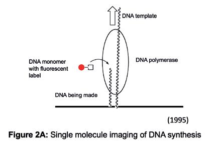
Figure 2A: Single molecule imaging of DNA synthesis.
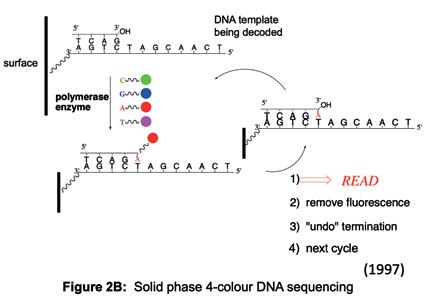
Figure 2B: Solid phase 4-colour DNA sequencing.
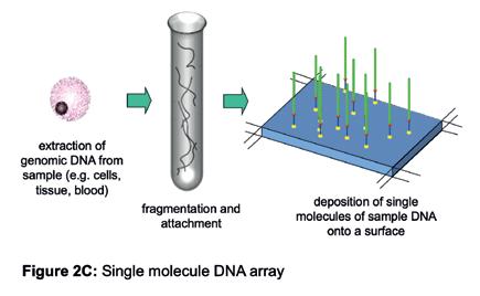
Figure 2C: Single molecule DNA array.
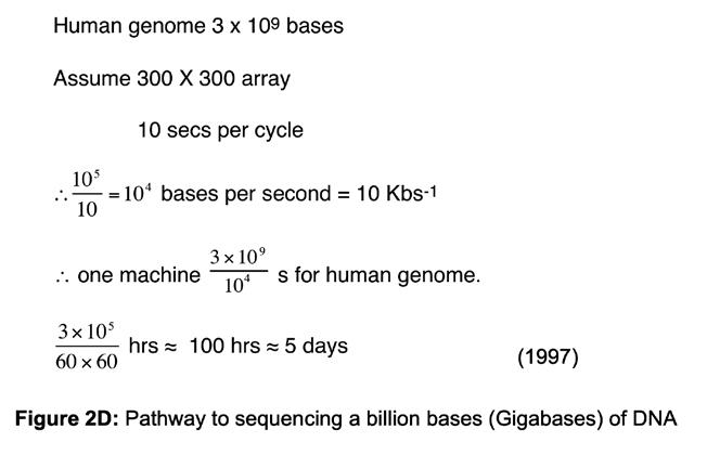
Figure 2D: Pathway to sequencing a billion bases (Gigabases) of DNA.
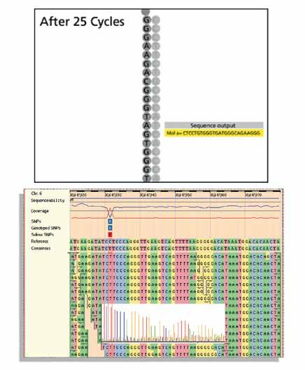
Figure 3: Sequencing and alignment of ‘reads’.
data science to develop an integrated working technology. There was much to be done that required the input of multiple disciplines. After starting to prepare another grant, we abandoned that approach and instead formed a company, which we called Solexa, as a vehicle to raise the substantial resources needed to support our development plan. We approached a London-based venture fund called Abingworth in 1997 and they decided to fund it. The venture world was new to me and certainly had its differences from academic grant funding. I recall one diligence question being, “Assuming this technology works, what is the market for human genome sequencing?”. I promptly replied, “That’s an easy one, it is zero.…”, which was certainly true at the time, although likely to change in what we saw as the future.
Solexa was formally founded in 1998 and for the first two years was incubated in our laboratories at the University Chemistry Department. By 2000, technical milestones had been hit and our investors were prepared to invest more so we moved the team and project to external premises near to the Sanger Institute to continue the technology development. I got used to regular drives down to the company whilst continuing my academic teaching and other research in the University. At Solexa a very talented team was built and we progressively reduced the various components to practise and integrated them together. One important part of the technology plan was changed. Directly sequencing single molecule fragments of DNA immobilised to a surface (Figure 2C) seemed elegant, but was practically challenging owing to stochastic events, particular to single molecule detection, that either gave false signal, or the absence of a signal. Single molecule optics were also expensive and occupied considerable space in the laboratory, whereas we were looking to package the system into an affordable, small, fridge-sized box to democratise its capabilities through enabling others. A solution was to amplify the single fragments of DNA on the surface to generate many identical copies at the same location on the surface. In a discussion with Sydney Brenner in 2002, he convinced me that this was a better way forward than continuing with single molecules. Solexa adopted a method for amplifying single DNA molecules on a surface that came from the work of Pascal Meyer and his colleagues at their biotech company Manteia. This technology was licensed and integrated well with the system developed at Solexa in 2004. By 2005 Solexa sequencing was deployed to decode a genome, albeit the small genome of bacteriophage phiX-174 (Figure 4). The first commercial sequencing system was launched in 2006 and was called the Solexa 1G Genome
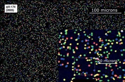
Figure 4: Image taken (at Solexa) during one cycle of sequencing. Each spot is hundreds of identical copies of a single DNA fragment. The colour indicates the base being decoded at each site in that cycle.
Figure 5: Solexa’s original technology and the present day Illumina system.
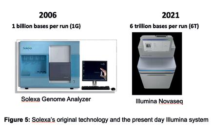
Analyzer, as it was able to read a billion bases (1 Gigabase) of DNA in a single experimental run. This was about 10,000-fold greater than the sequencing systems of 1997. In 2007, Illumina acquired the technology and continued to improve it. Present day systems that use the technology can generate several trillion bases of sequenced DNA per experimental run (Figure 5) at a rate of about 1 accurate human genome per hour on a single instrument, as compared to the decade or so taken to generate the human genome reference. An accurate human genome now costs approximately USD 600 to sequence and this cost is expected to fall further over the coming years.
What does faster, lower-cost DNA sequencing enable? A relatively small instrument sitting on a bench top in a laboratory can now provide the sequencing capacity equivalent to more than the global capacity of 20 years ago. As DNA and its sister molecule RNA are fundamental to all living entities, Next Generation Sequencing has been used very widely in life science and clinical research. The primary motivation for driving this technology was to enable improvements in health care through understanding our genomes, and so I will now focus on examples from three clinical areas.
Cancer Cancers are caused by changes (mutations) to your DNA sequence that either pre-exist at birth or are acquired post-birth. Given we each have a distinctive genome, every cancer will be genetically unique. Rapid DNA sequencing lends itself to building an understanding of common and less common genetic changes that occur in cancers and may provide insights into the mechanisms driving a particular cancer. There are already examples of particular cancers being classified by a characteristic mutation that can suggest a form of therapy likely to be more effective. For example, a mutation called the V600E in the BRAF gene, which changes the amino acid valine (V) to a glutamate (E) at position 600 resulting in overactive BRAF protein that signals uncontrolled cell proliferation to drive the cancer. Cancers diagnostically shown to carry the V600E mutation are being specifically targeted with a drug that acts by inhibiting the overactive BRAF. More complex patterns of cancer-mutations can be detected by whole human genome sequencing. A pioneering example came from a lab in Vancouver (Jones et al., Genome Biology 2010, 11:R82) where a patient with cancer of the tongue was being treated with the standard
of care therapy until the tumour had spread to the lung and became resistant to the therapy. Sequencing the normal genome of the patient, the primary tumour and the secondary tumour provided data that enabled a comparative analysis of mutations and how they might affect genes, their corresponding proteins and mechanisms that could drive proliferation of cancer cells. The information allowed the clinicians to hypothesise why the original treatment was no longer effective and make a reasoned decision to target an alternative pathway that was now driving the metastatic tumour, with a different therapy, which was able to shrink and manage the tumour for several months. Later on, the tumour again became resistant to the therapy and genome sequence analysis suggested yet another therapeutic that stabilised the tumour for a few more months. In this case, targeted treatment, informed by genome sequence information, extended the patient’s life. It also tracked the evolution of the genome of a tumour under the pressure of sequential drugs, leading to a suggestion that had it been known at the outset what was known at the end, it may have been logical to simultaneously prescribe the three different therapeutic agents in combination to manage the tumour and block pathways of resistance before they emerged. While there are promising examples, it is early days. Large-scale cancer sequencing studies are underway, such as the international Cancer Genome Atlas, and the sequencing of NHS cancer patients carried out by Genomics England. Over the next decade, vast data sets linked to patient histories will be generated and promise to reveal the extent to which genomic medicine will be helpful in routine cancer care.
Rare diseases Rare diseases collectively affect 1 in 17 people. They typically manifest in early life as a disorder that is difficult to diagnose, and are often genetic in their origin. A chain of many tests sometimes serves only to rule out known conditions, leading to continued suffering of the infant and distress to the parents, all after considerable time and expense. One approach being adopted in such cases is to sequence the whole genomes of a trio (mum, dad and child) then look to identify and interpret genetic changes unique to the child that may explain the disorder. A combination of rapid sequencing of DNA from a blood sample, computational analysis of the data and clinical interpretation by physicians is in many cases leading to an actionable diagnosis. A pioneer of this approach, Dr Stephen F. Kingsmore, is listed in the Guinness Book of Records for going from patient
sample to diagnosis in under 24 hours. In a recently reported case (N. Engl. J. Med., 384; 22, 2159, 2021) it took under 15 hours from blood sample to a clinical diagnosis of a thiamine (vitamin B1) metabolism dysfunction in a fiveweek-old infant who was then treated with a rational programme of medication, including vitamin B1, and subsequently discharged. In cases where a suitable drug is not available, identification of the underlying cause of the disease provides the basis to guide and inspire the development of a future therapy. Falling sequencing costs are making genome sequencing of rare diseases more practical and it is being implemented in paediatric clinics globally including the work of the NHS and Genomics England.
Infectious diseases Infectious agents (pathogens) have genomes made up of either DNA or RNA. They can be identified by sequencing their genomes and the sequence information will also reveal any variants of a given pathogen. An early example of the application of rapid sequencing to infectious diseases was carried out at Addenbrookes in Cambridge, led by Sharon Peacock (N. Engl. J. Med., 366; 24, 2267, 2012), where a hospital outbreak was traced to a strain of methicillinresistant Staphylococcus aureus (MRSA), a very serious pathogen that exhibits resistance to most antibiotics. The sequencing also provided valuable information about the transmission pathways of this outbreak, which was contained.
In early 2020, an RNA virus was identified as the agent responsible for the disease that would be known as COVID-19. A sample taken from the lung of a critically ill patient was subjected to rapid sequencing and though the sample was complex and comprised nucleic acids from a number of sources, a method termed metagenomic analysis, enabled the genome of the virus to be extracted and assembled computationally. The sequence information helped subclassify the virus: SARS-CoV-2. After the outbreak became a global pandemic routine sequencing of samples from patients diagnosed with COVID-19 at scale has helped identify variants of the virus and track the transmission of particular strains of the virus nationally and globally. Over four million cases of SARSCov-2 have been sequenced, with the information shared via a publicly available database (https://www.gisaid.org/). The information gives a global readout of the origins, evolution and tacking of the virus and provides data that can guide the development of optimised vaccines, particularly in response to emergent variants.
The Future I have provided a glimpse of how Next Generation Sequencing is contributing to the management of human health. We are in the early phase and I expect to see greater application of genomics over the next two decades that will reveal what is possible with greater clarity. Perhaps there will be a future where routine, asymptomatic sequencing of a blood sample will run an individual ‘system check’ to provide an early warning of various diseases, whilst also reporting on the changing environment of pathogens in which we coexist.
The opportunistic deviation from a curiosity-driven research collaboration that started some 25 years ago led to the unexpected development of a technology that has turned out to be useful. It is essential to support and carry out basic, exploratory research as it educates, informs and sometimes leads to useful outcomes. In this case, being in the Cambridge environment certainly helped.


