4 minute read
Better views
Innovative microscope system provides insights into COVID-19 spike protein
Influenza research is well-suited to physics research, because protein clustering is central to the virus life cycle. Measuring clusters in different ways and understanding how they’re evolving are critical in advancing treatment therapies.
In the last two decades, advances in super-microscopy led by the Sam Hess lab at the University of Maine have resulted in new insights — and sights — of influenza particle surface spikes. The surface structures of spike proteins are where the body’s immune response to the virus begins.
Now, in the SARS-CoV-2 pandemic, what Hess and his undergraduate and graduate students have learned about surface spikes on virus particles is contributing to our understanding of the COVID-19 spike protein. The spike protein from the influenza virus is hemagglutinin (HA); the spike protein from the coronavirus is called S. Basically, the spike proteins on the surface of the virus stick or bind to cells in the respiratory tract. Spikes also allow the virus to enter through a process called membrane fusion.
Membrane fusion depends very much on clusters of the spike protein. Hess has been studying that process of how the clusters form, looking at how the host cell might play a role in generating those clusters and investigating, ultimately, how to disrupt that process.
“These clusters are crucial for the infection process, yet nobody knows why they arise,” says Hess. “There were some theories at that time when I first came to UMaine about why clusters of viral proteins occur. Our data showed those theories to be wrong, which didn’t make me very popular. It did lead us to ask new kinds of questions.
“When the coronavirus pandemic started, I realized we’d probably have to find some new ways to fight the new virus,” he says. “There were some similarities and differences that I noticed between the SARS coronavirus and the influenza virus. I thought of using our molecular microscopes to look at similarities and differences between those two. We’ve been looking at two of the most important proteins involved in the beginning of infection — the spike proteins.”
In 2005, Hess and UMaine professor of chemical and biomedical engineering Michael Mason led the development of a breakthrough microscope system called FPALM (fluorescence photoactivation localization microscopy) to image cells with membranes that contain the HA spike protein. Prior to such super-microscopy, it wasn’t possible to create images of molecules on a small enough scale to test the biological models that predict how they may be organized. FPALM shattered the resolution limit of lens-based microscopes, known as the diffraction barrier, that had existed for more than a century.
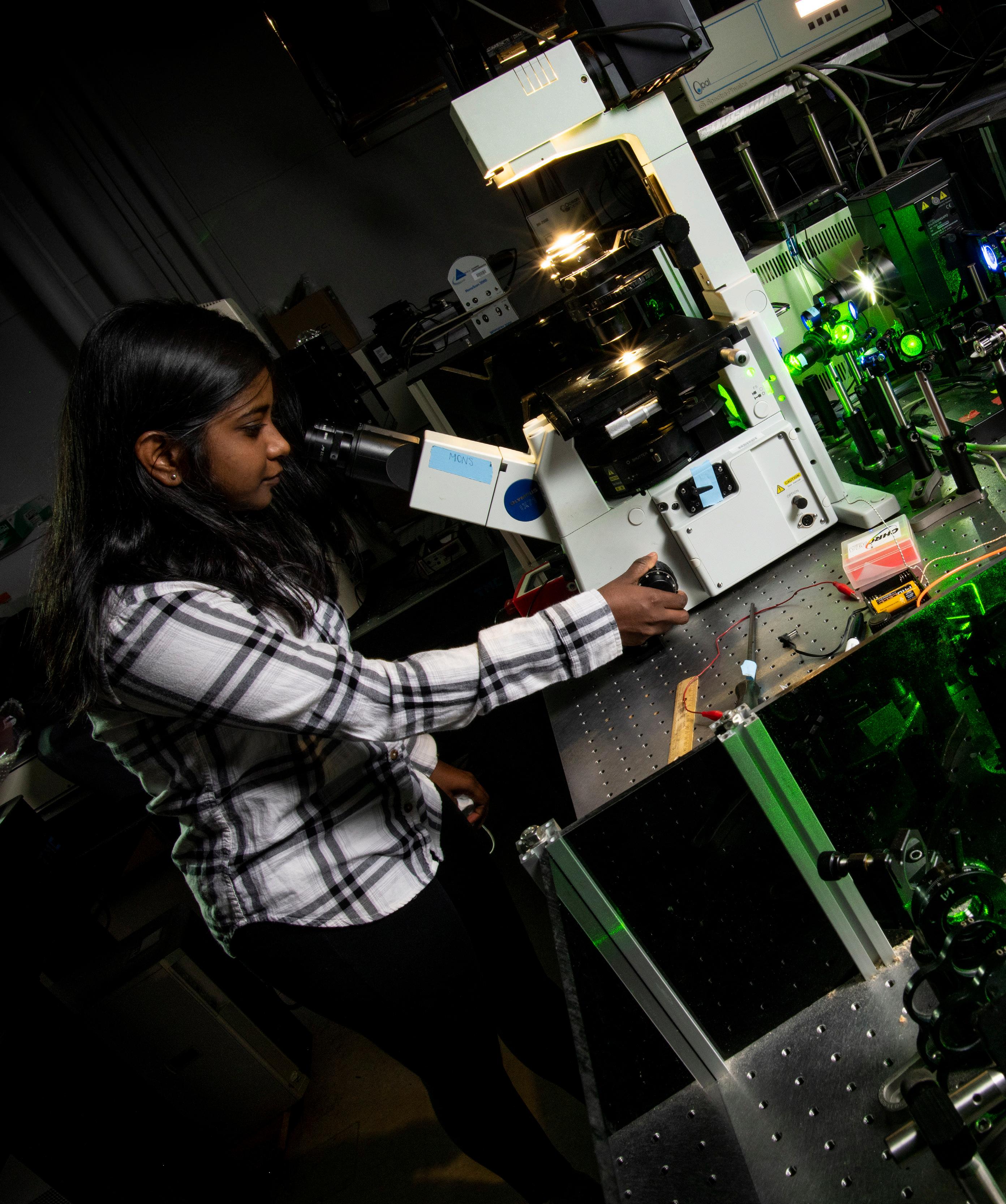
In the Hess lab, fluorescence photoactivation localization microscopy is enabling research advances in COVID-19 and influenza viruses.
The FPALM system, which uses photoactivatable dyes to identify individual molecules and separate them at the nanometer scale, was one of four groundbreaking advanced microscopy techniques that were able to achieve such singlemolecule imaging capabilities in the mid-2000s. Indeed, announcements of the 2014 Nobel Prize in Chemistry honored three recipients and cited other researchers involved in similar pioneering research, including Hess.
FPALM technology is now used in UMaine research in toxicology and muscular dystrophy. In virology, it has led to not only a better view of spike proteins, but also their cytoplasmic tail, which seems to interact with host cell components connected to signaling.
Parts of those spike proteins mutate fairly rapidly over time, interfering with the function of the immune system to recognize that structure as dangerous and attack it, says Hess. That’s one reason influenza vaccines have to be reformulated annually.
However, there’s a portion of the spike proteins — the tail inside rather than on the surface of the virus — that does not change very quickly. “That,” Hess says, “is what we’re going after.”
“People overlook this tail,” Hess says. “It’s inside the cell. It doesn’t seem to have any role in the lock and key mechanism of entry, but certain sequence elements are always there. We noticed when we expressed one of these spike proteins in a cell that there were some interactions between the tail and some of the host cell components. Then we started thinking about how we could disrupt that interaction — interfere with the function of the spike protein. Looking at that interaction and trying to figure out if there are drugs that could break that up, that’s been a thrust of our research for a few years.”
The result could be a new class of drugs that blocks this interaction between the spike protein and the host cell, disrupts those clusters of the spike proteins, and stops the virus from entering the host.
“The way viruses are mutating and the way sometimes you get breakthrough infections, having a backup (drug therapy) to help when an infection does occur is a real urgent need right now,” says Hess.


