7 minute read
Dr. Hsu’s Diverse Research
Dr. Hsu’s Diverse Research Attracts Major Funding
Center for Oral Biology
COB embraces an interdisciplinary approach.

– Wei Hsu, PhD
Wei Hsu, PhD, is surrounded by the two things he loves most. Dozens of photos of his wife and two daughters - from school portraits to smiling poses in tutus-- adorn every wall in his small office amidst the many piles of scientific journals, lab reports, paper reviews and textbooks that completely cover his desk.
Most of the piles are related to the five major projects Hsu and his team of seven are managing. Very simply, Hsu’s lab focuses mainly on the role of cells--the signals they send and the paths they take –that cause bone development and disease.
“Basically, we study the genes whose products-- the proteins-- control signal transduction pathways,” he said. “We are particularly interested in how these pathways control normal processes of mammalian development, from a single fertilized egg into a multi-organ organism-and how these pathways go amiss to cause human disease.
Hsu, whose work has attracted significant funding through the last 12 years when he first joined COB, has primary appointments in Center for Oral Biology and Biomedical Genetics, and a secondary appointment in the Wilmot Cancer Center. He is also a member of the UR Stem Cell and Regenerative Medicine Institute.
His interest in skull development first began as a young man in high school who enjoyed chemistry, and thought he would study it further in college. But the first two years of college-level chemistry in his native Taiwan was nothing like he experienced in high school. “I felt like I didn’t know chemistry anymore,” he remembered. “It felt very artificial and it didn’t make a lot of sense to me.”
Feeling depressed and unsure about his future, it wasn’t until his third year in college that things began to look brighter. “I took biochemistry and it changed my life,” he said. “The chemical reactions that naturally happen within the body were fascinating to me, and that class was a turning point in my life and the beginning of my career path.”
His interest in molecular biology and genetics landed him a full fellowship to Mt. Sinai Medical Center in NYC, where he earned his Master’s and PhD in Biomedical Sciences. Hsu did his post doc at Columbia for three years where he worked on mammalian genetics and development biology before he was promoted to research faculty. In 2002, URMC’s Center for Oral Biology recruited him for the work he did with characterization of key signaling regulators in mammalian development and cancer.
Some of his research at Columbia included studying a gene that has the same signaling pathway known to be very prominent in cancer. During his work in this area, he made a surprising discovery that the same gene also plays a role in craniofacial development and disease, which then started his current research program.
“COB was the one place I found that embraces an interdisciplinary approach,” said Hsu. “Our center has people from various disciplines, including microbiology, genetics, biochemistry, pharmacology and physiology, which creates a diverse and stimulating environment.”
He recently landed a three-year, $1.06 million grant from the New York State Stem Cell Board (NYSTEM) to continue his work related to the stem cell’s role in skull deformity. (Hsu is one of six UR School of Medicine and Dentistry scientists awarded $3.5 million in NYSTEM funding). Another grant, originally funded by NIH’s NIDCR in 2006, was competitively renewed for another five years in 2012 for $1.9 million, focusing on how the signaling pathways orchestrate to form normal or abnormal development during infancy.
He published findings in 2010 that provided a new explanation for the earliest stages of congenital skull deformity in newborns, known as craniosynostosis. Abnormal head shape due to craniosynostosis, affecting one in 2,500, can restrict normal brain growth and result in neurodevelopment delays and elevated intracranial pressure. The chief cause is a defect in osteoblasts, the type of cells most important for making bone.
“We found that when a certain type of stem cell goes awry, it leads to a new mechanism for craniosynostosis,” Hsu explained, whose study was published as the cover story in Science Signaling, and showed a new mechanism for craniosynostosis, a result of a disruption among the earliest forms of cells.
Initially, the bones that make up the cranium are individual plates of skull bone, and are separated by gaps called sutures. In humans the bone plates gradually fuse together, starting at birth and ending at about age 30. Two different types of bone formation processes take place during the first 18 months of life that are critical to the proper bone formation. The first type is responsible for final development of the skull bones, jaw bones and collar bones. The second type controls development of the long bones in the body. During the first event, a type of stem cell – the mesenchymal cell – must transform into bone forming osteoblast cells, which deposit the bone matrix. The majority of bone is made after the matrix hardens and entraps the osteoblasts.
Hsu’s group discovered when the signaling pathways that determine the fate of the mesenchymal stem cell are altered, they change to the second type of bone forming process, resulting in the skull sutures closing prematurely.
Since then, and now with the NYSTEM grant’s support, Hsu and his team have been working to identify and isolate this stem cell population and characterize it, and investigate how these stem cells contribute to craniofacial bone development and craniosynostosis.
The mesenchymal cell based therapy is a very big field, and scientists for the past 30 years have been trying to implement it in other areas of the body. “In the past, it has shown functional improvement, but unfortunately only short term or very limited,” Hsu explained. “They don’t survive and we think the wrong type of cells was used. Our hope is to find the cell that can replace the damaged tissue and directly regenerate new bone.”
If Hsu and his team are successful in identifying the right cell, it could have positive implications to improve conditions that affect the aging population, such as osteoporosis and bone fractures.
Hsu’s research strategies are also apply similar strategies in stem cell and signal pathways Apply research strategies in other areas, including breast cancer, neurodevelopment and degeneration diseases such as Alzheimer’s and Parkinson’s.

Dr. Hsu has received distinction by receiving funding through many sources, including National Kidney Foundation fellowship award, the Northeast Regional Developmental Biology award, the PHS grant awards from the National Institutes of Health, an Idea award from the Department of Defense, the Basil O’Connor award from March of Dimes Foundation, and New York State Stem Cell Science awards.
He serves on grant/external review panels for NIH, Dept of Defense, Alzheimer’s Assoc., Florida Dept. of Health, among others. He is invited to lecture at international conferences and leading research institutions, and serves on the editorial board for several scientific journals.
Dr. Hsu’s other projects include:
Breast Development and Tumorigenesis
The main focus is to investigate cellular signaling in normal development and neoplastic transformation of the mammary gland. We are particularly interested in roles of mammary stem cells and parity-induced mammary progenitors in these processes.
Stem Cell Biology
This is an integral part of our projects studying the genetic control of cellular signals and signal transduction mechanisms underlying development of lineagespecific stem cells and niches in skeletal, reproductive, skin and nerve systems.
Ubiquitin-like Modifiers

A multidisciplinary approach has been initiated to study SUMO modification in mammalian development and disease. Studies include SUMO-mediated regulation in embryonic and extraembryonic development, cardiovascular, skeletal and neurological disorders, and cancers. His project deciphers the regulatory mechanism underlying the making of Wnt and its signaling effects in signal-producing and signal-receiving cells. Our current focus is to investigate the reciprocal regulation of Wnt and Gpr177/mouse Wntless in development of various organs as well as pathogenesis of human diseases.


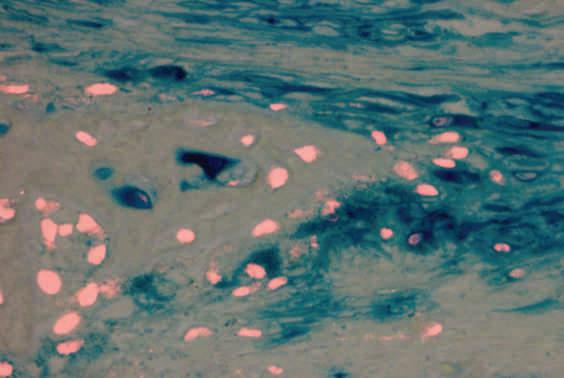
Undifferentiated mesenchymal cells are stained by beta-gal in blue. Red fluorescence is osteoblast cells identified by a specific marker. The blue cells (mesenchymal cells) will differentiate into the red fluorescence positive cells (osteoblast cells/bone forming cells) in this image.
Green fluorescence is GFP staining of undifferentiated mesenchymal cells within the skeletogenic mesenchyme. Blue fluorescence is DAPI counter staining to identify all cells.
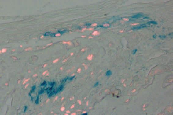
Similar to the image above, the blue represents the mesenchymal cells and will differentiate into the red positive osteoblast or bone forming cells.


