University of Rochester | Ernest J. Del Monte Institute for Neuroscience Vol. 24 - 2025
A WORLD AWAY
The island nation at the center of mercury and brain research
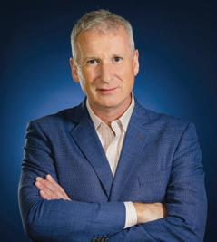
John J. Foxe, PhD
Kilian J. and Caroline F. Schmitt
Chair in Neuroscience
Director, Ernest J. Del Monte
Institute
for Neuroscience
Professor & Chair, Department of Neuroscience
FROM THE DIRECTOR’S DESK
As the daylight becomes longer again in Rochester, a new year begins, and I find myself reflecting on another incredible year—advancements in research, groundbreaking discoveries, and historical opportunities to transform clinical and translational work to improve lives and find the best answers for people with intellectual and developmental disabilities. This year marks the centennial year for the University of Rochester Medical Center (URMC), but to quote the CEO of URMC, Dr. David Linehan, “Let’s reflect a little bit on the past 100 years, but let’s focus more on what the next 100 years could be and how we’re going to be a part of the change in medicine.”
It seems a satire to have a white sand beach on the cover of our newsletter coming to you while Rochester is under a blanket of white snow. It's a juxtaposition that may seem almost cruel if icy air is nipping outside your door. But, is also an incredible reminder that our research stretches well beyond our labs and travels over continents and oceans to the most appropriate location for scientific discoveries. It is also a reminder that, as humans, we have so much to learn from each other, no matter the weather outside your front door.
I’m looking forward to all that 2025 will bring. The 2024/2025 Del Monte Institute Neuroscience Diversity Commission’s NEURO YES series kicked off, bringing postdoctoral fellows from across the country to Rochester for a mentorship opportunity before entering the job market. The Neuroscience Graduate Program retreat in April will fuel innovation and collaborations and bring together a community I am so proud to be a part of. And this is all just the beginning.
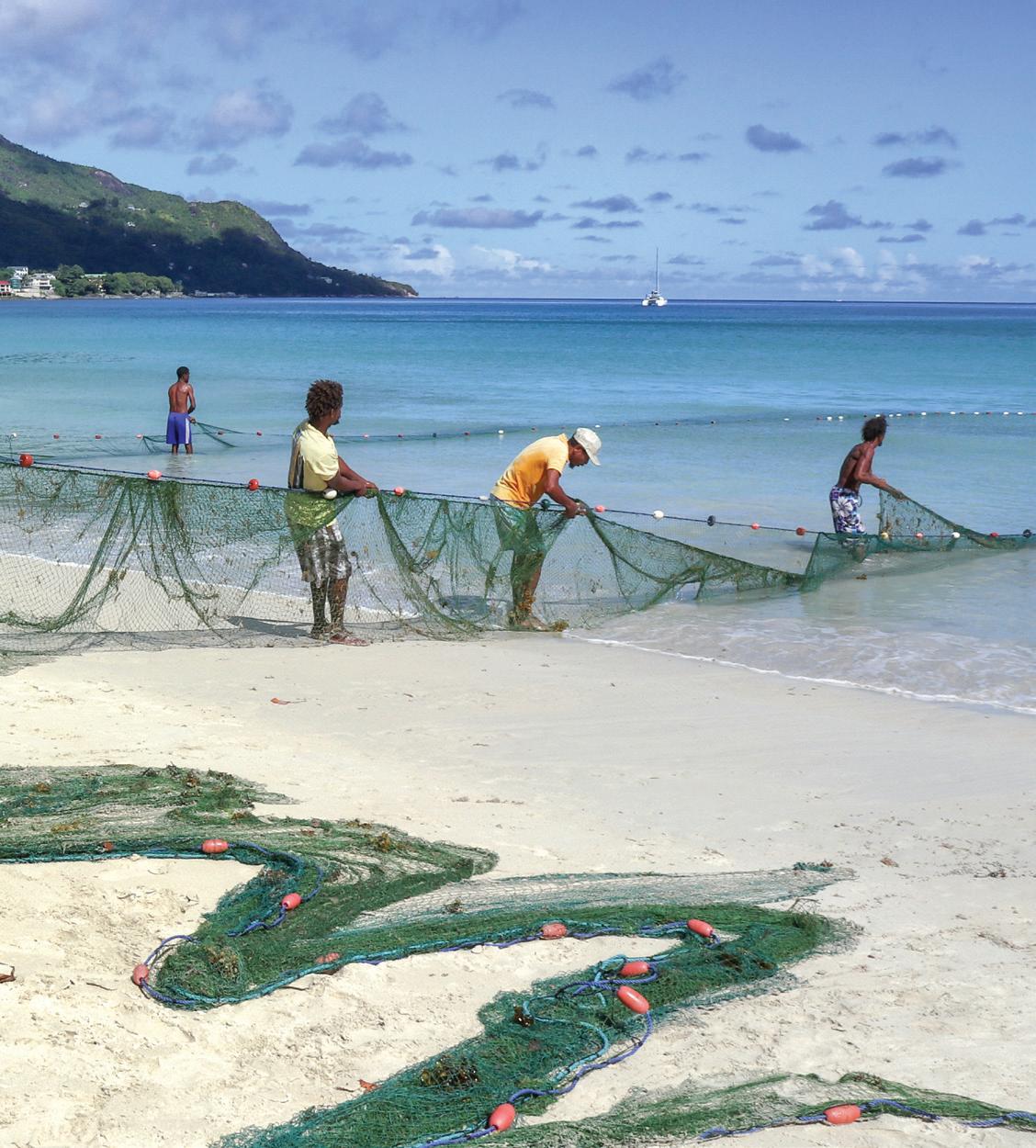
ON THE COVER:
People net fishing on a beach on Seychelles Island.
Photo by Conrad Shamlaye, MD
In Science,
John J. Foxe, PhD
Del Monte Institute for Neuroscience Executive Committee
John Foxe, PhD
Chair, Department of Neuroscience
Bradford Berk MD, PhD
Professor of Medicine, Cardiology
Robert Dirksen, PhD
Chair, Department of Pharmacology & Physiology
Diane Dalecki, PhD
Chair, Department of Biomedical Engineering
Jennifer Harvey, MD
Chair, Department of Imaging Sciences
Robert Holloway, MD, MPH
Chair, Department of Neurology
Paige Lawrence, PhD
Chair, Department of Environmental Medicine
Hochang (Ben) Lee, MD
Chair, Department of Psychiatry
Shawn Newlands, MD, PhD, MBA
Chair, Department of Otolaryngology
Webster Pilcher, MD, PhD
Chair, Department of Neurosurgery
Steven Silverstein, PhD
Professor, Department of Psychiatry
Duje Tadin, PhD
Chair, Department of Brain & Cognitive Sciences
Editor/Writer
Kelsie Smith Hayduk
Kelsie_Smith-Hayduk@ urmc.rochester.edu
Contributors
Mark Michaud
Maureen Malone
Feature Photography
John Schlia Photography
Conrad Shamlaye, MD
FISHERMAN ON SEYCHELLES ISLAND
NEUROSCIENCE
TABLE OF CONTENTS
2
NEWS BRIEFS
The latest in published research that is providing new insight into gaming addiction in teens, risk of psychosis, and how a common sleep aid may leave behind a dirty brain.
4
FEATURE: FISH, MERCURY, & THE BRAIN
For decades this large-scale study has helped informed researchers and policy makers on the right balance of fish consumption during pregnancy.
8
FACULTY Q&A
Karlo Lizarraga, MD, is an associate professor of Neurology, Center for Health and Technology, Neuroscience, and Neurosurgery. He studies the most effective treatments for the motor symptoms of Parkinson's disease.
9
STUDENT SPOTLIGHT
Aaron Huynh is a second-year in the Neuroscience Graduate Program. As a firstgeneration student, he aims to help develop better methods for movement recovery in stroke patients, while also being a mentor to the next generation of scientists.
Neuroscience Vol. 24 Winter 2025
LISTEN TO A NEW EPISODE OF NEUROSCIENCE PERSPECTIVES TODAY
Photo by Conrad Shamlaye, MD
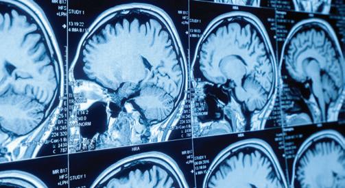
Researchers uncover possible new biomarker for psychosis diagnosis
A single five-minute scan could potentially improve the ability to predict which at-risk individuals will transition to a psychotic disorder. Brian Keane, PhD, assistant professor of Psychiatry, et al., identified a potential biomarker for psychosis, which could enable earlier diagnosis and personalized treatment. Analyzing MRI scans, they discovered that sensory regions in the cortex exhibited weaker interconnections and stronger connections to the thalamus—the brain's information relay station. These connectivity differences were specific to the somatomotor network, which is responsible for processing bodily movements and sensations, and a visual network that generates representations of objects and faces. The combined dysconnectivity patterns across these two networks form a "somato-visual" biomarker.
Gaming addiction in adolescents may be present in the brain before symptoms begin
Analyzing data from 6,143 video game users aged 10 to 15 over four years, John Foxe, PhD, director of the Del Monte Institute for Neuroscience observed that individuals exhibiting more symptoms of gaming addiction had lower brain activity in regions responsible for decision-making and reward processing during initial fMRI scans. The findings highlight that while gaming isn't inherently unhealthy, certain adolescents may be more susceptible to developing addictive behaviors. This research is part of the Adolescent Brain Cognitive Development (ABCD) Study, the largest long-term study of adolescent brain development.
Understanding the link between HIV and brain health
People with HIV, even those on effective treatment, are more vulnerable to cerebral small vessel disease (CSVD), especially as they age. This disorder can cause strokes, memory problems, and other cognitive difficulties. New research supported by the National Institute of Neurological Disorders and Stroke and led by University of Rochester Medical Center neurologist Giovanni Schifitto, MD, and cardiovascular biologist Jinjiang Pang, MD, PhD, will focus on Delta-like 4 protein (DII4) hypothesized to play a key role in the deterioration of the microscopic network of blood vessels that serve the brain.
The team believes that this research's findings could help scientists ultimately develop new treatments to prevent or treat CSVD in people with HIV.
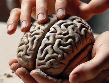
Sculpting the brain (without chisel or scalpel)
Researchers have developed a groundbreaking method to noninvasively "sculpt" brain activity patterns, potentially enhancing learning and treating neurological disorders. Led by Coraline Iordan, PhD, assistant professor of Brain and Cognitive Sciences and Neuroscience, the team employed real-time neuroimaging and neurofeedback to modify how the brain processes visual information. Participants in the study viewed abstract shapes while undergoing functional magnetic resonance imaging (fMRI). They were instructed to generate mental states that would stabilize the oscillation of these shapes, effectively aligning their brain activity with patterns predetermined by the researchers. Notably, participants achieved these neural adjustments without explicit awareness of the targeted patterns, highlighting the brain's capacity for implicit learning. These findings, published in the Proceedings of the National Academy of Sciences, suggest a promising future for noninvasive brain modulation in cognitive enhancement and rehabilitation.
(University of Rochester illustration / Prompting and editing by Sandra Knispel using DALL-E)
Brain immune cells may also be from Mars and
Venus
The function of microglia, the immune cells of the brain and central nervous system, may not be as similar across sex as once thought, that’s according to new research from the Majewska Lab at the Del Monte Institute for Neuroscience at the University of Rochester. This discovery could have broad implications for how diseases like Alzheimer's and Parkinson's are approached and studied, and points to the necessity of having gender specific research. It is already known that more women are diagnosed with Alzheimer’s and more men are diagnosed with Parkinson’s but it’s unclear why.
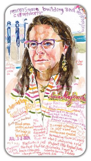
VISIT THE NEUROSCIENTIST IN COLOR
GALLERY-ANIA MAJEWSKA, PHD
“It is a fortuitous finding that has repercussions for what people are doing in the field, but also helps us understand microglia biology in a way that people may not have been expecting,” said Ania Majewska, PhD, professor of Neuroscience and the senior author of a study out today in Cell Reports that shows how microglia respond differently in adult male versus female mice when given an enzyme inhibitor to block its microglia survival receptor. “This research has a lot of ramifications for microglia biology and as a result all these diseases where microglia are important in a sex specific manner.”
Common sleep aid may leave behind a dirty brain
Research appearing in the journal Cell describes for the first time the tightly synchronized oscillations in the neurotransmitter norepinephrine, cerebral blood, and cerebrospinal fluid that combine during non-rapid eye movement sleep in mice. These oscillations power the glymphatic system—a brain-wide network responsible for removing protein waste, including amyloid and tau, associated with neurodegenerative diseases.
The study, conducted by a team from the University of Rochester and University of Copenhagen led by Maiken Nedergaard, MD, DMSc, co-director of the University of Rochester Center for Translational Neuromedicine, also explored whether sleep aids replicate the natural oscillations necessary for glymphatic function. The team focused on zolpidem, a sedative marketed under the name Ambien, which is frequently prescribed to treat insomnia. They found that while zolpidem effectively induced sleep in the mice, it also suppressed norepinephrine oscillations, disrupting the glymphatic system and impeding the brain’s waste-clearing processes, a finding that raises concerns about its long-term use.
How does what we see enhance what we hear?
Researchers are exploring how visual cues enhance the brain’s ability to understand speech in noisy environments. Edmund Lalor, PhD, associate professor of Biomedical Engineering and Neuroscience, received a $2.3 million grant from the National Institutes of Health that will support this research. It builds on a project originally started by a pilot award from the Del Monte Institute for Neuroscience. Lalor’s team will monitor the brainwaves of people who are deaf or hard of hearing and who use cochlear implants because the auditory system is especially noisy for them. Researchers hypothesize that someone who got a cochlear implant later in life may use more visual cues to get information than those who received the implant as an infant. A better understanding of how the brain processes audiovisual information could lead to better technology that aids with hearing.
Biotin may shield brain from manganese-induced damage, study finds
High levels of exposure to manganese can be toxic, particularly to the central nervous system. Chronic manganese exposure may result in a condition known as manganism, characterized by symptoms resembling Parkinson's disease, including tremors, muscle stiffness, and cognitive disturbances.
New research led by Sarkar Souvarish, PhD, an assistant professor in the Departments of Environmental Medicine and Neuroscience, employs model systems and human nerve cells to show the mechanisms by which manganese inflicts damage to the central nervous system. The study, published in Science Signaling, also suggests that the vitamin biotin may have a protective effect, potentially mitigating manganese-induced damage.
Portrait by Charmaine Wheatley
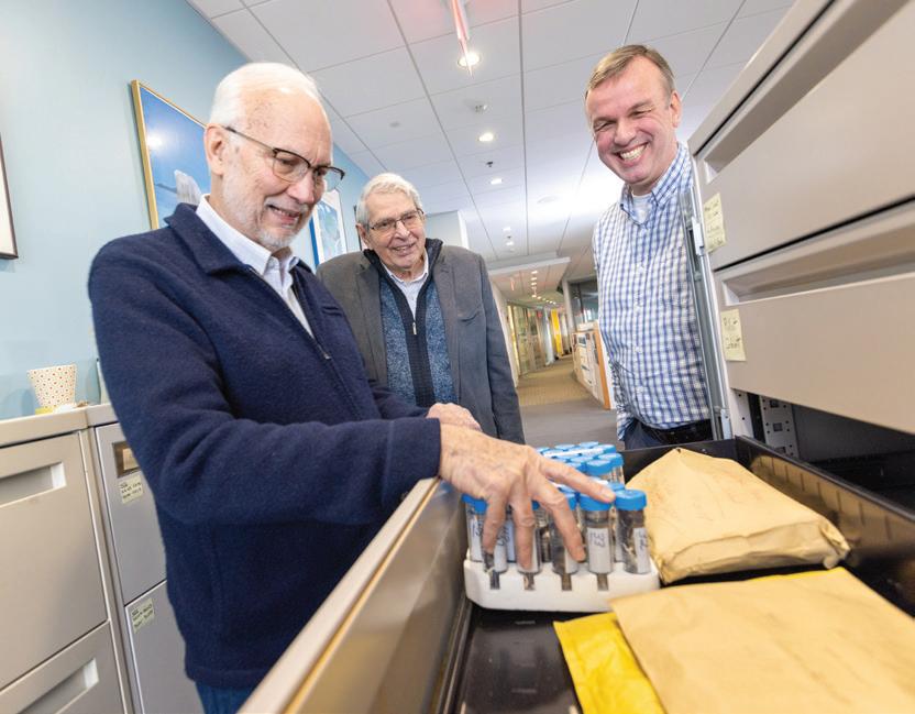
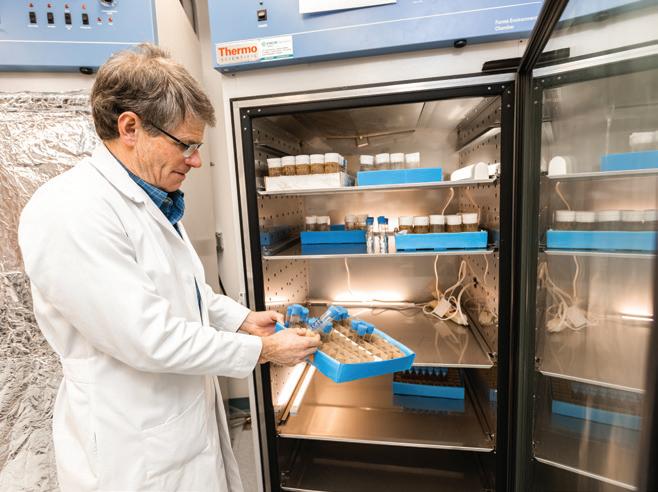
making it a robust resource for researchers studying mercury exposure.
Gene Watson, DDS, PhD (left), looks through hair samples from participants in the Seychelles Child Development Study with Gary Myers, MD, (center), and Edwin van Wijngaarden, PhD. Mercury is known to deposit in hair,
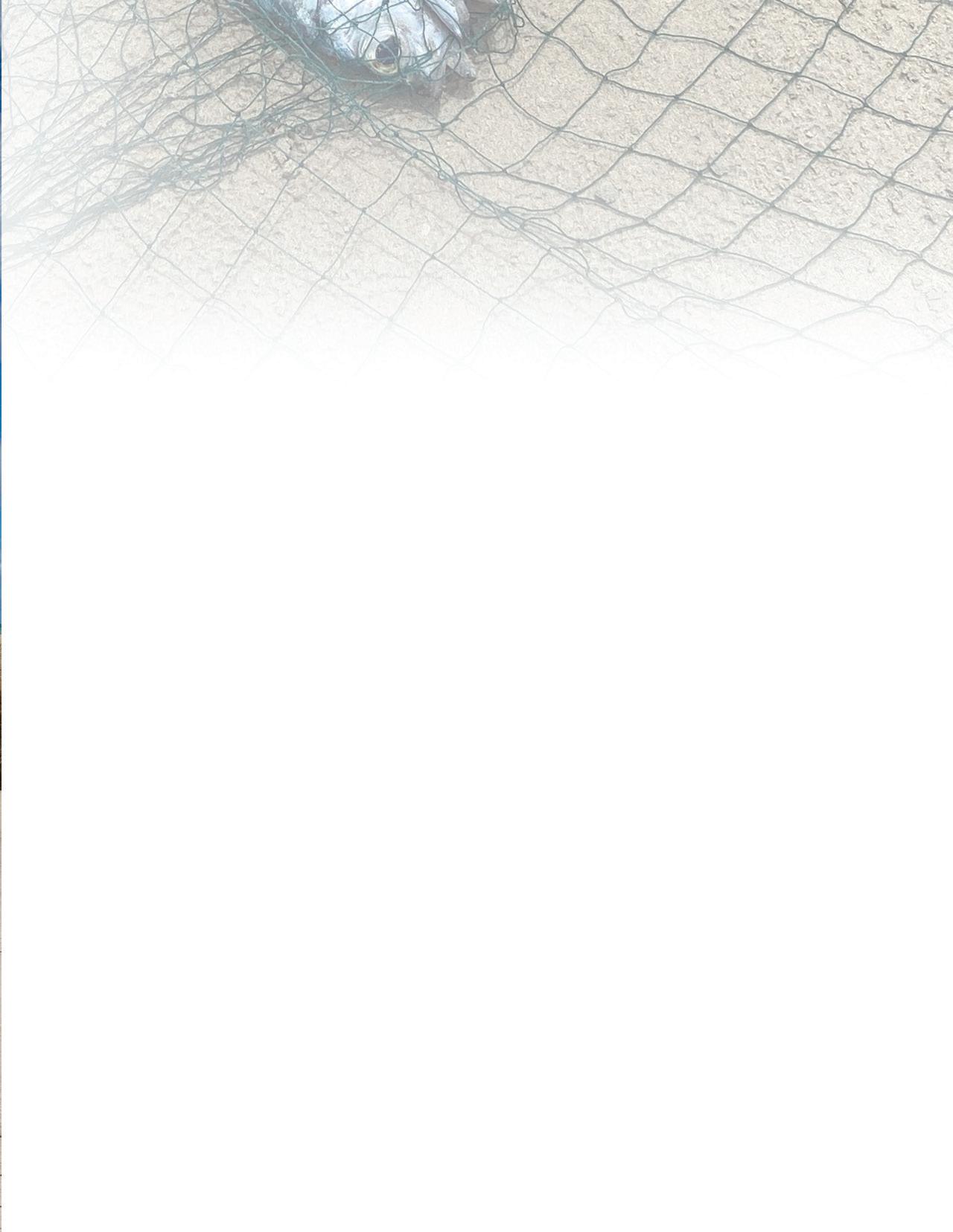
Seychelles beach. Photo by Conrad Shamlaye, MD
Fish, Mercury, and the Developing Brain DECADES OF INSIGHTS FROM THE SEYCHELLES CHILD DEVELOPMENT STUDY
By Mark Michaud
There is a growing understanding of the role of chronic low-level exposure to environmental toxicants in human diseases. Mercury, a known neurotoxicant at a high level of exposure, is among the top chemicals identified by the World Health Organization as a “major public health concern.” The presence of mercury in fish has led the U.S. Food and Drug Administration (FDA) and Environmental Protection Agency (EPA) to recommend that mothers limit fish consumption during pregnancy.
However, more than three decades of research in Seychelles, whose residents eat ten times more fish than in the U.S., has found no evidence of neurodevelopmental harm linked to mercury exposure via fish. In fact, the study suggests that the omega-3 fatty acids and other micro-nutrients found in fish, which are critical for brain development, may counteract any potential adverse effects of mercury.
“The associations between lowlevel mercury exposure, nutrients, fish consumption, and child neurodevelopmental outcomes are complex. Tackling these questions, which have global health and economic implications, has required an international team,” said University of Rochester Medical Center (URMC) epidemiologist Edwin van Wijngaarden, PhD, the principal investigator of the Seychelles Child
Development Study.
Origins in the Middle East
The story of the Seychelles study begins in Iraq. In the early 1970s, the country imported grain treated with a methylmercury-based fungicide used to prevent spoilage during storage and transport. While intended solely for planting, a combination of a severe famine and misunderstood labeling led to it being consumed, poisoning thousands. The symptoms, like ataxia, sensory impairment, tremors, and seizures, pointed to the neurotoxic effects of the chemical. Prenatal exposure caused devastating effects on unborn children, leading to outcomes such as cerebral palsy.
The Iraqi government reached out to Tom Clarkson, PhD, an expert in the toxicology of mercury at URMC, to help assess the extent of contamination and develop strategies to mitigate its impact. Clarkson was already studying mercury’s basic properties and effects, making him one of a handful of global authorities in the field. Alongside URMC neurologist David Marsh, MD, and others, Clarkson would visit Iraq twice a year for the next decade, collecting data and conducting evaluations. The team analyzed mercury levels in hair and blood samples, identified families affected by prenatal exposure, and studied the neurological impacts on infants and children born to exposed mothers.
Their findings provided further evidence that mercury exposure disproportionately affects the developing fetus, with mothers often asymptomatic while their children exhibit severe neurological impairments. The data also highlighted the need to differentiate between mercury exposure from acute poisoning events—like Iraq and an earlier episode in Minamata, Japan—and chronic, low-level exposure from dietary sources like fish. And while fish is an important source of protein for much of the world, this was not the case in Iraq.
The perfect partnership
Joined by URMC pediatrician Phil Davidson, PhD, and neurologist Gary Myers, MD, the team began searching for a location for a new large-scale study that would help researchers better understand the risk-benefit balance of fish consumption during pregnancy. Mercury is deposited in the oceans through pollution or natural events such as volcanoes, forest fires, and erosion. Bacteria transform it into methylmercury, which eventually works its way up the food chain to fish. The consumption of fish is the primary way humans are exposed to mercury. At the same time, fish are a rich source of nutrients essential to early brain development, including polyunsaturated fatty acids, selenium, iodine, and vitamin D.
The team scouted locations in South America and considered the Maldives.
By 1986, they settled on the Republic of Seychelles, an archipelago of 115 islands in the Indian Ocean, off East Africa. “The reason for going to Seychelles is because fish is consumed daily. We figured that if you’re going to find adverse effects from eating fish, it ought to appear in this population, compared to the U.S., where fish is consumed a couple of times a month,” said Myers, who served as study liaison with Seychelles from 1986 to 2019, and is now an emeritus professor of Neurology at URMC and still involved in the study.
A population with access to free universal care, national healthcare and education databases, limited industrial sources of pollution, and easy international access all made Seychelles an ideal location for researchers to recruit and follow study participants over the length of the study. This was a long time, as it would turn out. Some volunteers have been enrolled for more than three decades and counting.
Most importantly, the study had a champion in Conrad Shamlaye, MD, an epidemiologist who worked closely with the Seychelles Minister of Health. From
the start, the Seychelles government was a full partner and would be involved throughout the research process, from the study design through its implementation to the dissemination of study findings.
“The partnership meant that the study would have access to the expertise and experience of Seychelles professionals, and the results would benefit the population,” said Shamlaye. Health and Education Ministries members helped craft the study instruments and translate protocols and instruments into local language and context. The collection of additional data important to the government was integrated into the study design.
Building one of the largest longitudinal studies
The URMC team arrived in Seychelles in 1986 and started recruiting a pilot
cohort, which would ultimately consist of 759 children. Data from this cohort was the basis for a National Institutes of Health award in 1990, a grant that has since been continuously funded. Data from the pilot cohort suggested that, while there might be evidence of mercury risk, other fish nutrients may be covering up the adverse effects.
In 2000, a second group of volunteers was recruited, called the first nutrition cohort, to be followed by a larger second nutritional cohort in 2008. The researchers collected hair, blood, urine samples, and cord blood from infants, and child development was assessed regularly and over several years. In total, the Seychelles study includes 3,000 mothers and children across multiple cohorts, creating a large and unique set of longitudinal data. The oldest cohort is still being tracked after more than three decades, providing researchers with insights across the lifespan.
As the study expanded, so did the need for additional research expertise in nutrition, genetics, cardiovascular, and mental health outcomes. Teams from Ulster University, led by JJ Strain, PhD, and Karin Broberg, PhD, with the Karolinska Institute at Lund University in Sweden, and Pascal Bovet, MD, MPH, from the University of Lausanne in Switzerland, joined the study, along with the growing ranks of researchers at the University of Rochester, including biostatistician Sally Thurston, PhD, dental researcher Gene Watson II, DDS, MS, PhD, cardiologist Wojciech Zareba, MD, PhD, and audiologist Mark Orlando, PhD, MBA, among others.
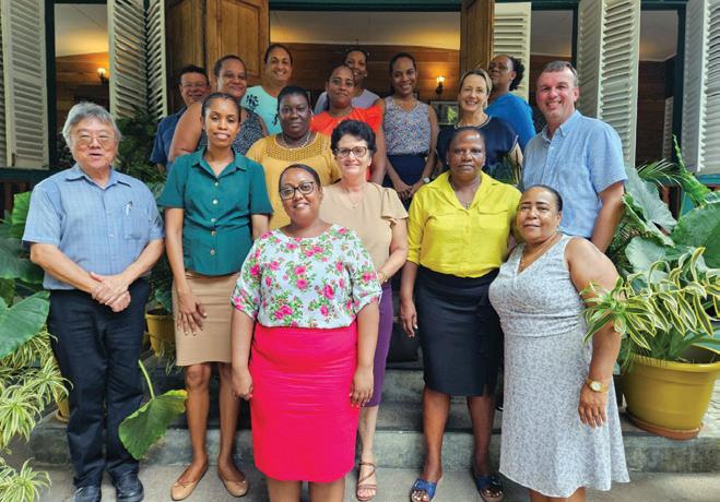
“Seychelles has never issued any advisories restricting fish consumption because of toxicological concerns."
Drs. Conrad Shamlaye (front left), van Wijngaarden and Emeir McSorley (middle right) with members of the Ministry of Health in Seychelles, including research nurses and administrative staff who are part of the study team.
Genetic and basic science insights
Matt Rand, PhD, at the time with the University of Vermont, became involved in the Seychelles study after being invited to participate in a National Institute of Environmental Health Sciences grant application in 2012. This collaboration would expand the project’s scope. Rand, who moved to the URMC Department of Environmental Medicine later that year, and his colleagues are studying the mechanisms of mercury toxicity and its effects on development, leveraging model organisms like Drosophila.
The Seychelles study has upended assumptions about mercury’s toxicity and pointed to evidence of either protective genetic factors, dietary components, or both. Researchers in the Rand Lab have been trying to untangle these questions. Their work has identified rare genetic variations that may increase mercury toxicity risk. More recent findings indicate the gut microbiome plays a role in mercury elimination, suggesting a potential target for interventions to mitigate mercury exposure risks.
“Our findings underscore the importance of considering genetic variability when assessing the risks associated with methylmercury exposure from fish consumption,’ said Rand. “They also highlight the need for personalized approaches in public health advisories, as genetic predispositions can influence individual susceptibility to environmental toxins by several-fold.”
Challenging conventional wisdom
Decades of research in Seychelles have shown no evidence of neurodevelopmental harm from prenatal mercury exposure from fish consumption. Yet, the FDA and EPA’s guidelines continue to advise expecting mothers to limit fish intake based on mercury neurotoxicity.
“Seychelles has never issued any advisories restricting fish consumption because of toxicological concerns. The overall study findings support the continuing consumption of a large variety of ocean fish, which is not only a traditional way of life but also has health benefits,” said Shamlaye.
Myers agrees and believes fish consumption advisories can be “overly cautious and counterproductive” and have been shown to discourage fish consumption despite its nutritional benefits, especially to the developing brain. In his opinion, the scientific evidence from the Seychelles study does not support their need, especially at the mercury exposure levels encountered through typical fish consumption. “Given that billions of people across the globe depend on fish for daily nutrition and malnutrition is a global problem, the wisdom of advisories is questionable.”
Lasting impact and future directions
Shamlaye points to how the study exposed the country’s health and education professionals to colleagues
in the US and Europe. The project also permitted Seychelles to contribute to international forums, including the World Health Organization and the United Nations. “Health programs for children have benefitted from additional input from the study, for example, in defining standard growth charts for Seychellois children and screening tests for child development. Study findings have also supported health, social, and education programs in Seychelles.”
The work in the Seychelles continues, and researchers plan to expand beyond mercury to explore other environmental exposures, such as endocrine disruptors, PFAs, and PFOAs. Researchers also plan to examine the role of dietary factors and exposures in aging, neurodevelopment, and epigenetics. During the course of the study, Seychelles faced rapid social and economic development, leading to changing consumption patterns towards more meat and less fish, increased calorie intake, and less physical activity. This new research will further our understanding of lifespan health outcomes linked to fish consumption and other environmental factors.
Matt Rand, PhD, checks drosophila samples in lab at URMC.
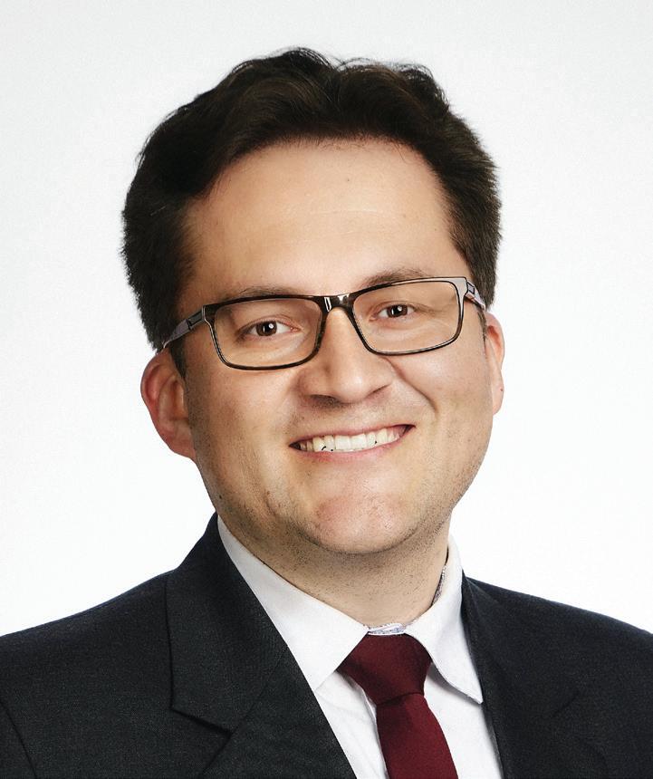
Q&A with Karlo J. Lizarraga, MD, MS
Karlo J. Lizarraga, MD, MS, is an associate professor of Neurology and founding director of the Motor Physiology and Neuromodulation Program. He also has appointments in Neuroscience and Neurosurgery. He completed his MD at the Universidad Nacional de San Agustin and a Master of Science in Clinical Research at the Medical University of South Carolina. After completing his residency in neurology at the University of Miami, he completed fellowships in clinical neurophysiology at the University of Miami and in movement disorders at the University of Toronto. Lizarraga is board-certified in neurology and clinical neurophysiology by the American Board of Psychiatry and Neurology.
Please summarize your research.
The most effective treatments for the motor symptoms of Parkinson’s disease are the dopaminergic drug levodopa and deep brain stimulation (DBS). In addition to their motor benefits, these treatments affect cognition and gait in ways that we do not fully understand. Treatment effects on cognition and gait could contribute to disabling symptoms such as freezing of gait and falls. My research uses high-density EEG techniques to advance the understanding of the effects of levodopa and DBS on cognition and gait, with the goal to personalize treatments and improve outcomes for the growing number of people living with Parkinson’s disease.
How did you become interested in your field?
I was privileged to witness how people affected by movement disorders could go from using a wheelchair to being able to walk, run, and even dance after DBS. This exposure solidified my interest to pursue an academic career in neurology. I went on to complete residency in neurology and fellowships in clinical neurophysiology and movement disorders, focusing on motor physiology and DBS. As a resident, I helped conduct a study that suggested that bilateral and right subthalamic DBS have similar effects on gait in people with Parkinson’s disease. As a fellow in movement disorders, I designed and conducted a study that suggested that levodopa combined with left subthalamic DBS could be harmful for gait but not for cognition in this population.
What brought you to the University of Rochester?
Dr. Robert Griggs, who is an icon in neurology, approached me while I was presenting a poster. I was
starstruck. Besides his multiple personal accomplishments, I was really impressed by the NIH-funded training program in Experimental Neurotherapeutics he had built at the University of Rochester School of Medicine and Dentistry. I was attracted to the academic culture, which combines research, education, and patient care and includes several international collaborations.
Who are you collaborating with, and why?
I am collaborating with Ed Freedman, John Foxe, and the team in the Cognitive Neurophysiology Laboratory at the University of Rochester. This laboratory conducts basic science and translational experiments using high-density EEG techniques. Our cross-disciplinary collaboration aims to develop a non-invasive biomarker of the effects of levodopa and DBS on cognition and gait in people with Parkinson’s disease. The first participant in our study displayed a unique EEG pattern associated with notable gait improvement after DBS. We have obtained preliminary data thanks to funding from the Schmitt Program in Integrative Neuroscience (SPIN) Pilot Project Program through the Del Monte Institute for Neuroscience. We are currently applying for extramural funding.
What is your favorite piece of advice?
My favorite advice comes from “The Book of the Five Rings” by Miyamoto Musashi: “No matter how hard you labor, if your heart is not aligned, your course, no matter how seemingly right, won’t be the true one. Without aiming for a genuine path, even a slight mental detour can cause significant deviation. Reflect on this.”
STUDENT SPOTLIGHT
Aaron Huynh
Aaron Huynh is a second-year student in the Neuroscience Graduate Program at the School of Medicine and Dentistry. He received his undergraduate degree in Brain and Cognitive Sciences from the University of Rochester. He was a student in the biomedical research training program Post-baccalaureate Research Education Program (PREP) at the Medical Center. Huynh currently works in the lab of neurologist Ania Busza, MD, PhD, where his research focuses on ways to assess arm movement recovery in stroke patients over time.
“We’re particularly interested in the acute setting,” said Huynh. “We have an ongoing study that tracks the progress of stroke patients admitted to Strong [Memorial Hospital]. We use wireless sensors to collect electrical activity of muscles involved in simple movements from when they are admitted through six months post-stroke. We are using clinical measures and novel clinical and translational methods to identify ways to evaluate and hopefully improve motor recovery, specifically in the upper extremities. If we can find ways to develop individualized treatment, we hope that can improve people’s quality of life and overall functioning.”
Having the opportunity to work in several different labs during his time in Rochester, Huynh realized that translational research was the area of neuroscience that he was most interested in pursuing. While he was a PREP student, his cousin was diagnosed with an aggressive form of brain cancer, so being able to work under the mentorship of the Joan and Gary Morrow Endowed Distinguished Professor of Supportive Care in Cancer Michelle Janelsins, PhD, MPH, and AnnaLynn Williams, PhD, assistant professor of Surgery, Cancer Center, and Public Health Sciences, was the right place for Huynh. “I was fortunate to have Michelle’s and AnnaLynn’s mentorship. They gave me the support I needed to keep pushing myself to figure things out on my own,” said Huynh, who would go on to first author his first research paper published in Brain, Behavior, & Immunity that found a neurologic biomarker that increased in patients with cancer and preliminarily related to changes in cognitive function.
Outside the lab, Huynh has become one of the newest Del Monte Institute for Neuroscience Diversity Commission members. “As a first-generation student of immigrants, the expectation for my parents was never to really go to college. I never thought I would be an undergraduate, let alone a graduate student. But I think I’m here because of the mentorship and guidance I had from high school and beyond. I think the programs the Neuroscience Diversity Commission offers allow me now to be a mentor.”
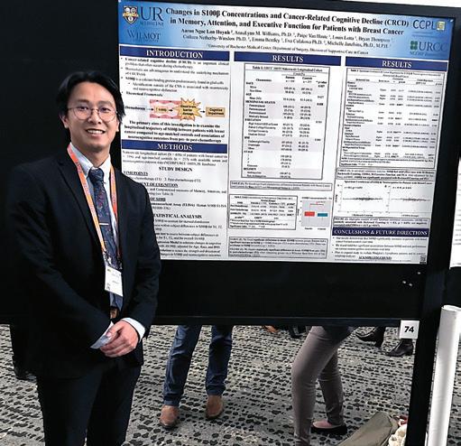
Huynh stands with his research poster at the 2023 International Cognition and Cancer Task Force (ICCTF) in San Diego. The research presented in this poster was the initial data for Huynh's first published research paper.
University of Rochester Medical Center
601 Elmwood Avenue, Box 603
Rochester, New York 14642
Scan to receive the Neuroscience Newsletter
Visit us online: delmonte.urmc.edu
Follow us @URNeuroscience Subscribe @URNeuroscience
giveto.urmc.edu/delmonte
CELEBRATING GILBERT GOTTFRIED
Comedy stars unite in NYC to support muscular dystrophy research
An event to celebrate the release of Gottfried’s long-anticipated vinyl album, Gilbert Gottfried Still Screaming, also raised awareness and support for the Gilbert Gottfried Myotonic Dystrophy Type 2 (DM2) Research Fund. Chad Heatwole, MD, director of URMC’s Center for Health + Technology presented updates about DM2 research during the event.