12 minute read
Equine Athlete


Hock Injuries
Trauma, stress, disease and conformation can be factors
By Heather Smith Thomas
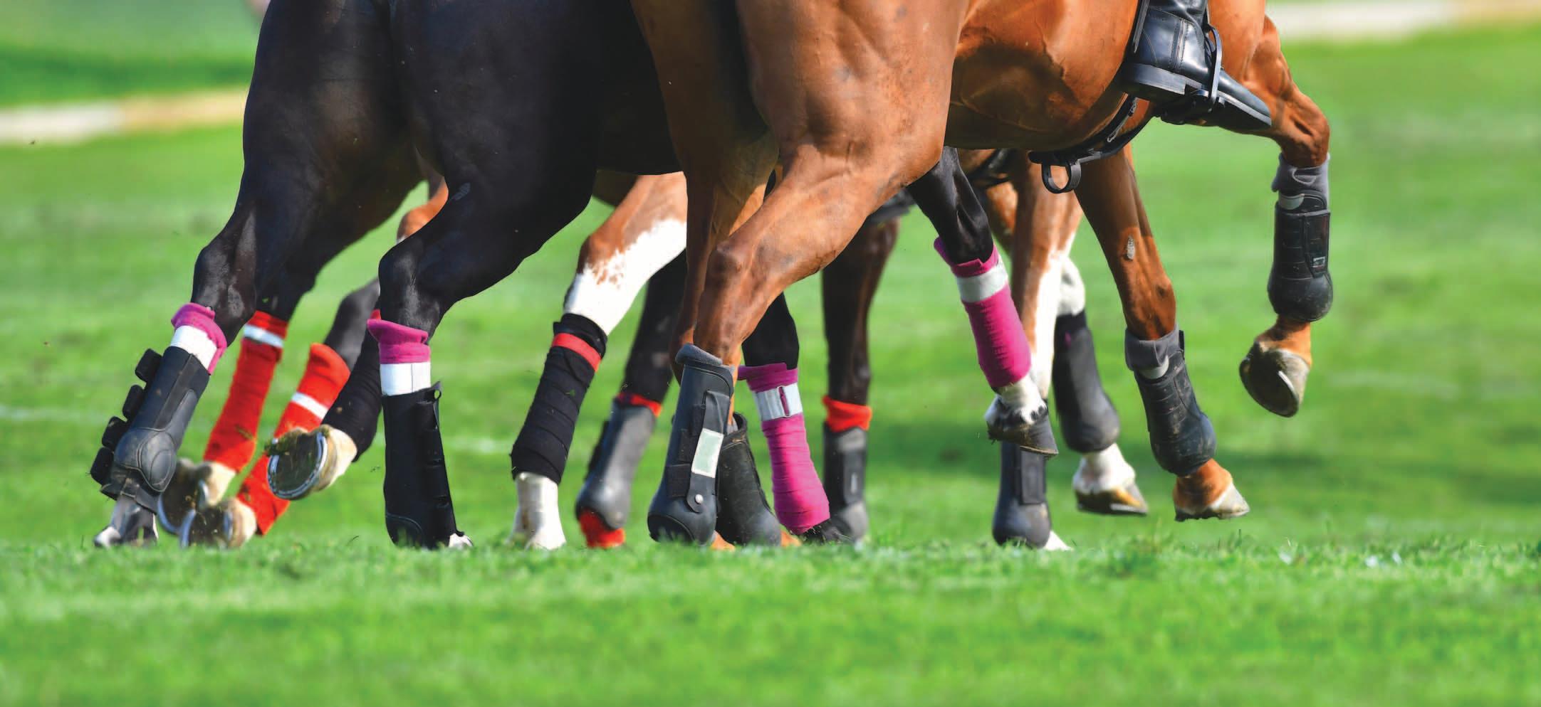
The hocks of performance horses are often under a lot of stress, which can lead to soreness.
The hock is one of the largest, most complex and hardest worked joints in the horse’s body so it must be strong and sturdy to avoid injury. The hock is often under great stress because of the activities we ask the horse to do, and it may be injured during high speed performance.
Dr. Olivia Rudolphi (Rudolphi Veterinary Services, in Noble, Illinois) says some of these injuries may be due to developmental/congenital lesions such as an Osteochondritis dissecans (OCD) within the joint— and these might not show up until the horse is put into training and doing more exercise. “Causes of hock lameness can vary between the young horse and the more mature horse. In the young horses we may see a puffy hock or a foal with some sort of insult to the joint. As young horses start training, we may see the hock develop swelling if there is an OCD lesion, and there may be lameness,” she says.
Jillian Mills, DVM, DACVSMR (Presidio Equine Sports Medicine & Rehabilitation, Encinitas, California), says hock injuries are interesting because this joint is a combination of high- and low-motion joints.“These joints can undergo different pathological changes that can lead to clinically relevant disease,” she explains.
“With hocks we will see either acute traumatic injuries, DOD (developmental orthopedic disease) or fatigue-related injuries. Acute injuries often involve fractures, which may include the tibia and cannon bones, as well as collateral and intra-articular ligament injuries,” says Mills.
“The upper joint (the high-motion joint) more often has developmental issues such as OCD fragments and subchondral bone cystic lesions. If there is an OCD fragment, the horse may need surgery to have the fragment removed, however this is not always necessary,” she says.
“We know that the joint environment is not as healthy as it would be if an OCD lesion was not present. On a pre-purchase exam, for instance it can be difficult to determine whether surgery may be indicated in the horse’s future to remove the osteochondral fragments. For a young horse that hasn’t had the opportunity to prove itself, if we find an osteochondral fragment in a joint that carries excess fluid, and where the horse trots off lame following flexion testing, there’s a higher probability that it will need surgery to remove that chip. The presence of the OCD fragment is an indication that the joint is less healthy, and the horse is more prone
to developing arthritis,” says Mills.
“By contrast, if the horse is 10 years old and has already proven itself at the buyer’s prospective level of competition, the OCD lesion on a pre-purchase exam might not be an issue,” she says.
“Fatigue-related problems might include subchondral bone disease and a breakdown of the joint’s cartilage. When that happens we have to manage these horses for arthritis. We sometimes see arthritis in the upper joint, but this is significantly less common than in the lower joints and often results in debilitating lameness. Usually when we’re talking about fatigue-related injuries in the hock we are dealing with arthritis in those lower hock joints rather than the upper joint.”
If there are changes in the upper joint, these clinical signs generally include a significant increase in joint fluid, which creates a pronounced swelling. “If there is an osteochondral fragment in there, there is often joint swelling and the horse may be positive to flexion testing. However, when a horse reacts positively to hock flexion, this is not a specific finding; the pain could be originating from either the high- or low-motion joints.” says Mills.
“With the lower joints, sometimes we can palpate thickening of the joint capsule, but you cannot palpably detect an increase in joint fluid. It’s more of a clinical assessment, especially if we are not doing nerve blocks. In cases of under-performance, we might look at a horse and see that it is a little bit short-strided in both hind limbs and a bit worse on the inside versus the outside of a circle, and positive to flexion tests. The veterinarian might have a discussion with the owner about medicating the joint as a diagnostic therapeutic, which is essentially a treatment trial to see if the horse improves. Bilateral hind-limb asymmetry is most common in an arthritis type of presentation versus something acute, where you might detect pronounced soft tissue swelling. In those situations, we often see a unilateral lameness, meaning only one limb is affected.” One hock may have suffered traumatic injury.
“Between the talus and the calcaneus, within the high-motion tarsocrural joint, there is another articulation called the talocalcaneal joint. This is a low-motion articulation that can also develop arthritis. When this occurs, the pain can be quite debilitating and extremely difficult to manage, but this is also relatively rare,” says Mills.
Duncan Peters DVM, DACVSMR, ISELP Certified Member (East-West Equine Sports Medicine, Lexington, Kentucky), says we often see hock soreness in performance horses due to strain and stress. “In young horses (weanlings, yearlings) we may see developmental orthopedic disease, such as OCD or bone cysts in the hock joints. These defects are usually discovered early on, and many are taken care of surgically. Occasionally some of those are not evident until the young horse starts training and then gets a little osteochondral fragment (usually in the tibial tarsal joint) and some associated swelling and lameness within the joint,” he says.
“Most of those chips in the joint occur early on in training or when the horse starts racing or doing more extensive exertion in competition. The horse then has to stop training/competing and have the chip removed surgically. Some of those horses can get back to work and training within 90 days or so and don’t miss very much time,” says Peters. If the problem is discovered early and taken care of, the outcome is usually good.
“More commonly, the hock problems we see in young horses are just a soreness. As the horse starts working and asked to do more, the hock joints get sore, just because of the mechanics of that area of the leg. The hock is unique because the upper joint is a high-motion joint and the lower joints are low-motion but must endure a lot of concussion. Those lower joints are held together with many small ligaments—holding those two rows of small bones together.” The hock has seven

When a horse responds positively to a hock flexion test, it could be originating from either the high- or low-motion joints.
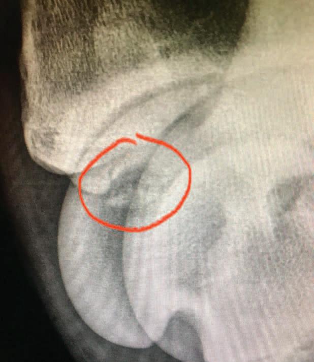
Asymmetrical distention of the hock may mean there is potential for OCD fragments, joint chips, etc. These radiographs reveal bone chips in the joint.
With some hock problems, it is wise to monitor the changes by taking radiographs annually. bones and is similar to the human ankle.
If there is too much strain on the hock, there will be inflammation associated with the ligaments holding those small bones together, as well as inflammation within the joint itself--which causes synovitis in the joint. “Depending on what we find, it may require a different course of treatment,” says Peters.
“The other thing we might see in some of those young horses in training is some bone soreness related to concussion in the lower joints. This may depend on the surfaces they are working on, the level of activity, and the maturity and quality of the bone.” Some of these horses will experience bone bruising and bone soreness.
Dr. Mark Cassells (Homestead Veterinary Hospital, Villa Ridge, Missouri) says his practice deals with a lot of sport horses and in these disciplines he doesn’t see many hock problems in young horses, compared to what might be seen in racehorses, because horses in these other disciplines don’t start training quite as young and are not asked to do extreme exercise with immature joints. “We see hock problems later in the dressage horses, barrel horses, polo horses, etc. when the hocks are already broken down from the exertion they did 10 years earlier,” he says. “When I see a hock problem in a fairly young horse I look at symmetry of the hock, making sure there’s no joint distention on one side compared to the other. If there is asymmetrical distention, this may mean there’s potential to find OCD fragments, joint chips, etc. so we check to see if that’s an issue. There is some debate as to whether these joint chips truly cause problems as the horses get older. Many horses continue to perform at a high level of activity with a joint chip,” says Cassells. Whether that chip causes problems may depend on if it ever moves around and gets caught in the joint. If it is embedded in the joint capsule and not floating around in the joint, it may not be an issue.
“I think the bigger problem is what that chip represents. If it’s due to a defect in the cartilage, this may be more of an issue than the chip itself. With those horses I usually try medical management first, if they are having a problem. The medical management usually involves injection of the joint with regenerative therapies. I try to avoid corticosteroids in these horses because the cartilage is already impaired. I think the regenerative therapies such as Pro-Stride, IRAP, stem cells, etc. are superior to using corticosteroids in these joints, to try to reduce any cartilage damage or at least keep it from getting worse,” Cassells explains.
“I am a big believer in some of the supplemental systemic treatments like Adequan or Pentosan. Anything we can do proactively that will reduce the likelihood of a problem due to joint inflammation will be helpful. Cosequin is a feed-through product that works very well, as does Platinum Performance CJ, for joint health. There are a number of products that people use, including Legend and polyglycan.” The veterinarian can give advice on what to use. It may depend on the situation and whether the owner is comfortable giving shots or whether the horse will or won’t eat medication in the grain. What works on one horse may not necessarily work on another.


“For me, if the horse does not have a lameness issue, then I try to manage it with systemic joint supplementation. If the horse starts developing lameness issues, then surgery becomes higher on my list of things to do, and medical management with regenerative therapies versus corticosteroids,” says Cassells.
If there is a questionable area on a hock that’s noted on radiographs, he suggests taking subsequent radiographs over time, such as every 6 months to a year. “This can show us any progression of the condition, or lack of progression of a disease condition. If it is maintaining well, and the horse is performing well, then maybe nothing else needs to be changed.”
Peters says synovitis and bone trauma can lead to arthritis in the lower joints. “This can progress and become serious if not taken care of early on. Usually we will use injections in those joints (steroids, hyaluronic acid, IRAP, stem cells, platelet rich plasma, amnionic fluid isolates, etc.) in an effort to reduce inflammation and curb development of arthritis. We’ll also back off on exercise and training for a while. If there is bone soreness and trauma— perhaps some micro-fractures associated with the bone bruising—this may take four to six months before the horse can go back to work.”
If the joint has synovitis, many of those horses will respond favorably if their routine is changed a little, backing off on the level of work, and treating the joints themselves with injections. “Those horses may only lose two to four weeks of work and then can get right back to it,” says Peters.
Several of these problems may show up with similar signs, but may need a completely different course of treatment and a different length of time off, in terms of how they respond, so it is important to have a good diagnosis.
“Another thing that’s interesting about the hock is that sometimes there is involvement with the proximal suspensory ligament. There may be some pain-causing damage to that ligament where it originates at the back of the hock. This may actually be the primary problem that leads to secondary hock problems. We find this on MRIs more than with any other type of diagnostic tool,” Peters says.
“These horses won’t be performing well; they have a soft-tissue problem related to the origin of the suspensory ligament, either at the back of the cannon bone (just below the hock) or even into the plantar ligament—since some of those fibers attach up into the plantar ligament at the back of the hock. That whole area functions as one unit. There may be general hock soreness that includes a soft tissue component. The high suspensory area may need to be addressed or explored to make sure we are taking care of all the problems that are causing the soreness,” says Peters.
There are also some unusual aspects sometimes encountered with hock joint soreness. “There may be collateral ligament injuries. These are interesting because the hock has two sets of collateral ligaments. One set is under tension during flexion and the other set is under tension during extension of the hock joint. Occasionally we see ligament damage in one or the other set, depending on what kind of trauma occurred. Collateral ligament injury can certainly occur but is not quite as common.”
Depending on where the injury is located—which bone and surface within the hock—the horse might need arthroscopic surgery. “There might be interference with joint articulation,” says Rudolphi. Some cases are more involved than others. “If we see a problem in one hock we definitely want to check the other one because these conditions can be bilateral, just at different extents,” she explains.
“If an OCD fragment is removed, then afterward we may put in a biologic product like IRAP to help heal those—to help the joint surface improve and ultimately have fewer problems on down the road,” says Rudolphi.
The x-ray on the left shows a normal hock with its many bones. The x-ray on the right shows a fusion of the talocalcaneal joint (see arrows), which is the curved juncture between two of the hock bones—the talus and calcaneus. It also shows a fusion (see bottom arrows) of the distal intertarsal joint on the lower part of the hock.


