1 minute read
Medical Physics Students Organize Wright Middle School Field Trip
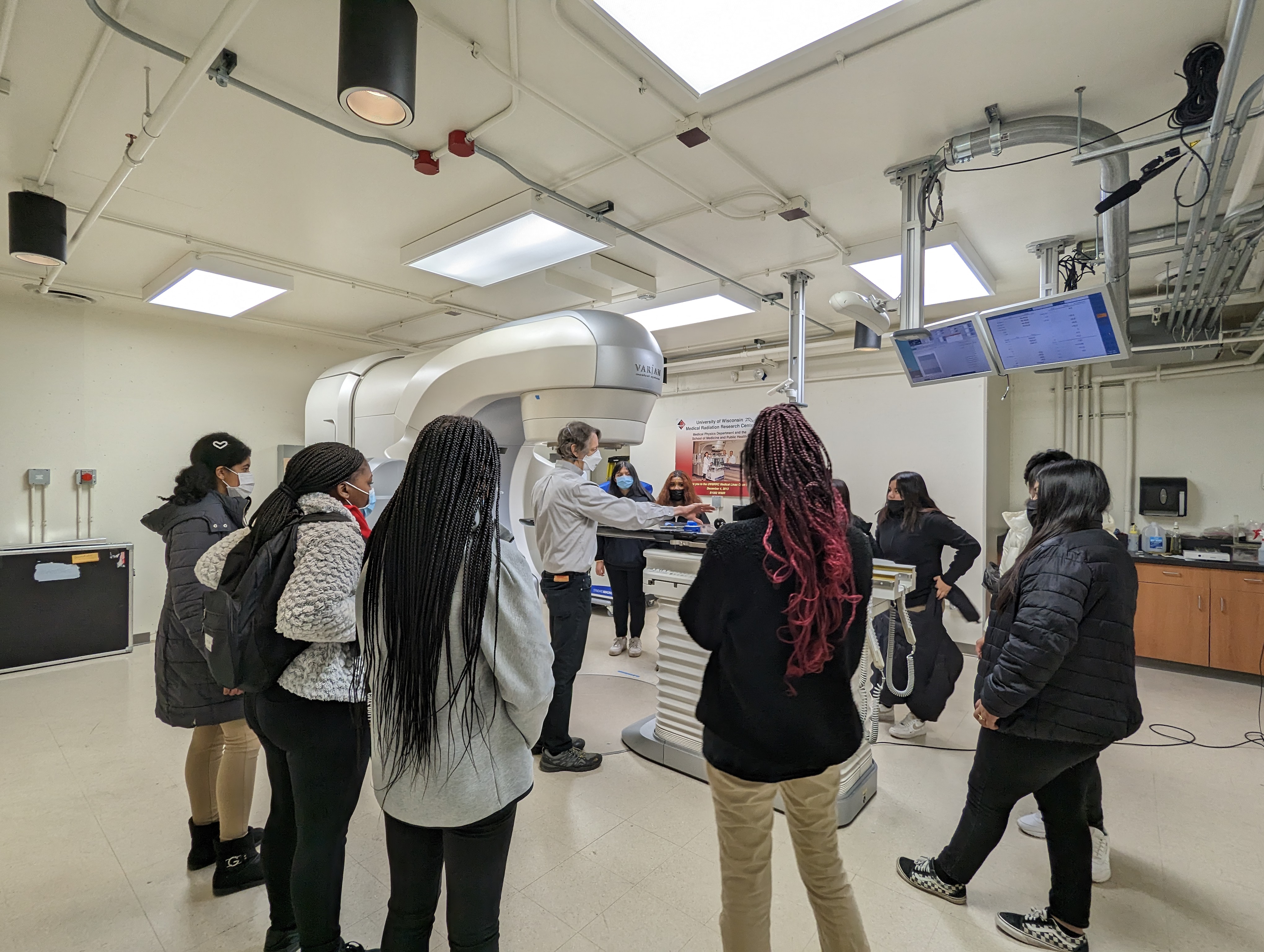
The educational and career opportunities of medical physics and radiology are often unknown to young students. In an effort to increase the visibility of these fields, members of the Departments of Radiology and Medical Physics hosted a field trip on March 24, 2023, for the students from James C. Wright Middle School. The event, which was held in the Wisconsin Institute for Medical Research (WIMR) building, was designed to introduce the students to both key concepts in medical physics and career pathways.
“We thought this field trip would be a great opportunity to develop interactive activities that showcased medical physics using equipment we typically don’t have available for other outreach events,” said Lucky Volety. Volety, a graduate student in the Department of Medical Physics, organized the event along with other members of the Medical Physics Outreach Committee.
“We wanted to inspire the students to think about their future career paths and explore options they might not have considered before,” she added.
During the day, which included a number of engaging presentations and activities, the students had an opportunity to observe s histotripsy. This is an emerging treatment that using noninvasive ablation technology, guided by real-time imaging, to mechanically destroy tumors or blood clots. Ayca Kutlu, MD, an Honorary Research Fellow in the Department of Radiology, and Joseph Whitehead, MS and Grace Minesinger, two Graduate Research Assistants in the Department of Medical Physics, led a demonstration: using waves of ultrasound on a gelatin “phantom” to illustrate the process.
After teaching the basics of radiation therapy, Daniel Anderson and Jeff Radtke led the students through various hands-on projects in the Calibration Laboratory.
Dr. Szczykutowicz and two Imaging Physics residents – Kricia Ruano-Espinoza, PhD and Jonathan Troville, PhD – led an interactive CT activity. Students scanned fruit, vegetables, and other objects to learn the principles involved in creating an image.
Finally, Lucky Volety and Dr. Ruano-Espinoza guided the students through an ultrasound scanning activity. In groups, the students scanned a gelatin phantom containing olives or grapes to simulate tumor detection.
The close collaborative relationship between the Departments of Medical Physics and Radiology allowed the students to be introduced to both research and clinical possibilities of these exciting fields.
“Throughout the activities, students were very engaged, asked insightful questions, and expressed interest for a career in this area of medicine,” Volety said.


