Radiology News
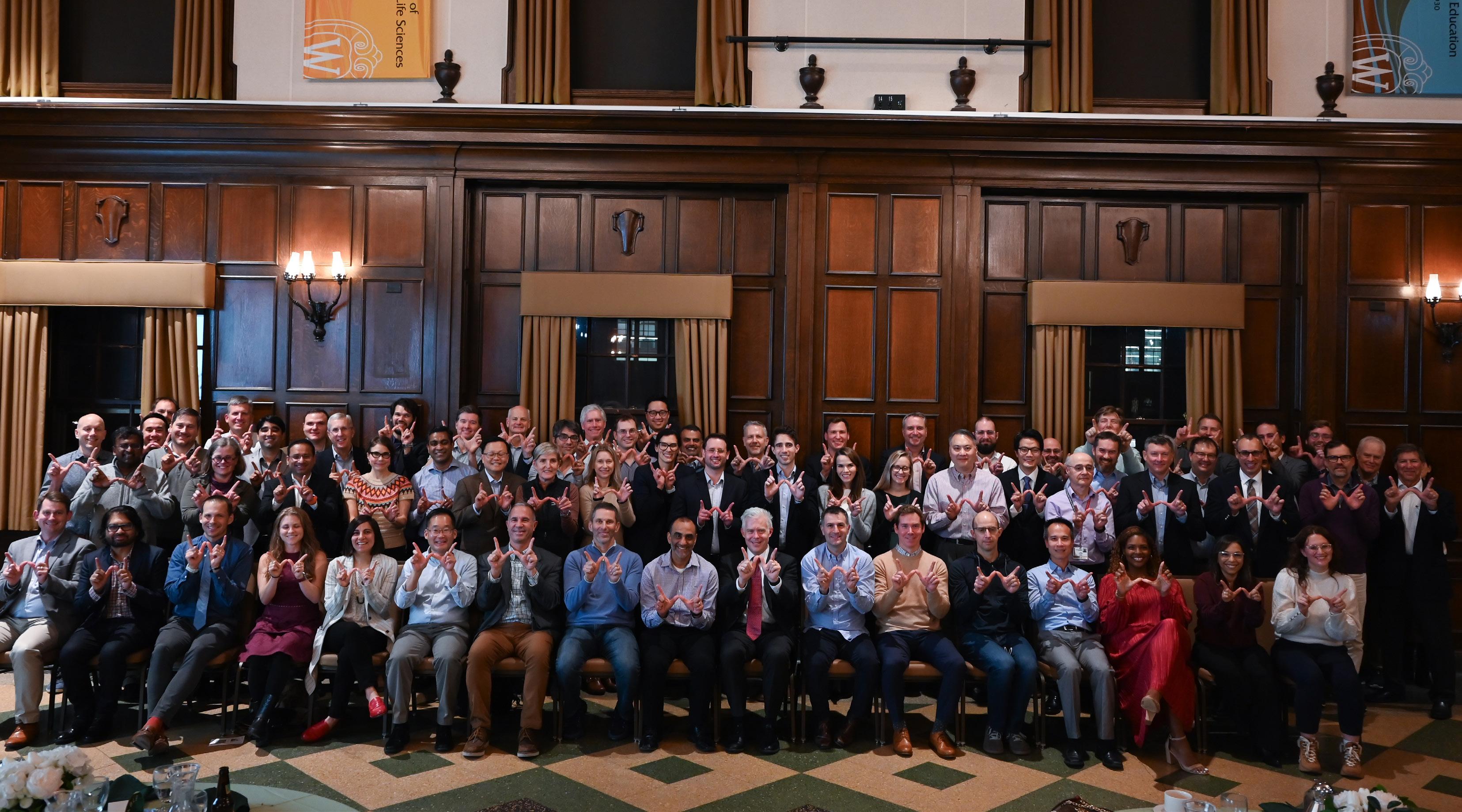

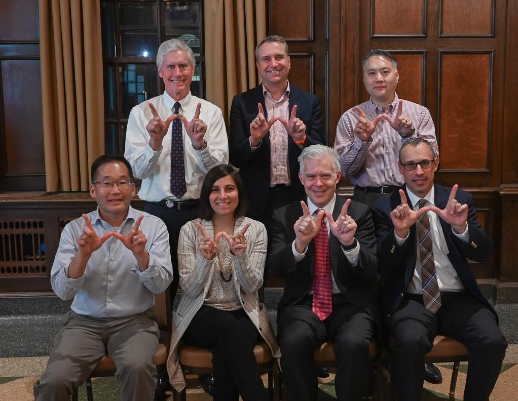
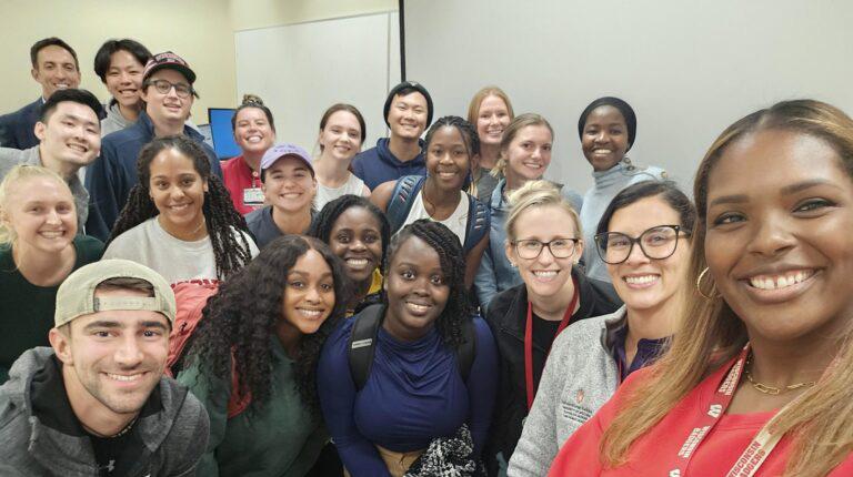
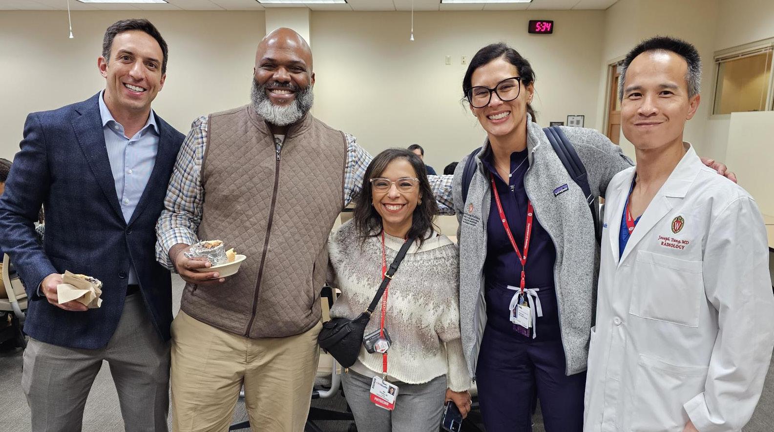
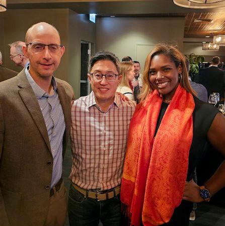
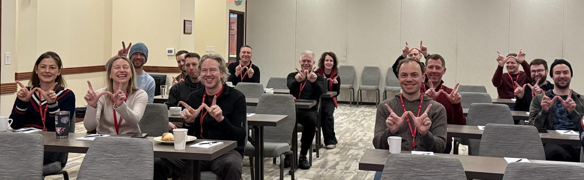
Photo from the Faculty Dinner

With the start of a new year – and my second year as chair – I find myself reflecting on our past, present and future.
In 2027, we will celebrate our centennial anniversary, and I appreciate how the foundation of past accomplishments empowers us to reach new heights as we move into our second century. Our former colleagues wrote foundational textbooks that educated past generations of radiologists and medical physicists. They invented and introduced the first commercial prototypes for digital subtraction angiography. They developed microwave ablation technology that revolutionized clinical care. And so much more.
A quick look at the “2024 by the Numbers” graphic and articles in this issue proves our present is just as impressive. Four faculty members received Gold Medals in 2024. The opening of Eastpark Medical Center enables us to treat more patients with cutting-edge treatments like theranostics. Our researchers are using AI to detect the extent of tumors, provide a “biological age” to help guide patients’ decisions, and decrease the time a child needs to hold still for an MRI. Our faculty are launching online courses to provide education across the globe.
Our future will prove even brighter. We’re welcoming more faculty and trainees every year. We’re improving coordination of physics resources to support our clinical mission by establishing the new Clinical Imaging Physics section. We’re establishing new traditions like the University of Wisconsin Radiology Summit.
I encourage you to celebrate our past, embrace our present, and dream big for our future together.
On, Wisconsin!
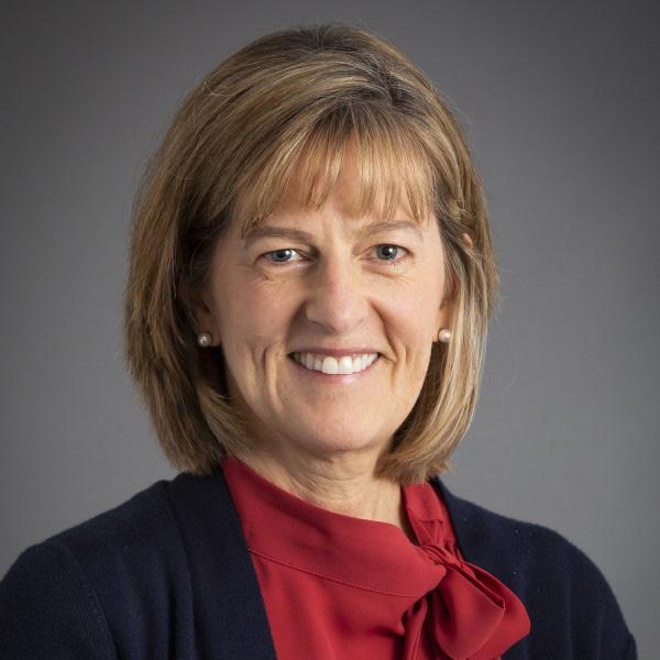
Scott B. Reeder, MD, PhD
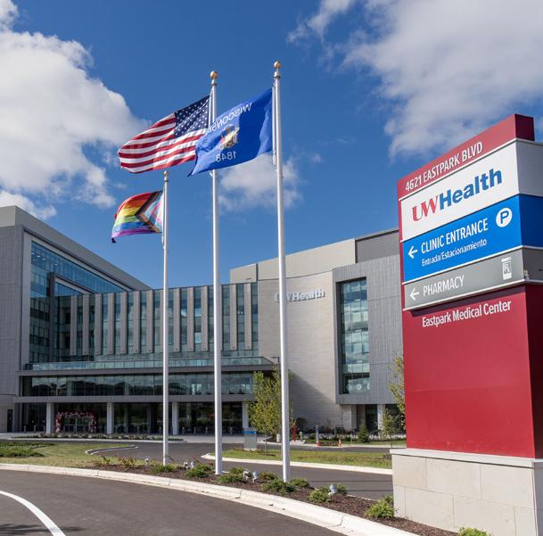
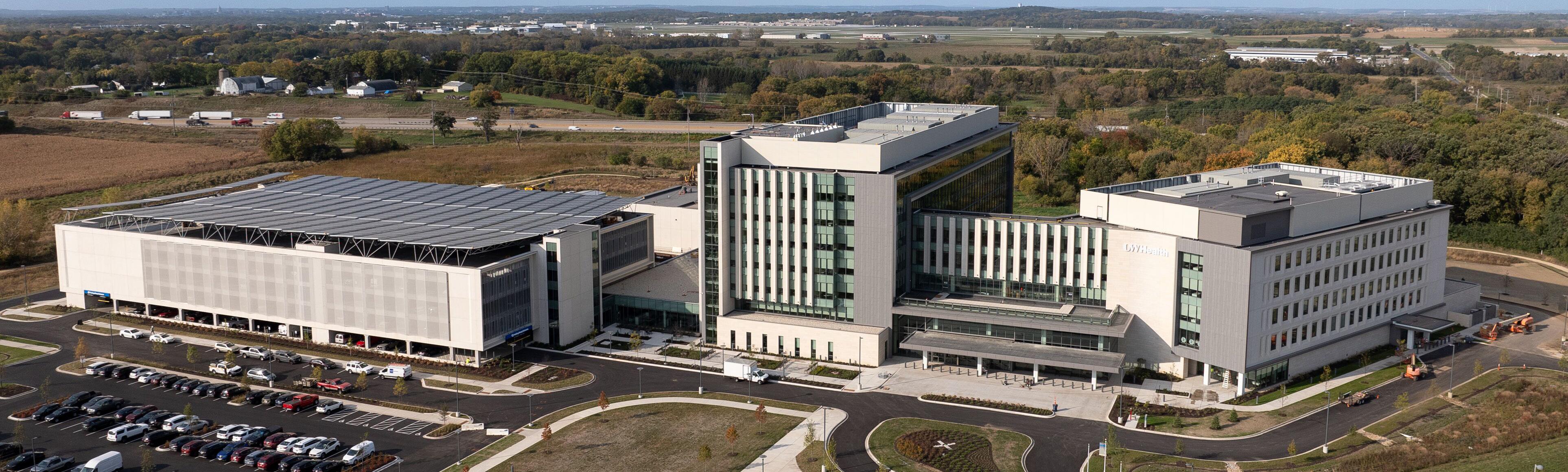
The UW Health Eastpark Medical Center began welcoming patients on October 28, 2024, for diagnostic radiology services, interventional radiology procedures, and nuclear medicine imaging conducted by UW Department of Radiology faculty and staff. Soon after, cancer care was fully ramped up and includes breast imaging services, radiation oncology and theranostics.
Most of radiology services are located on the first level with the waiting room located in the north wing. In addition to state-of-the-art scanners, Eastpark features a new approach for the MRI scanner and patient prep rooms.
“The design for patient flow in and out of the MRI scanners has already proven to save time with only three minutes between the last sequence of one patient and the first sequence of the next patient,” Senior Vice Chair of Clinical Operations Michael Tuite, MD explained. “In addition, the patient experience has improved as they are rolled into the scanner room instead of having to approach the sometimes intimidating machine.”
The only National Cancer Institute-designated comprehensive cancer center in Wisconsin, the newest location for the UW Health | Carbone Cancer Center includes the Breast Center and Clinic on the fourth level. Patients will also access novel treatments such as theranostics at Eastpark.
The UW Departments of Radiology and Medical Physics have established the new Section of Clinical Imaging Physics to provide technical expertise and coordination of physics resources in support of our clinical mission. Alisa Walz-Flannigan, PhD joined the university in October 2024 to serve as section chief.
“This new section demonstrates the continued legacy and commitment of UW–Madison to the high standards in medical imaging required to provide optimal patient care,” Chair Scott Reeder, MD, PhD noted. “We are fortunate to have Alisa Walz-Flannigan serve as section chief as she brings a wealth of leadership, technical and educational experience.”
Section members optimize imaging protocols, troubleshoot image degradation problems, conduct acceptance testing of equipment, and perform routine quality assurance testing. They also help drive innovation and support clinical translation.
“Our imaging physicists partner closely with modality chiefs as well as other radiologists and technologists,” explained Dr. WalzFlannigan. “Through these relationships, we better understand the workflow and needs in the evolving enterprise of modern medicine and help advance patient care.”
A key element of effective teamwork is knowledge sharing, an area Dr. Walz-Flannigan is well-versed in. “I’m passionate about education and workforce development. I’ve previously worked with the Mayo Clinic’s residency programs and Marshfield Clinic’s School
of Radiography.” In addition, she currently serves as the Imaging Physics Residency program director within the UW Department of Medical Physics.
Dr. Walz-Flannigan and her section members are developing a resource website to share information about equipment, testing, accreditation, and quality control. In addition, they will develop training for radiology staff and residents.
Many of the new section members are also members of the Section of Imaging Sciences and conducted similar work prior to the new section’s foundation. The Imaging Sciences section differs in its primary focus, which is research. However, the two sections will work closely together.
Section members engage in activities, such as:
• Ensuring patient and occupational safety with ionizing and non-ionizing radiation as well as acoustic energy
• Optimizing imaging protocols, reconstruction, and postprocessing
• Standardizing and managing imaging protocols
• Supporting clinical translation and development for innovation
• Evaluating and selecting equipment
• Conducting regulatory and accreditation testing
• Developing policies, procedures, and workflows
• Site planning, siting and shielding
• Providing accreditation support
• Training and providing educational resources
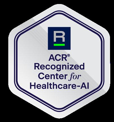
UW–Madison Department of Radiology recognized by American College of Radiology for responsible AI practices
UW–Madison Department of Radiology proudly practices safe, responsible integration of artificial intelligence (AI) into clinical, research, and educational endeavors. Due to this unwavering dedication to best practices, the American College of Radiology (ACR) has identified the department as an ACR Recognized Center for Healthcare-AI (ARCH-AI) organization.
The ARCH-AI program aims to help radiologists provide safe and effective implementation of AI products to provide better patient care. Criteria to become an ARCH-AI site include:
• Establishing an interdisciplinary AI governance group.
• Maintaining an inventory of AI algorithms with detailed documentation.
• Ensuring adherence to security and compliance measures.
• Engaging in diligent review and selection of AI algorithms.
• Documenting use cases and training procedures.
• Monitoring algorithm performance, including safety and effectiveness.
• Participating in the Assess-AI national AI registry for performance benchmarking.
AI is just one technological advancement the department leverages to provide enhanced quality and care for patients and increased workflow effectiveness.


UW and Mass General Brigham enter in collaboration with Microsoft to advance AI in medical imaging
On July 24, 2024, Microsoft announced collaborations with the University of Wisconsin School of Medicine and Public Health and UW Health, and Mass General Brigham to further advance AI in medical imaging to drive clinician efficiency and enable better health outcomes.
Department Chair Scott Reeder, MD, PhD highlighted UW–Madison’s reputation for embracing technical innovations to transform the field of radiology with new scientific discoveries, leading to an improvement in clinical care.
“We are excited to collaborate with Microsoft on the development, validation, and thoughtful clinical investigation of generative AI in the medical imaging space” Dr. Reeder shared. “Our focus is to bridge the gap within medical imaging from innovation to patient care in ways that improve outcomes and make innovative care more accessible.”
News of the collaboration was widely reported on, including features on AuntMinnie.com, PR Newswire, and Radiology Business.
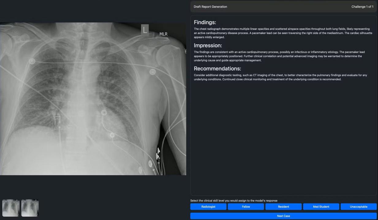
UW Department of Radiology partners on launch of the Healthcare AI Challenge
The UW Department of Radiology joins host Mass General Brigham in the Healthcare AI Challenge. The first-of-its-kind interactive virtual environment enables healthcare professionals to experience the world’s most advanced AI healthcare solutions.
Participating healthcare professionals can explore cutting-edge AI solutions designed for specific medical tasks, such as medical image interpretation, within a simulated environment. Professionals with relevant healthcare credentials can provide feedback on the performance and utility of these solutions, resulting in publicly available insights and analytics.
The feedback gathered through the initiative will be used to benefit stakeholders and patients worldwide.
The Healthcare AI Challenge chose radiology as the first specialty because it has been an early adopter of AI solutions.
“Medical imaging provides many types of data, and up to 95% of healthcare data collected is unstructured, non-text data,” said Richard Bruce, MD. “AI has the potential to interpret and distill that data at a new scale and speed, but what we need is the ability to quickly test and compare different AI solutions. The Healthcare AI Challenge will offer a platform to evaluate and compare tools across various clinical situations.”
Organizations participating in the Healthcare AI Challenge Collaborative also include Emory Healthcare and the Department of Radiology at the University of Washington School of Medicine.
In addition to healthcare institutions, the American College of Radiology (ACR) has joined the Healthcare AI Challenge Collaborative as a founding member, ensuring its 42,000-member community can access the Healthcare AI Challenge.
Mass General Brigham made the announcement through a press release. The announcement was also featured in the American Hospital Association and Becker’s Hospital Review.
Ali Pirasteh, MD received a 6-month, $900K grant for “Novel Early Detection of Lung Cancer by PET Imaging” from Earli, Inc.
Vivek Prabhakaran, MD, PhD received a $600K grant for “Optimizing Brain Health in Transgender Patients using Gender Affirming Hormone Therapy” from the Wisconsin Partnership Program (WPP) and the Partnership Education and Research Committee (PERC).
Apoorva Safia, PhD received a 3-year, $200K grant for “Multimodal AI based Predictor of Alzheimer’s Disease (MAPAD)” from the Alzheimer’s Association.
Prashant Nagpal, MD received a $95K grant for “Cardiac Image Quality with PIQE” from Canon Medical Systems USA.
Oliver Wieben, PhD received a 1-year $80K grant for “Anatomic Imaging Derived 4D Hemodynamics using Deep Learning” from the National Institutes of Health/Third Coast Dynamics, Inc.
Labros Meimetis, PhD received a $50K Piloting Research Innovation & Market Exploration (PRIME) grant for “Toll-Like Receptor 7/8 Agonists for Targeted Cancer therapy” from Discovery to Product (D2P).
Tim Szczykutowicz, PhD received a 1-year, $293K grant for “Clinical Feasibility and Evaluation of Silicon Photon Counting CT” from GE HealthCare.
Alan McMillan, PhD received an $85K grant for “Context Aware Multimodal Information Retrieval Systems” from Applied Research Laboratory for Intelligence and Security (ARLIS)/University of Maryland.
Tyler Bradshaw, PhD received a 2-year, $37K grant for “Torch Recon: An Innovative Reconstruction Software for Increased Throughput and Improved Low-Count Quantitative SPECT Imaging” from the National Institutes of Health/Voximetry.
Kevin Johnson, PhD received a five-year, $3.5 million grant for “Pediatric Cardiopulmonary MRI using RF Navigators and High Dimensional Deep Learning” from the National Institutes of Health/National Heart, Lung, and Blood Institute. This project aims to develop MRI technology to make it easier to image the cardiac and pulmonary systems in pediatric patients without sedation or radiation.
Imaging the cardiac and pulmonary systems in pediatric patients presents challenges. MRI scans are highly sensitive to motion, and young patients often struggle to remain still for the duration of the scan. As a result, captured scans may be unclear and contain motion artifacts like blurring or ghosting. To obtain accurate imaging in a clinical setting, pediatric patients are often sedated before the scans; however, this is not always a viable option.
Dr. Johnson is collaborating with colleagues at UW–Madison; Stanford University; University of California, Berkeley; and University of California, San Francisco to develop a dual-pronged method to solve the challenge of capturing pediatric MRI scans. The first involves fast imaging techniques to reduce the overall time it takes to capture scans.
The second approach measures motion during the scans. The team developed a unique method that transmits a Wi-Fi (5 GHz) signal through the body, tracking interference caused by the patient. The signal changes pattern with bulk motion, breathing, and beating of
the heart; effectively measuring the amount of motion during a scan.
This information is then harnessed in a high dimensional deep learning approach, which applies 2D and 3D artificial intelligence (AI) technologies to high resolution 4D applications. Ultimately, this allows scans that have been disturbed by motion to be reconstructed.
Dr. Johnson noted “We hope that this grant will push MRI closer to a more push-button method, closer to that of CT. This particular project will target the imaging of congenital heart disease, providing extremely valuable information for treatment and monitoring.”
Current cardiopulmonary evaluations typically require multiple exams to evaluate the cardiovascular and pulmonary systems; which can involve complex logistics and delay patient care. The combination of increased MRI speed and ability to reconstruct images via deep learning would expedite the treatment and reduce stress for the patients.
While this project is using imaging techniques for pediatric patients with congenital heart disease, the increased MRI speed and image reconstruction methods could potentially be expanded to capture accurate imaging for other motion-related imaging issues.
Pallavi Tiwari, PhD received a $3.4 million, multi-institutional grant from the National Institutes of Health/National Cancer Institute for the project “Radiomic spatial maps for identifying viable tumor extent on multi-parametric MRI for Glioblastoma.” This novel project will be the first to utilize artificial intelligence (AI) to detect hard-to-see glioblastoma tumor edges during a clinical trial.
Glioblastoma is an aggressive cancer with an average 15-month life expectancy. Surgery is the main treatment due to its fast-acting nature; however, tumors often reach microscopic niches in the brain tissue that are not visible during surgery. As a result, most surgical procedures don’t completely remove the tumor and it continues to spread.
Microscopic niches pose another problem – existing methods to determine tumor edges, like biopsies, may miss these areas and lead to inaccurate results. These methods are not routinely done as part of a clinical practice due to the expense and need for patients to travel for the procedure.
The project’s overarching goal is to use AI to accurately predict and map out where the tumor is, resulting in complete removal during surgery. Dr. Tiwari explained how the research group will evaluate the effectiveness of AI in determining where the tumor ends and where healthy brain tissue begins in the clinical trial phase.
“We use spatial radiomaps (i.e. “virtual biopsy maps”), which act as a sort of GPS map. We take a simple MRI scan of the patient’s brain, and then apply the AI analysis to create the radiomap. From there,
we look to see where the map says the tumor is, and we perform a biopsy to check for accuracy and see if that tissue is cancerous.”
This is a multi-institutional endeavor, with Dr. Tiwari serving as primary investigator along with Manmeet Ahluwalia, MD from the Miami Cancer Institute who leads the clinical trial. Researchers from the Cleveland Clinic Lerner College of Medicine and the H. Lee Moffitt Cancer Center & Research Institute are also contributing.
Multi-institutional research projects can be challenging, especially when sharing patient data, but Dr. Tiwari emphasized that the collaborative nature is one of the key strengths of this project.
“When training AI models, diversity of data is key; and the multiinstitutional approach provides an advantage. Having a diverse set of studies from multiple sources helps ensure that accurate results from the AI analysis are reproducible from one case to another, across institutions.”
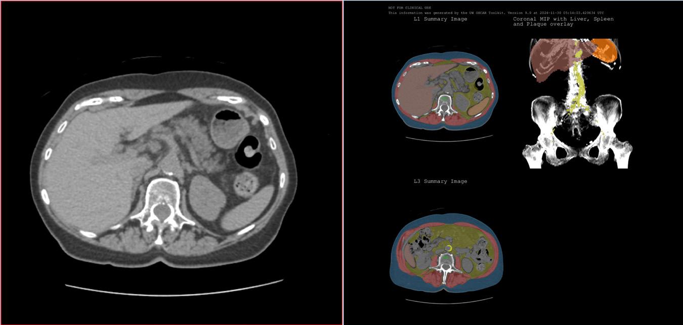
Having the AI analysis use imaging information, with corresponding pathology, and genomic information, to create biologically inspired image-features that drive the analysis is a cornerstone of the project.
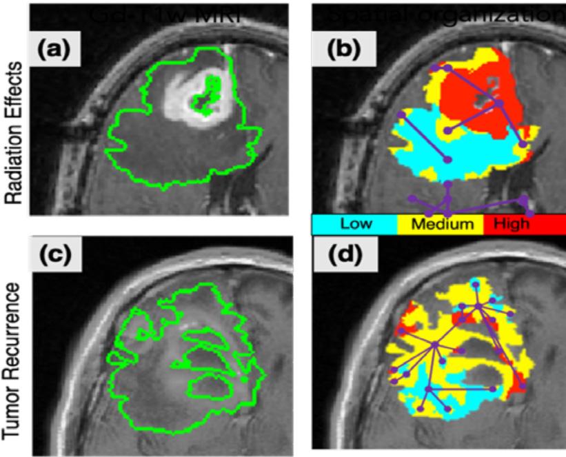
Preliminary results for lesion complexity features. (b), (d) demonstrate undirected subcompartment graphs for (a) treatment effects, and (c) tumor recurrence. Graph complexity measures (density, transitivity, assortative) will help create a lesion complexity signature on a per-voxel basis.
Perry Pickhardt, MD and John Garrett, PhD are among the finalists for the 2024 Wisconsin Alumni Research Foundation (WARF) Innovation Awards for their work, “Innovative Screening Tool for AgeRelated Diseases,” which aims to provide a “biological age” for a patient that may differ from their actual age and guide improvements in health. Dr. Pickhardt and Dr. Garrett collaborated with the late Michael Kattan, PhD, MBA, from the Cleveland Clinic Department, who was an innovator in modern statistical prediction methods for medical decision making.
Drs. Pickhardt, Garrett, and Kattan developed a set of AI learningbased tools that measure and assess different characteristics of a patient’s CT scan, such as aortic plaque, muscle density and distribution, and organ volumes and densities. Using this information, a predictive model compares the patient’s data against people who share the same age, genetic background, and other factors that can impact health. The resulting analysis can predict how the patient is aging compared to other individuals who share similar traits and provides a “biological age.”
With the insight this technology could provide in clinical settings, health care professionals and patients could better understand what may be causing a higher biological age, such as high aortic plaque and fat. Specific medical and lifestyle changes could be identified and tailored to reverse these conditions and lower the advanced biological age.
In a video about this work, Dr. Garrett says, “one of the things I love about this field of opportunistic screening is that one of the goals is
to do more than just get somebody back to baseline; but to find a way to help them live their healthiest, best life and to prevent health issues from happening in the first place.”
The WARF Innovation Awards recognize some of the best inventions at UW–Madison. An independent panel of judges selects the winners from a field of six finalists drawn from hundreds of invention disclosures submitted to WARF over the past year.
“Each year, our Innovation Awards shine a spotlight on the most exciting early-stage discoveries on campus,” says Erik Iverson, CEO of WARF. “We’re pleased to celebrate the nominees and all UW–Madison innovators working to discover and translate research with the power to impact lives.”
Earlier this year, Dr. Pickhardt was honored with a WARF Named Professorship. This award recognizes faculty who have made major contributions to the advancement of knowledge, primarily through research, but also through their teaching and service activities.
This fall, Women Professionals in Radiology (WPR) co-leaders Erica Knavel Koepsel, MD; Jade Anderson, MD, and Leslie Nelson, DO hosted Radiology Roundtable: Jeopardy & Mentorship Night. The event was in partnership with the UW School of Medicine and Public Health’s Radiology Interest Group (RIG), which Venkata (Vinny) Meduri, MD and Matthew Lee, MD lead.
Medical students engaged with the mentors in small groups on the following topics:
• A Day in the Life of a Radiologist with Edwarda Golden, MD
• Embracing the Future/AI with Joseph Tang, MD
• Radiology Subspecialties with Erica Knavel Koepsel, MD
• Global Radiology Opportunities with Mai Elezaby, MD
• How to be a Great Radiology Resident/Optimize Radiology Residency Apps with David Kim, MD
• Private Practice vs Teleradiology vs Academics with Jason Stephenson, MD and Shawyon Shadman, MD (Madison Radiologists)
About 40 students attended the event, which also included a “speed networking” component for the faculty to share their experiences and insights with the medical students. Residents Allison Couillard, MD and Matthew Elissa, MD also participated, sharing their perspective and advice for the students as current residents.
Following dinner and the mentorship sessions, the entire group gathered to play a case-based Jeopardy game, where prizes for the final team included a gift card. The students enjoyed competing against each other and learning more about the field of radiology!
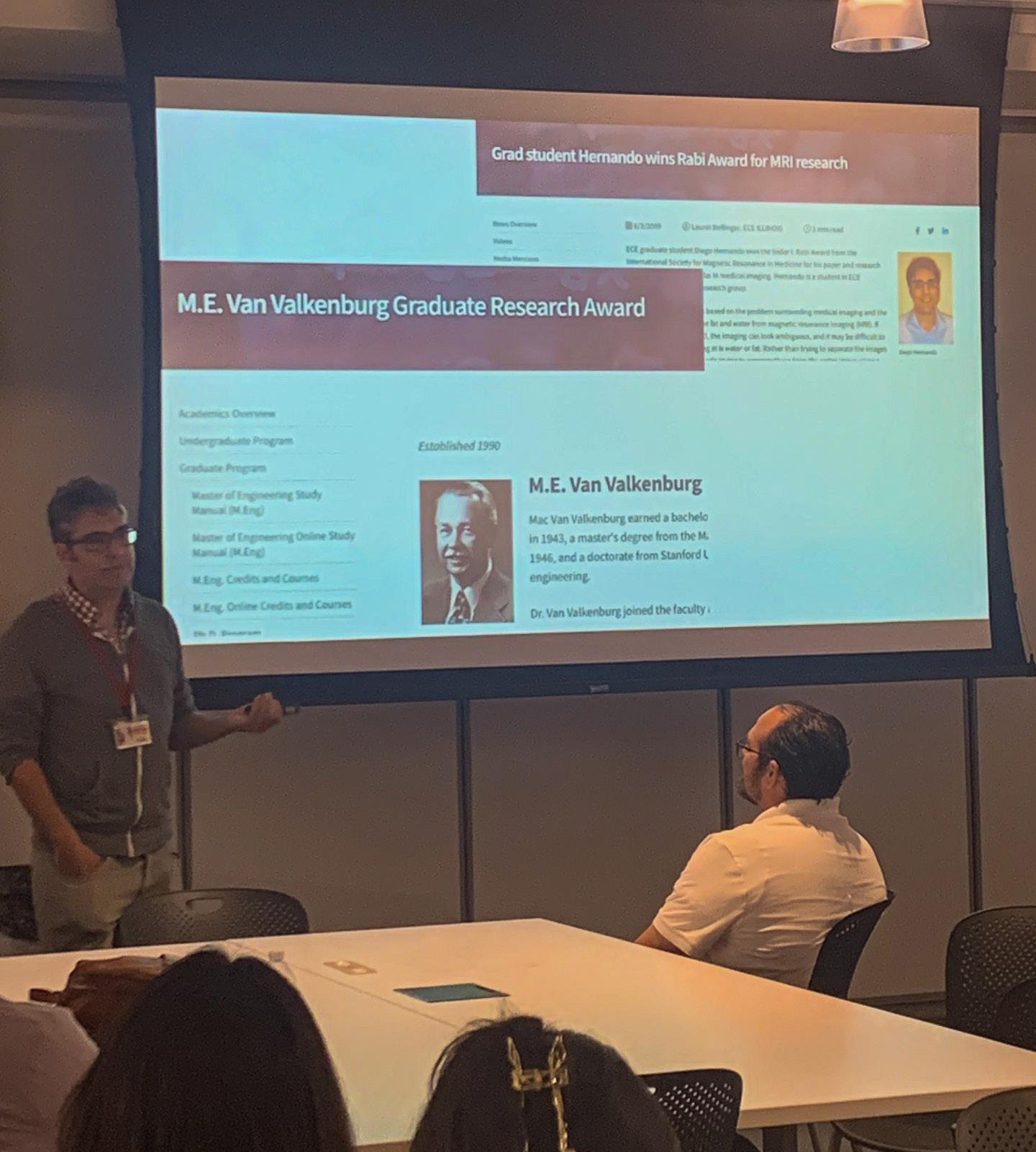
Diego Hernando, PhD shared his career journey with medical students during the UW School of Medicine and Public Health’s Office of Multicultural Affairs (OMA) monthly dinner series in September 2024.
Dr. Hernando enjoyed telling his story and connecting with the students.
“I was excited to meet from different cultures, people that have different perspectives about what it means to be in Madison and at the university. I also benefit from the conversation and questions; and hearing other people’s thoughts and experiences and sharing my own.”
For students interested in pursuing a doctorate degree, Dr. Hernando emphasized the importance of mentorship. “Who your mentors are has such a massive impact on your life and career; and not just in terms of ‘are they a famous doctor’ or ‘are they a great researcher.’ Are they someone with whom you can have a real mentorship relationship, and who is going to be supportive for many years?”
He encouraged students to keep their options open as they advance in their careers, and make sure they think about what they want in life as a whole.
“I love working hard on research and collaborating with really smart people to solve difficult problems. But I also think about when I’m in my 70s, sitting on my porch, am I going to regret not publishing more papers or getting more grants, or will I wish that I had spent
more time with my family? Make sure that what you’re doing is aligned with your priorities and your long-term goals.”
The OMA dinner series hosts a large diversity of faculty speakers from across the School of Medicine and Public Health. The overall goal for the event is for the faculty to connect with students through stories of their journeys to medicine. Previous speakers from the department include Ivan Rosado Mendez, PhD; Anand Narayan, MD, PhD; Mai Elezaby, MD; Erica Knavel Koepsel, MD; Jade Anderson, MD and Jason Stephenson, MD
Michael Hartung, MD collaborated with Radiopaedia to launch a continuing medical education course, Abdominal CT Essentials, which covers abdominal CT protocols, search pattern, acute abdomen and cancer imaging.
Through interactive cases and video lessons, Dr. Hartung addresses common diagnoses ranging from appendicitis and pancreatitis to liver tumors and cancer mimics.
The course features two and a half hours of video lessons and over 150 teaching cases for participants to hone and test their skills as they progress at their own pace. Dr. Hartung is no stranger to delivering radiology lectures and embraced the challenge of producing a video course as an exciting way to reach radiology trainees worldwide.
“It was a tremendous amount of work and organization to pull it all together with a complex organization process, and producing the video content at a high level was challenging compared to the more informal nature of in-person teaching.” He also noted that he focused on ways he could make the content lively and engaging for viewers.
Available on-demand, Radiopaedia offers the course free of charge to practitioners in 125 low- and middle-income countries. This accessibility is one of the key factors that motivated Dr. Hartung to partner with Radiopaedia as it aligns with his passion for enhancing global health and radiology education.

Through his global health volunteer work, Dr. Hartung helped establish partnerships with Tenwek Hospital in Bomet, Kenya and Diospi-Suyana Hospital in Curahausi, Peru, creating a global health elective for radiology residents at UW Hospitals and Clinics. Dr. Hartung has also served as a mentor for international residents to prepare and submit cases to the Penn Radiology Global Health Imaging Case Competition.
In addition to the course on Radiopaedia, Dr. Hartung provides free educational materials on his Learning Abdominal Radiology website.
At the annual Radiological Society of North America (RSNA) meeting, David Bluemke, MD, MSB, PhD and Perry Pickhardt, MD were honored for their outstanding contributions to radiology education; each receiving a Lifetime Honored Educator Award from the society.
This prestigious award recognizes individuals who further radiologic education through the creation of high-quality educational content.
Dr. Bluemke has been an active member of RSNA, participating on grant and education review committees, delivering scientific and educational lectures, and serving as a former editor of Radiology. He previously accepted RSNA’s Honored Educator Award in 2021, 2022, and 2023.
“I am deeply honored to receive this from the RSNA,” says Dr. Bluemke. “The RSNA is the leading force in our field in advancing state-of-the-art science and medical advances. I have truly enjoyed helping to translate these advances by being part of the many educational opportunities sponsored by the RSNA.”
During the RSNA meeting, Dr. Bluemke presented “Cardiac Imaging as a Surrogate Marker In Drug Trials” as a keynote speaker.
Dr. Pickhardt’s commitment to advancing the field of abdominal imaging – and his work has led to over 500 scientific publications and book chapters, as well as multiple textbooks.
He has been a dedicated educator at UW–Madison for over two decades and serves as the chief of gastrointestinal imaging for the department. He previously accepted RSNA’s Honored Educator Award in 2014, 2018, and 2019.
During the RSNA meeting, the educational exhibit Dr. Pickhardt co-authored, “Acute Mesenteric Ischemia (AMI): Understanding the Imaging Findings and Making a Critical Diagnosis,” received a Certificate of Merit.
Earlier in 2024, both faculty members received gold medals, honoring a lifetime of achievements. The Society for Cardiovascular Magnetic Resonance awarded Dr. Bluemke with their Gold Medal in January. The Society of Abdominal Radiology presented Dr. Pickhardt their Gold Medal in April.
Many of our faculty have received the annual Honored Educator Award from RSNA. In addition, Meghan Lubner, MD received the Lifetime Honored Educator Award last year.
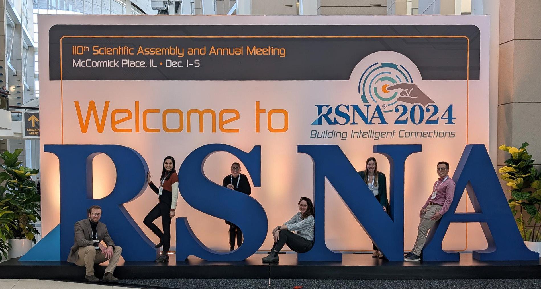
Over three dozen faculty members, trainees, and medical students braved the chilly weather in Chicago for the Radiological Society of North America (RSNA) annual meeting December 1-5, 2024. Our department’s expertise garnered wide recognition, with members receiving honors and awards across various areas of the field.
For the second year in a row, Mark Schiebler, MD received an Honored Educator Award. The award acknowledges those most invested in furthering radiologic education through the creation of high-quality educational content in their field of study.
In recognition of their outstanding medical imaging research, Diego Hernando, PhD; Kevin Johnson, PhD; and Pallavi Tiwari, PhD were inducted into the Academy for Radiology & Biomedical Imaging’s Council of Distinguished Investigators Class of 2024 during the meeting.
Matthew Lee, MD was featured on RSNA’s Daily Bulletin for the education exhibit “CT Imaging Biomarkers for Phenotypic Biological Aging: Overview and Comparison with Other Radiological and Non-Radiological Approaches.” John Garrett, PhD and Perry Pickhardt, MD were co-authors on the exhibit.
Over 60 faculty members, alumni, and colleagues from across the country came together to reconnect at the social mixer hosted by the department. The event also served as a celebration for those who were honored during the larger RSNA meeting.
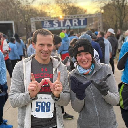
Running from meeting to meeting wasn’t enough for Tim Szczykutowicz, PhD and Meghan Lubner, MD! They participated in the RSNA 5K Fun Run with research partners Eugene Liu, Mark Frontera, and Jean-Baptiste Thibault from GE HealthCare. Funds collected from the run support the RSNA Research & Education Foundation.
Faculty, trainees, and students received the following accolades at RSNA 2024:
• Alison Gegios, MD; Amy Fowler, MD, PhD; Ryan Woods, MD; Roberta Strigel, MD; Thomas LoDuca, MD; Lonie Salkowski, MD, PhD; Mai Elezaby, MD and Anand Narayan, MD, PhD received a Certificate of Merit for the education exhibit “MultiModality Review of Vascular Conditions of the Breast.”
• Matthew Lee, MD; Perry Pickhardt, MD; Meghan Lubner, MD; Giuseppe Toia, MD and David Kim, MD received a Certificate of Merit for the education exhibit “Acute Mesenteric Ischemia (AMI): Understanding the Imaging Findings and Making a Critical Diagnosis.”
• Elizabeth Sadowski, MD received a Magna Cum Laude award for “A Picture Is Worth Thousand Words, But Optimized Imaging Is Priceless: Expert Guidance On How To Fine Tune Your Us Images For O-Rads Us Assessment.”
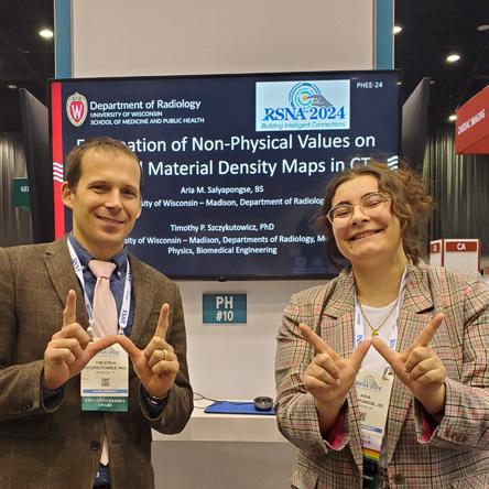
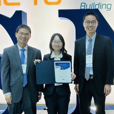
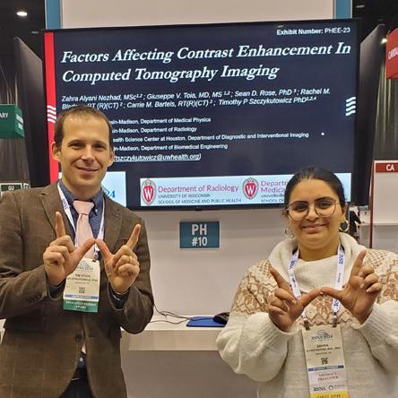
• Zahra Alyani Nezhad, a PhD student (advisor: Tim Szczykutowicz, PhD) won a Certificate of Merit for “Factors affecting contrast enhancement in computed tomography Imaging.”
• Aria Salyapongse, a PhD student (advisor: Tim Szczykutowicz, PhD) won a Cum Laude Award for “Explanation of non-physical values on spectral material density maps in CT.”
• Linying Zhan, a PhD student (advisor: Guang-Hong Chen, PhD) received the RSNA Trainee Research Prize for “Performance Evaluation in Photon Counting CT (PCD-CT): Measuring DQE and Detector Dead Time Using an ImageBased Framework.”
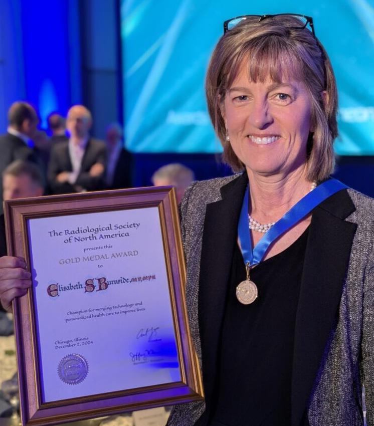
Elizabeth Burnside, MD, MPH, MS, FACR, accepted the prestigious Gold Medal from the Radiological Society of North America (RSNA) at the annual meeting. Dr. Burnside is the first radiologist from the UW, and the 22nd woman, to receive this honor since the award’s inception in 1919.
The Gold Medal is RSNA’s highest accolade and awarded to those who have rendered unusual service to the science of radiology. A renowned specialist in breast cancer, Dr. Burnside is engaged at the cutting edge of patient-centered research to improve screening mammography. Her unique contributions include developing innovative approaches at the intersection of breast cancer screening, biomedical informatics, and population health.
Dr. Burnside has shown her commitment to advancing patient care and the medical imaging field through her participation in national organizations and her leadership roles. Currently, Dr. Burnside serves as associate dean of team science and interdisciplinary research at the medical school, as well as the executive co-director of the Institute for Clinical and Translational Research.
We interviewed Dr. Burnside about the Gold Medal and her career. Read her full interview here.
Can you share what the receipt of this award means to you?
I am greatly honored and am proud to be the first awardee at UW–Madison to receive this special recognition. Notice that the RSNA Gold Medal is given for “unusual service” to radiology. I do consider myself somewhat unusual, because I have a combination
of interests, which include imaging technologies that accurately diagnose disease, public health applied across populations in effective and sustainable ways, and biomedical informatics to enable computers and humans to work together to deliver innovations for the benefit of healthcare and human health in general.
I am the first woman to be tenured in radiology at UW–Madison and I am proud that several other women have followed me since. I have always had an eye toward ensuring that women are represented in radiology and in other areas of health and medicine in which they are underrepresented. Perhaps my elevation of women in the specialty is another contribution that could be characterized as unusual. We are making progress.
Looking back on your career, what are some of the most significant or impactful moments that you’ve faced?
During my residency at University of California San Francisco (UCSF), I realized I was missing a key component of the future when I considered that computer science and machine learning were revolutionizing other areas like communication and finance.
Thanks to the insightful and empowering mentorship I received from Ron Arenson, MD, then chair at UCSF, I decided to attend Stanford to pursue my master’s degree in biomedical informatics between residency and breast imaging fellowship. This was a landmark move for my career, because it enabled me to harness the potential that biomedical informatics had for both radiology and medicine in general. San Francisco as a leader in this transformation and the academic institutions in the Bay Area leading the computer science revolution were key ingredients to my future career.
My unconventional training and expertise fueled another key moment for me when I was honored to deliver the Monday RSNA
Plenary Address in 2023 entitled “Leading Through Technology: Valuing Artificial and Human Intelligence.”
I endeavored to demonstrate why, as we embrace artificial intelligence (AI), we must intentionally preserve core manifestations of human intelligence, especially those at the heart of patient care, including judgment, transparency, trust, and communication. We must foster lifelong learning across the entire radiology workforce to identify and advance the opportunities and recognize and call out the pitfalls of generative AI. We must employ leadership models like functional leadership, servant leadership, and transformational leadership designed to maintain a culture of stakeholder empowerment, collaboration, and continuous learning to reap the benefits of this fast-paced AI-informed phase for which our specialty is poised.
What advice would you give to young radiologists, or students who are interested in entering the field?
As it happens, radiology was scheduled as my last rotation in the third year of medical school training before the deadline for specialty applications. By the time I began radiology, I had in mind my future would be in a surgical subspecialty for which I had already turned in my applications!
But, as I experienced my radiology rotation, I had the feeling that I had found my future career – largely, because of the people. I immediately felt what I would call professional kinship; I just really liked the people. Since then, I have contemplated: why? Maybe it is because I am fortunate to be a lifelong learner, and I found radiologists to be constantly curious, always striving to understand a disease process and figure out how to diagnose or treat better, faster, and more efficiently. Yes, the imaging was fascinating; yes, I knew I would never know everything; but really, I felt like I had found
individuals who I admired, found inspiring, and really liked “hanging around with.”
As an athlete participating in team sports throughout my education, I felt like I was at home on the diagnostic radiology team, which was constantly expanding to include members from other specialties to take care of patients. So as far as advice goes, I would emphasize finding an area to pursue in which you are surrounded by people who you like and who drive and inspire you to be your best.
Is there anything else that you’d like to share?
I firmly believe that an “unusual” career is often what a specialty needs to advance in unique, important, and sustainable ways. I have been grateful for those who have recognized that the combination of imaging, public health, and data science (informatics, computer science, biostatistics) will help the powerful tools that diagnostic imaging brings to adapt to the future of healthcare.
I also am grateful to those who perpetuate the importance of interdisciplinary collaboration, an underpinning value of diagnostic radiology. In many ways, radiologists are specialists who create bridges between other specialties (e.g., medicine and surgery). This role is an important one as we strive to see patients in a holistic way and not as a collection of organ systems or a series of interventional procedures.
I hope and believe we all can continue to keep these values of interdisciplinarity, teamwork, and patient-centered care at the forefront of our specialty so we advance the health of individual patients and the population as a whole.
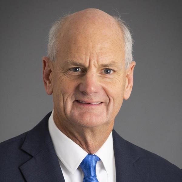
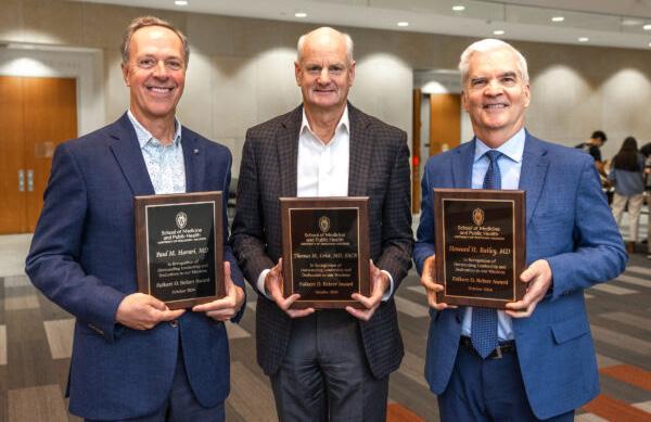
Recognizing his profound contributions to the field of radiology, Thomas Grist, MD received two significant honors this fall.
The UW School of Medicine and Public Health named Dr. Grist a recipient of the Folkert O. Belzer Award alongside Howard Bailey, MD and Paul Harari, MD. Additionally, the GE HealthCare Foundation gave funds to establish a professorship in his name that will support the UW Department of Radiology’s research initiatives.
Dr. Grist accepted the Folkert O. Belzer Award, the highest honor bestowed by the medical school, during the fall faculty and staff meeting on October 28. The award, named after the former Department of Surgery chair whose work helped revolutionize organ transplantation, recognizes the school’s most outstanding leaders who have significantly impacted the school and academic medicine.
During the ceremony, Dean Robert Golden, MD emphasized that while they practice in different medical disciplines, each recipient is a “quadruple threat” — an outstanding clinician, renowned researcher, deeply respected teacher and mentor, and an effective leader.
Dean Golden also highlighted the impact that Dr. Grist made in the radiology department during his time as chair.
“Our department of radiology rose to the very highest levels of national prominence, garnering significant recognition for its training and research programs. At the same time, the department became renowned for its highly successful partnership with industry leader GE HealthCare.”
The partnership with GE HealthCare continues to thrive, having recently been renewed for another decade. Dr. Grist played a key role in developing this partnership. His work with Charles Mistretta, PhD, on the Time-Resolved MR Angiography (TRICKS) technique and its application to GE HealthCare machines sparked the idea for university researchers and GE HealthCare engineers to collaborate.
Since that pivotal moment, the partnership has led to many more innovations that have improved public health. To celebrate his legacy of innovation in clinical care, imaging research, and education; the GE HealthCare Foundation has given $3 million for the Thomas Grist, MD/GE HealthCare Foundation Distinguished Chair in Radiology Research.
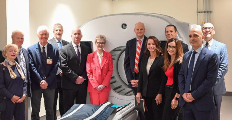
As part of the celebration, GE HealthCare members and members of the radiology department toured the Eastpark Medical Center to view the radiology service areas, including the StarGuide scanner. Left to right: Jeanne Grist, Jamey Weichert, Michael Tuite, James Pipe, Adam Holton, Catherine Estrampes, Thomas Grist, Erin Angel, Tim Szczykutowicz, Manuela Govin, Ertan Cetingul, Chad Rowland.
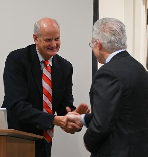
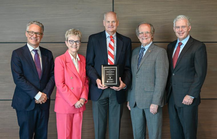
On November 7, GE HealthCare board members celebrated the professorship at the newly opened Eastpark Medical Center. Catherine Estrampes, president and CEO, GE HealthCare in U.S. and Canada; and Dean Golden presented Dr. Grist with a plaque during the celebration to mark the occasion.
Reflecting on his long history with the company, Dr. Grist expressed his gratitude for everyone who helped foster the partnership between UW–Madison and GE HealthCare.
“This is all about building bridges between academics and industry and it’s transformational. It’s about how we cross that divide and get across that bridge; and it has clearly been what has made
my professional career so exciting and meaningful.”
Dr. Grist concluded with an inspiring message for the future. “For those of you who are going to continue this collaboration, go forth and cross that bridge. I think that together we will do amazing things. We’ll see great things in the future to improve the health of our patients, and that’s what this is really all about.”
On July 1, 2024, Erica Knavel Koepsel, MD assumed the role of Interventional Radiology section chief and began a fiveyear term as the Andrew B. Crummy Professor in Radiology.
Dr. Knavel Koepsel completed her training at the University of Wisconsin Hospital and Clinics, where she made department history as the first woman elected as chief resident.
Just one year into her career as a faculty member at UW–Madison, Dr. Knavel Koepsel launched the iMRI program; a collaborative effort with the Department of Urology to perform biopsies and treat prostate cancer with cryoablation. Her work on this program garnered statewide attention, including a feature on Wisconsin TV station, WKOW. The iMRI program is the only one of its kind in Wisconsin and one of six locations nationwide where patients can undergo these procedures.
Earlier this year, Dr. Knavel Koepsel led a team of interventional radiologists in performing Wisconsin’s first clinical histotripsy-based ablation, also marking the first time a radiology department in the U.S. performed this procedure.
Beyond her innovative clinical work, Dr. Knavel Koepsel is a dedicated mentor. As co-chair of the Women Professionals in Radiology (WPR) group, she supports her peers and provides guidance for women who are early on in their careers.
Additionally, she mentors through the UW School of Medicine and Public Health’s Building Equitable Access to Mentorship (BEAM) program. The BEAM program pairs faculty with students from underrepresented backgrounds in medicine to enhance their professional development and academic success.
Dr. Knavel Koepsel shared that she’s honored to be appointed as the Andrew B. Crummy Professor. “Dr. Crummy was one of the founders of Interventional Radiology; and was vital to the advancement of the field with the development of Digital Subtraction Angiography. His contributions to UW–Madison and the field of radiology are spectacular. With this professorship, I hope to continue Dr. Crummy’s mission by supporting innovation, research, and educational opportunities in the section while always striving for ways to improve patient care.”
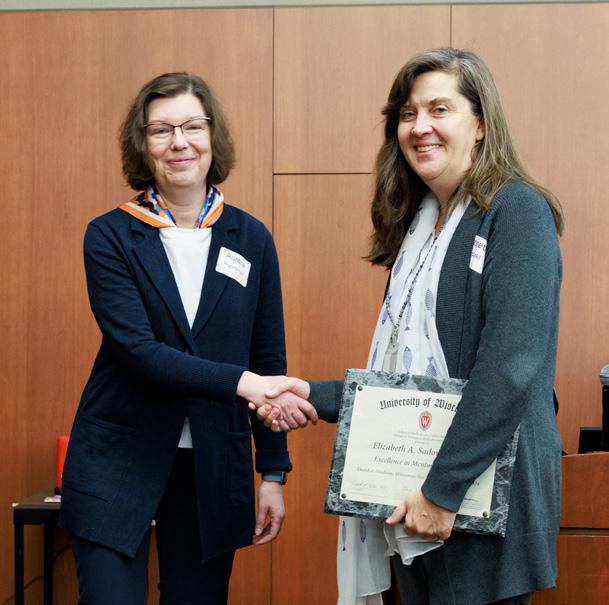
Elizabeth Sadowski, MD received the 2024 Excellence in Mentorship Award from the UW School of Medicine and Public Health’s Group on Women in Medicine and Science. She was nominated by Jessica Robbins, MD, who received the award last year. “I can unequivocally say that I would not be where I am today in my academic clinical career without Dr. Sadowski’s mentorship, coaching, and sponsorship,” says Dr. Robbins.
In 2001, Dr. Sadowski joined the department as a fellow in abdominal imaging and continued as faculty in the Abdominal Imaging and Intervention section as well as serving as the director of gynecological imaging.
Throughout her career, she has focused on advancing women’s health through imaging and presenting her work internationally. She is a founding chair and member of multiple national committees focused on gynecologic imaging, including the Society of Abdominal Radiology (SAR) Disease-focused Panel on Uterine and Ovarian Cancer and the American College of Radiology (ACR) Committee on Ovarian-Adnexal Reporting & Data System (O-RADS). In addition, she led committees focused on diversity and inclusion, including the ACR Committee on Women and Diversity as well as the SAR Diversity and Inclusion Committee.
Dr. Sadowski has been a remarkable mentor to faculty in radiology and has guided trainees and early-career faculty locally and nationally, shaping the next generation of leaders.
We interviewed Dr. Sadowski to learn more about her long history as
a mentor that led to this award. Read the full interview here.
Can you share what the receipt of this award means to you?
I am honored and humbled to receive this award. You may not always know how your interactions impact others and I feel fortunate to work with all those who I have mentored – feeling they have taught me as much as I have taught them. So to be recognized for mentoring makes me feel happy and that perhaps I’ve made a difference in other people’s lives and careers.
Are there any key mentors you had who made a significant or positive impression on your career?
Some of my key mentors early in my career were Drs. Mark Kliewer, Parvi Ramchandani (PENN), Susan Rebsamen and Deborah Rubens (University of Rochester). They empowered me to think outside the box and reach out to others who work in the field of gynecologic imaging to help me build my career in this area. Their insightful views humanized the academic world, which can sometimes be cold and harsh. No one talks about these aspects of academics, yet many experience this. I’ve made it my mission to talk to my mentees about building a diverse network of colleagues, mentors and sponsors within and outside of their institution, to support them.
Why is mentorship important to the field of radiology?
We can all mentor one another in our work to improve the lives of our patients. You can learn from everyone – those junior, senior and at your level. Never be afraid to collaborate and choose projects you are passionate about. As the Greek proverb says: “A society grows great when (people) plant trees whose shade they know they shall never sit in.” When we all work together and promote one another, we move forward in a positive direction, even if we never know the true impact of our work.
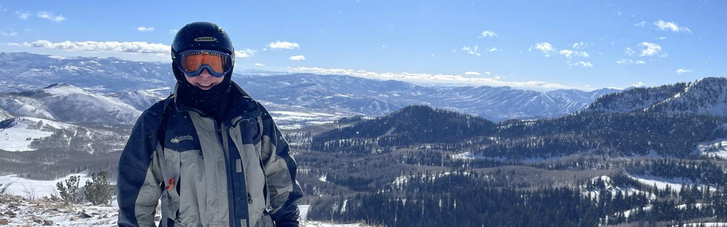
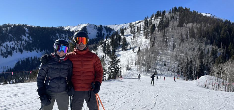
The Department of Radiology established a new tradition this year by hosting the first, annual University of Wisconsin Radiology Summit. During the week of January 20, 2025, radiologists from across the country convened to learn, connect and ski the beautiful mountains in Park City, Utah.
Summit Co-Directors Tabby Kennedy, MD and Roberta Strigel, MD, MS developed a program that featured 27 sessions, ten speakers, and two exhibits by trainees. Topics ranged from facial trauma to the fundamentals of ankle MRI. In addition, Benjamin Walker, MD from the Department of Anesthesiology provided techniques for minimizing opioid use in children.
“Our department is home to experts in every sub-specialty and we were pleased so many could join us as speakers this year,” Dr. Kennedy notes. “Plus, the informal setting created a collegial spirit that followed attendees out to the slopes!”
In addition to downhill skiing, participants could be found cross country skiing, snowshoeing and more. When not enjoying what some call the greatest snow on earth, many could be found sharing a meal in Park City’s historic downtown.
“The chance to spend quality time with colleagues was invaluable,” explains Dr. Strigel. “Unlike at conferences where you may only have a few moments to catch up with a former trainee or colleague from across the country, we had opportunities for rich conversations.”
As pleased with the first year as the co-directors are, both are eager to grow the summit. “By the end of the week, everyone was talking about next year,” notes Dr. Kennedy.
“We encourage everyone to update their contact information so they don’t miss out on next year,” Dr. Strigel says.
To confirm that the department has updated contact information, people can send an email to radnews@uwhealth.org or contact the co-directors directly. Next year’s dates will be announced on the UW Radiology Summit webpage.
See you in 2026!
Faculty Speakers
• Kathleen Fink, MD
• Tabassum (Tabby) Kennedy, MD
• Erica Knavel Koepsel, MD
• Perry Pickhardt, MD
• Scott Reeder, MD, PhD
• Andrew Ross, MD
• Roberta Strigel, MD
• Jonathan Swanson, MD, MBA
• Tim Szczykutowicz, PhD
• Benjamin Walker, MD
Trainee Exhibitors
• Alex McDonald, MD, Abdominal Imaging Fellow
• Erik Winterholler, MD, Diagnostic Radiology Resident
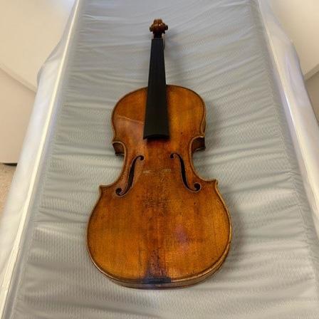
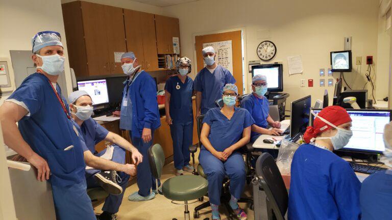
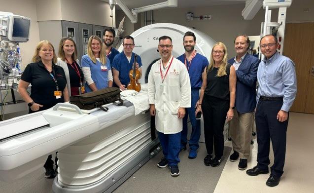
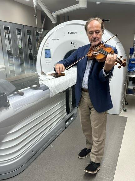
Imagine holding a piece of history in your hands — a violin crafted over 300 years ago by the legendary Antonio Stradivari. Now imagine peering so deeply inside that you can count the rings on the wood pieces used.
This is exactly what our radiologists and research staff experienced when they had the rare opportunity to scan a Stradivarius violin.
Antonio Stradivari, a master luthier from Cremona, Italy, crafted an estimated 1,100 instruments during his 71-year career. Only about 650 of these instruments have survived, including 450 to 512 violins. Stradivarius instruments are incredibly valuable, with some worth millions of dollars. For instance, the Lady Blunt Stradivarius violin sold for a record $15.9 million in 2011.
But why are these violins so sought after?
Stradivarius instruments are renowned for their fine symmetry, beautiful details, and unique tonal qualities. Theories about their exceptional sound include the special density of the wood used during the Little Ice Age and the varnish or chemical treatments applied to the wood.
The condition of a Stradivarius violin is a critical factor in determining its value. Even the most expertly crafted violins are not immune to the ravages of time. These instruments are particularly susceptible to low humidity, which can cause the wood to crack and the traditional hide glue to become brittle, leading to open seams.
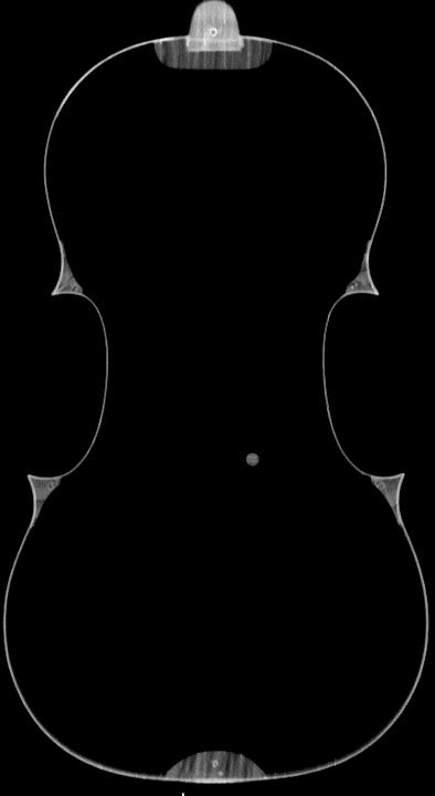
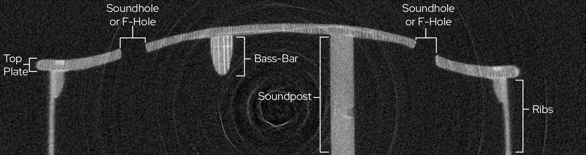
The violin scanned at UW–Madison had undergone repairs in the 1800s, but the specifics of these repairs were unknown. This is where the expertise of the UW Department of Radiology team came into play. The CT scans provided a non-invasive approach to viewing
the inner construction of the violin, revealing details that cannot be seen on the surface.
Gabriel Ben-Dashan is a former professional musician and the owner and director of Bein & Fushi, Inc., a firm specializing in the sales and restoration of fine and rare stringed instruments. He reached out to our team about having the violin scanned.
Fred Lee, MD – an amateur violinist himself – was happy to oblige. He along with Guiseppe Toia, MD; Tim Szczykutowicz, PhD;
Timothy Ziemlewicz, MD; fellow Troy Tenbrunsel, MD; as well as research analysts Rachel Bladorn and Kelsey Schluter used multiple scanners, including the Generation 3 Silicon-Based Photon Counting Scanner. Dr. Szczykutowicz developed the protocols for calculating how the scans could be accomplished.
While the scans may not unlock all the secrets of Stradivari’s craftsmanship, they provide a deeper appreciation for the complexity and beauty of these instruments.
It was a reminder of the intersection between art and science, and the privilege of contributing to the preservation of a priceless artifact.

Aaron LeBeau, PhD was featured on an NBC 15 news segment and article “Can sharks cure cancer? UW-Madison research shows positive advancements” on October 20, 2024. The associate professor in the UW Departments of Radiology and Pathology & Laboratory Medicine discussed his work developing shark antibodies for cancers. Read more and watch the clip.

Radiology professor Jamey Weichert, PhD, co-director for the Initiative for Theranostics and Particle Therapy (ITPT), spoke about the exciting field of theranostics with The Capital Times during their Idea Fest Session held in September 2024. He explained that theranostics had a lot of promise because clinicians do not need the precise location before using it as a treatment. Read more.

Pallavi Tiwari, PhD spoke to Wisconsin Public Radio (WPR) for the article “UW–Madison researchers use AI to identify ‘sex specific’ risk factors in brain tumors” published January 1, 2025. Dr. Tiwari and fellow UW–Madison researchers developed an AI model to identify why glioblastoma tumors are more aggressive in men than women. Read more.

Pallavi Tiwari, PhD presented “Are We Ready for an AI Oncolologist? How AI Can Help Doctors Personalize Cancer Care” at TEDxOshkosh 2024 on November 16, 2024. In her talk, she noted “With Al, and with our help, we can make this future a reality where no one has to be terrified of the words ‘you have cancer.’” Read more and watch the talk.
Lisa Schmaltz, MD, member of the Community Radiology section, retired in October 2024 after three decades with the UW Department of Radiology.
After earning her medical degree at the University of Missouri–Kansas City, Dr. Schmaltz completed her residency and fellowship at UW Hospitals and Clinics. She joined the faculty in 1996 upon completing her training.
Dr. Schmaltz served in leadership roles throughout her career. She held the position of chair of the School of Medicine and Public Health’s Clinician-Teacher Track Promotions Committee. Before her appointment as chair, Dr. Schmaltz served on the committee for seven years.
As medical director for Group Health Cooperative of South Central Wisconsin (GHC-SCW), Dr. Schmaltz implemented new clinical programs and technology training to improve patient care. These programs also expanded accessibility, allowing patients to receive care at GHC-SCW locations instead of an outside institution.
Dr. Schmaltz leaves a legacy of commitment to patients and dedication to improving healthcare access. Her colleagues in the Community Radiology section, and the entire department, will miss her and wish her the best in her retirement.
Radiologic Tech Lead for MRI Thomas McKinlay retired in January 2025 after working at UW Health for 31 years.
Tom started his career at UW Health as a diagnostic radiology technologist. After a year, he went to CT for three years before finishing his career supporting MRI.
He focused on patient care, construction, equipment, and MR/OR ablation and deep brain stimulation exams.
Beyond a talented MR technologist, Tom was known for known for creating a welcoming environment. A colleague noted that “Tom is a friend to everyone.”
Another colleague emphasized his skills and said “when Tom enters a room, you feel safe as he is confident in his abilities and work.”
People may still seeing his smiling face after his official retirement as he will be working as a PRN in MRI.
Nicholas Burris, MD joined the Section of Cardiovascular Imaging as an associate professor (TT) in January 2025. Prior to joining the department, Dr. Burris was a tenured associate professor at University of Michigan Health System with a dual appointment in Radiology and Biomedical Engineering.
Monica Cooley, MD joined the Section of Musculoskeletal Imaging and Intervention as an assistant professor (CHS) in September 2024. Her interests include interventional oncology and education.
Matthew Larson, MD, PhD joined the Section of Nuclear Medicine as an assistant professor (CHS) in September 2024. He also works in the Section of Neuroradiology. His interests include molecular imaging of neurodegenerative disease and neuro-oncologic processes.
Sarv Priya, MD, MBBS joined the department as an assistant professor (CHS) in September 2024 and will split his time between the Sections of Pediatric Radiology and Cardiovascular Imaging. Most recently, he served as a clinical assistant professor at the University of Iowa Hospitals & Clinics.
Veena Nair, PhD joined the Section of Clinical Imaging Physics as a professor (CHS) in January 2025. Prior to her faculty appointment, she worked in the department as a scientist for a decade. Her interests include human brain plasticity associated with aging and disease.
Michio Taya, MD joined the Section of Abdominal Imaging & Intervention as an assistant professor (CHS) in September 2024. His interests include oncologic imaging, population health, and resident education.
Jordan Fenner, MD joined the Section of Musculoskeletal Imaging and Intervention as an assistant professor (CHS) in September 2024. Her interests include teaching, sports imaging and intervention.
Matthew Niemeyer, MD joined the Section of Interventional Radiology as an associate professor (CHS) in September 2024. His interests include interventional oncology, portal hypertension, and large animal model translational research.
Alisa Walz-Flannigan, PhD joined the department in October 2024 as an associate professor and serves as the section chief for the new Section of Clinical Imaging Physics. She previously worked for the Marshfield Clinic Health System and the Mayo Clinic in Rochester.
In November, Sara John, RT (R) began in her new role of associate director of research. Sara has been with the department since 2009.
Leslie Nelson, DO has been appointed assistant block leader for the Phase 2 Acute Care Course.
Alisa Walz-Flannigan, PhD has been appointed the section chief for the new Section of Clinical Imaging Physics.
Timothy Ziemlewicz, MD has been appointed modality chief of ultrasonography.
Alan McMillan, PhD has been appointed a co-director of the UW Institute for Clinical & Translational Research’s Pilot Awards Program.
Zachary Borden, MD has been reelected to serve on the UW Medical Foundation’s Retirement Plan Committee.
Michael Tuite, MD has been reelected to serve on the UW Medical Foundation’s Compensation Development Committee.
Jessica Robbins, MD has been elected an At-Large member of the UW Health Medical Board.

Lonie Salkowski, MD, PhD has been appointed as the Vice Chair of the American College of Radiology’s Appropriateness Committee for Breast.
Jonathan Swanson, MD, MBA has been reelected to serve on the UW Medical Foundation’s Finance Committee.
Pamela Propeck, MD has been appointed to the American College of Radiology Breast Ultrasound Accreditation Committee for a four-year term.
Tyler Bradshaw, PhD has been selected to serve as the cochair of the Artificial Intelligence Task Force for the Society of Nuclear Medicine and Molecular Imaging for a two-year term.
Jade Anderson, MD has been appointed as a new Young Professional Society Member for the Wisconsin Radiological Society Board. She has also been appointed as an RSNA Delegate to the American Medical Associate (AMA) House of Delegates.
The highest honor a national society gives, gold medals recognize an individual’s outstanding contributions to the field. For the first time ever, the UW Department of Radiology had four gold medalists in a year.
David Bluemke, MD, MSB, PhD Society for Cardiovascular Magnetic Resonance
Elizabeth Burnside, MD, MPH, MS Radiological Society of North America
Fred Lee Jr., MD Society of Interventional Radiology
Perry Pickhardt, MD Society of Abdominal Radiology
Nicholas Burris, PhD has been appointed to the David A. Bluemke Professorship of Radiology.
Ran Zhang, PhD has been accepted into the Academy for Radiology & Biomedical Imaging Research’s Council of Early Career Investigators in Imaging.
Sean Golden, MD has been accepted into the 2025 Association of Academic Radiology (AAR) Faculty Development Program. This program brings together junior radiology physician faculty members early in their academic careers for a oneday event of education, mentorship, and networking.
Reinier Hernandez, PhD has been selected as an Early Career Innovator by the Office of the Vice Chancellor for Research.
Edwarda Golden, MD has been accepted into the SMPH Centennial Scholars Program and began her appointment on January 1, 2025.
A number of faculty received accolades at the Society for Advanced Body Imaging (SABI) annual meeting. Awards include:
• Giuseppe Toia, MD received the Resoundant Innovation Award for “Preliminary Experience Using ArtificialIntelligence for Auto-Protocoling CT and MRI Exams in a Large University Hospital Setting.”
• Matthew Smith, MD, PhD received an Outstanding Scientific Award, Summa Cum Laude for “Lung Perfusion Imaging Without Contrast: Clinical Feasibility Following Acute Blood Flow Occlusion.”
• Matthew Lee, MD received a Cum Laude Poster Award for “AI-driven CTbased biomarkers predict mortality risk in a large heterogeneous cohort of over 130,000 adult patients.”
• Jitka Starekova, MD was inducted as a society fellow
• John Garrett, PhD; Timothy Szczykutowicz, PhD; Greg Avey, MD and Meghan Lubner, MD were coauthors on “Factors Contributing to CT Trauma Scan Times at a Tertiary Center: Improving Emergency Department Trauma Imaging Workflow Through Targeted Interventions,” which won the Best SABI Authored Paper in the Journal of Computer Assisted Tomography (JCAT) for 2023.
Laura Eisenmenger, MD; Ali Pirasteh, MD and Scott Reeder, MD, PhD were highlighted in the UW Health Operational Update for their work to expedite MRI for emergency department patients.
Sean Golden, MD and Matthew Niemeyer, MD performed the first thyroid artery embolization for a thyroid goiter in Wisconsin!
Allison Grayev, MD and Andrew Ross, MD have been named Distinguished Reviewers for 2024 by the American Journal of Roentgenology.
Brian Mullan, MD received the Journal of Thoracic Imaging Editors’ Recognition Award for Reviewing in 2024.
Leslie Nelson, DO; Matthew Lee, MD; John Garrett, PhD; Joshua Warner, MD, PhD and Perry Pickhardt, MD received the Best of American Journal of Radiology (AJR) Award in the Multispecialty section for “Intra-patient Changes in CT-Based Body Composition After Initiation of Semaglutide (Glucagon-Like Peptide-1 Agonist) Therapy.”
Amy Fowler, MD, PhD and Anand Narayan, MD, PhD have been named as a Distinguished Reviewer in the Journal of Breast Imaging 2023-2024 Editor’s Recognition Awards.
Mark Schiebler, MD has been recognized as one of the top scholars worldwide (0.5%) by ScholarGPS for his strong publication record, the impact of his work, and the notable quality of his scholarly contributions.
Ke Li, PhD; Ran Zhang, PhD; Thomas Grist, MD; Guang-Hong Chen, PhD; Yinsheng Li; Zhihua Qi; Adam Budde and Xin Tie received the Moses and Sylvia Greenfield Award from the American Association of Physicists in Medicine for “A quality-checked and physics-constrained deep learning method to estimate material basis images from single-kV contrastenhanced chest CT scans.”
Anand Narayan, MD, PhD’s publication “Fostering Organizational Excellence through Inclusive Leadership: Practical Guide for Radiology Leaders” was featured in The Imaging Wire.
Weibo Cai, PhD was named the top-rated expert in Molecular Imaging worldwide from 2013-2023 by Expertscape.
Alejandro Roldán-Alzate, PhD was featured in the Quarterly article “From Summer Scholars to Physician-Scientists.”
Perry Pickhardt, MD was a 2024 semifinalist for Most Influential Radiology Researcher. He was also selected as an Evens Society Honoree for Mallinckrodt Institute of Radiology’s Alumni weekend. In addition he delivered the Etta K. Moskowitz lecture at Stanford University
Pallavi Tiwari, PhD Appointed as Vilas Distinguished Achievement Professor
This professorship recognizes UW–Madison faculty members whose distinguished scholarship has advanced the confines of knowledge, and whose excellence also includes teaching or service.
She will receive $75,000 in flexible funding and keep the Vilas Distinguished Achievement Professorship title for the duration of her career.
4
SCMR | SIR | SAR | RSNA DAVID BLUEMKE | FRED LEE | PERRY PICKHARDT | ELIZABETH BURNSIDE
UW–MADISON NAMED A NEW IVY BY

UW–MADISON RANKED #6 IN THE NATION FOR RESEARCH EXPENDITURES
NATIONAL SCIENCE FOUNDATION
$15 MILLION IN INTRAMURAL AND EXTRAMURAL GRANTS
28 NEW GRANTS AWARDED
$247,519 AWARDED FOR RESEARCH & DEVELOPMENT GRANTS
33 PATENTS RECEIVED
379 HOURS OF RESIDENT TEACHING CONFERENCES
42 RESIDENTS IN 3 PROGRAMS DIAGNOSTIC | NUCLEAR MED. | INTERVENTIONAL
11 OVERALL RADIOLOGY DEPT. FOR NIH FUNDING
11 FELLOWSHIP PROGRAMS
352 PUBLICATIONS AUTHORED BY FACULTY AND 309,229 CITATIONS
Sharon D’Souza, MD, MPH is a former Magnetic Resonance Imaging and Breast Imaging fellow. Currently, she works at Tulsa Radiology Associates in Tulsa, Oklahoma, where she combines her clinical practice with a passion for patient advocacy.
Read our full interview with her here.
What career milestones have you achieved since your time at UW?
I’ve kind of carved my own path a bit. I work in a private practice and am involved with political activism and advocacy, and physician grassroots efforts. It’s not quite private practice and not quite academic, it’s a little bit hybrid.
I’m a founding member of Physicians for Patient Protection, and very active within that group. Initially, we were physicians working independently to advocate for physician-led care; and we found each other and connected on social media. In 2018, we organized a trip to Washington DC to speak with legislators, and in 2023 we became an official 501(c)(3) nonprofit organization.
I’m currently the president for the Oklahoma State Radiological Society, and on the board for the Oklahoma State Medical Association and the Tulsa County Medical Society. I think it’s very important to be involved in all of these groups. Through this involvement, and my committee council activities with ACR [American College of Radiology] and SBI [Society of Breast Imaging], you tend to hear early about things brewing and can help translate that to physician involvement, getting the word out and tapping different connections to coordinate what’s needed — here’s where we need to write letters, here’s where we need to show up.
As physicians, we’re bombarded with responsibilities and there’s so much that there’s often not enough time for everything. I think it is important to try to make a little time because as much as we help patients in the reading room, we need to step out and look at the big picture. That’s what radiologists do, we look at the big picture. We’re the doctor’s doctor. We’re responsible for seeing everything on our image and can’t just focus on the one thing that they ask, you’ve got to look at everything.
How would you say your training has helped you in your career? I got excellent training, so that helped me take care of patients in that way. I made a lot of wonderful, lifelong friends here. I attend conferences and see folks from UW–Madison and we meet up routinely.
I also had wonderful mentors and resources, like the Women Professionals in Radiology group that was started by Elizabeth Sadowski, MD; Susan Rebsamen, MD; Lynn Broderick, MD; Donna Blankenbaker, MD and Elizabeth Burnside, MD, MPH. That was a really great way to make lifelong friends and find mentorship. Those connections make visiting Madison feel like coming home in a way.
Andrew B. Crummy, Jr., MD passed away on October 14, 2024 at the age of 93. In addition to being an innovator in our field, he was a beloved and respected colleague to many of us.
Dr. Crummy completed an internship and residency at the University of Wisconsin Hospital and Clinics in 1961. After completing his fellowship at Yale-New Haven Medical Center in 1963, he returned to Madison and joined our department. He immediately made his mark by introducing cutting-edge arteriogram techniques to cardiovascular and neuroradiology, as well as championing ultrasound as a diagnostic tool. Later, he collaborated closely with Charles Mistretta, PhD and other Medical Physics colleagues on the development and translation of digital subtraction angiography, one of the most important advances in medical imaging in the 20th century.
During his 33 years in radiology, the field expanded from the vascular arena to the broad-based specialty of interventional radiology. He helped establish the subspeciality and was a founding member of the Society of Interventional Radiology (SIR), which awarded him a Gold Medal in 2011. He also received the 2013 Flaherty Lifetime Achievement Award from the Wisconsin Radiological Society for his extraordinary contributions to the practice of radiology in the state.
UW Health Interventional Radiology Manager Jaime Nodolf passed away on November 19. 2024.
Jaime was part of the UW Health Radiology team for 24 years and was well known for her leadership of the Interventional Radiology team. She advocated for her team and ushered numerous equipment planning and construction projects to completion. She will be remembered most for the care she took of countless patients, her colleagues, and her team.
Laura Gerber, a radiology technologist, passed away on October 11, 2024.
Laura was a UW Health employee for 37 years, working in General Imaging and Mammography.
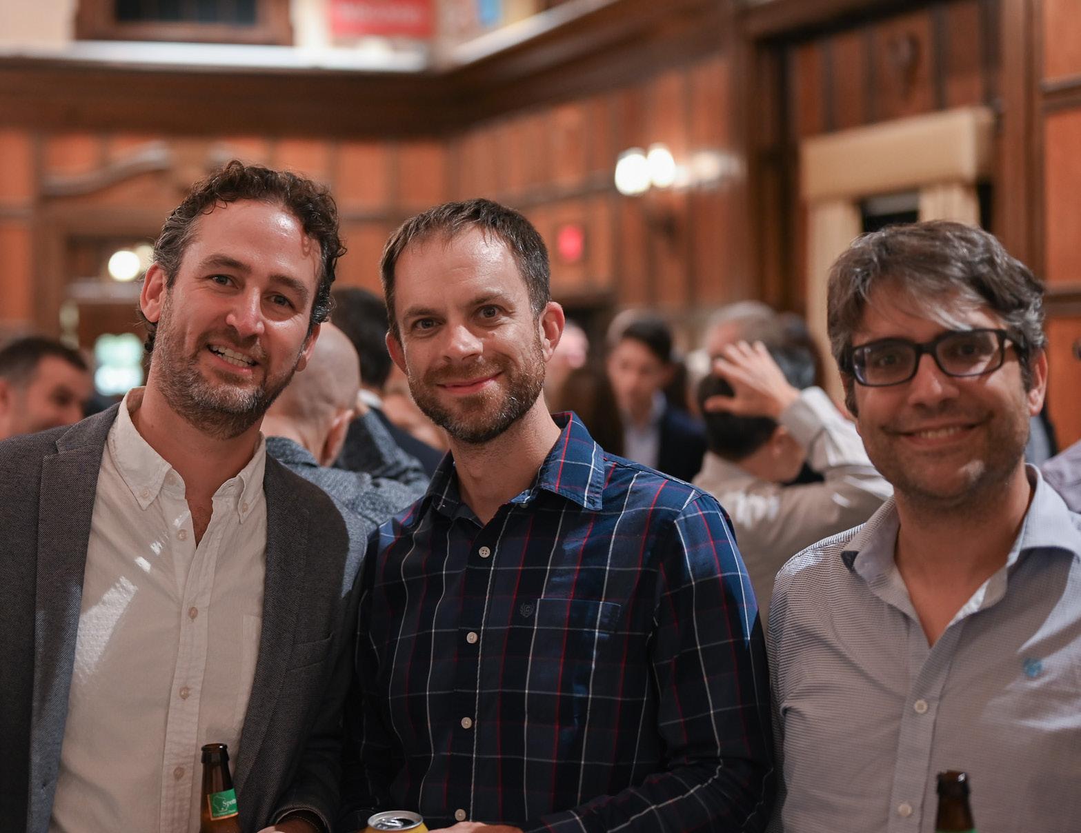
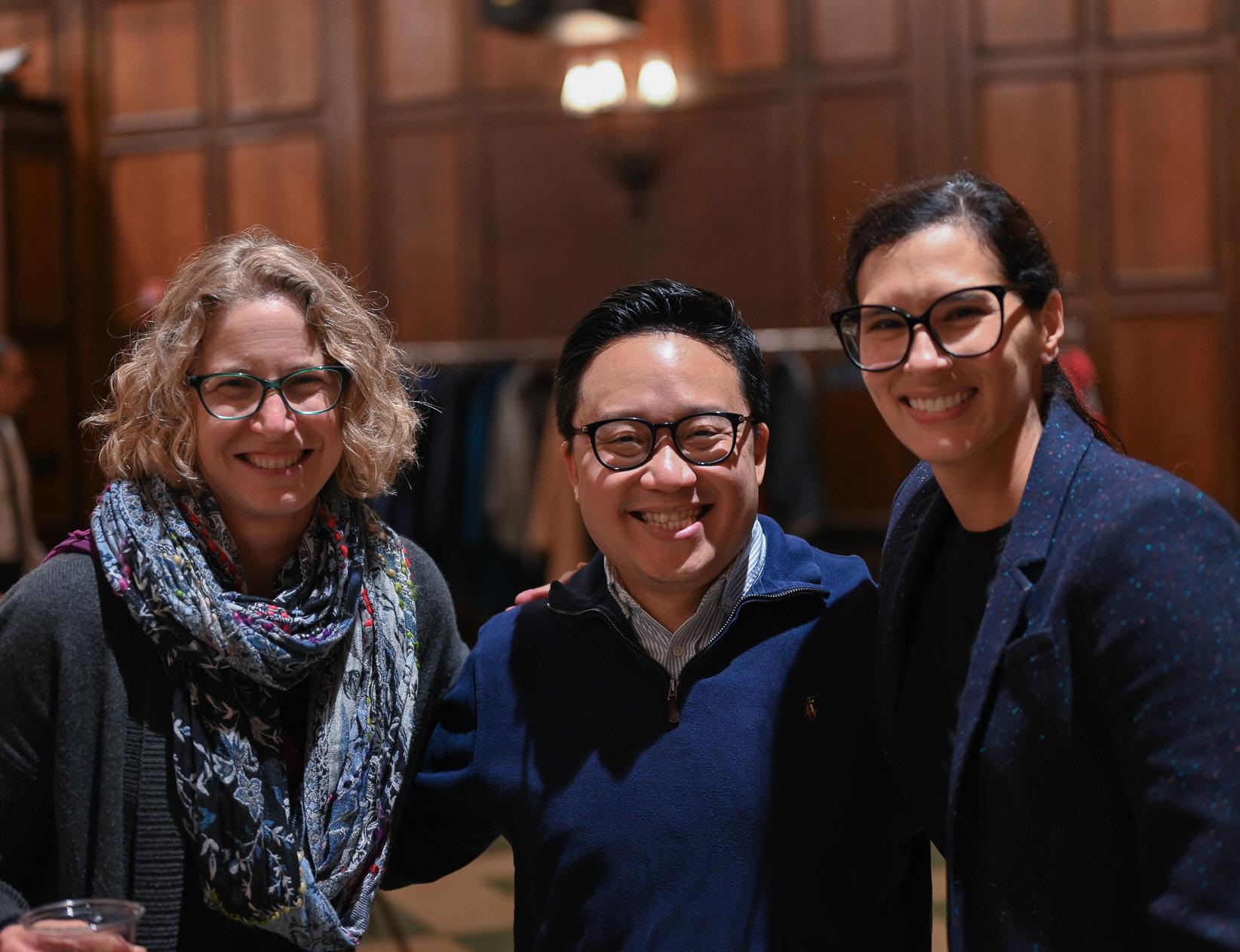
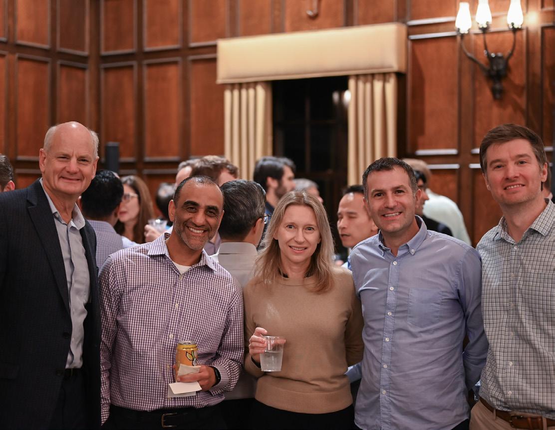
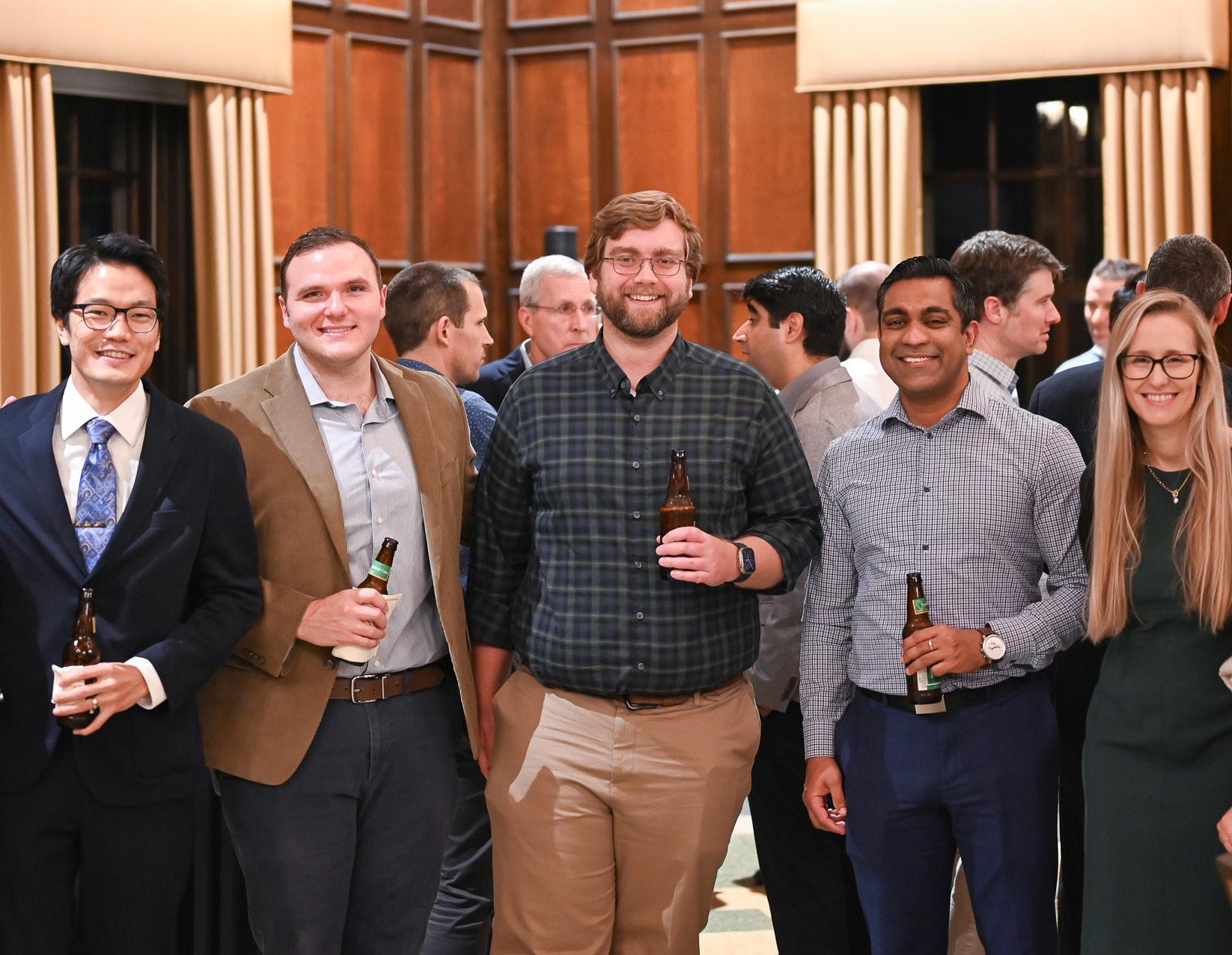
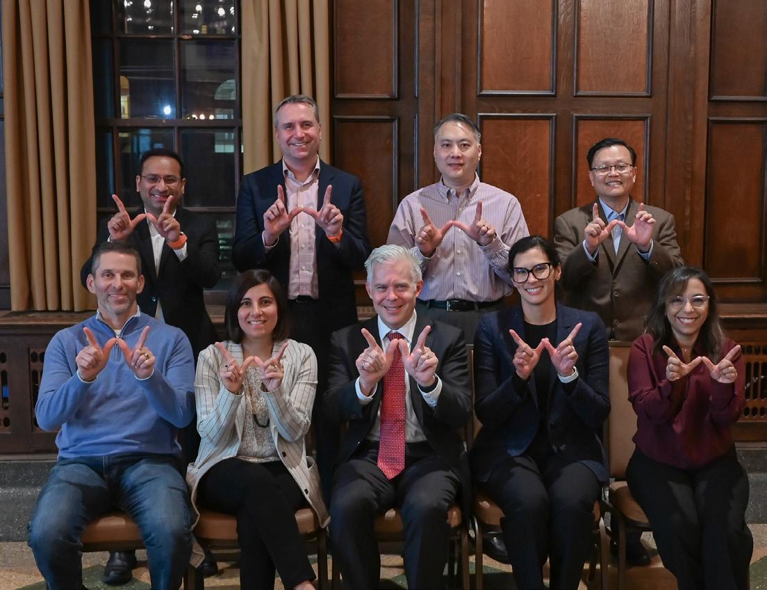
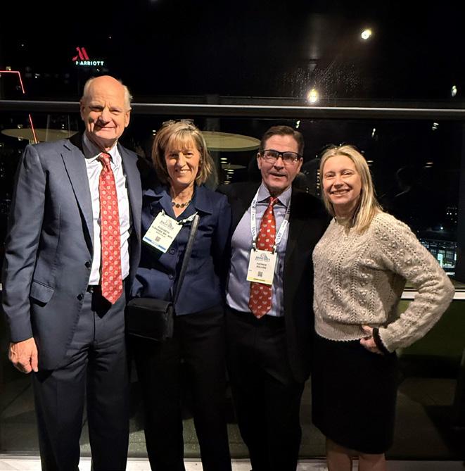
Photos from the annual Faculty Dinner held in November 2024 in Memorial Union.