37 minute read
Duchenne muscular dystrophy


Clinical course
Duchenne muscular dystrophy (DMD) is a fatal and rare muscle wasting disease with an incidence of approximately 1 in 5000 new-born boys.1, 2 The most frequently reported symptoms before diagnosis are gross motor delay, muscle weakness, difficulty walking, running and stair climbing, and frequent falls, whereas a proportion of patients show delay in cognitive and language development.3 Most boys without a family history are diagnosed before five years of age.4 Children with DMD suffer from a proximal to distal gradient of muscle weakness. The ambulatory phase of the disease can be divided into four clinical stages: early ambulatory, late ambulatory, early non-ambulatory and late-non-ambulatory.4 Clinical signs in the early ambulatory stage that can be observed are: delays in achieving developmental milestones, difficulties with running and jumping, a Gowers’ sign, frequent falls and keeping up with peers regarding gross motor functions. The late ambulatory stage is characterized by a significantly reduced walking speed, fatigue and pain after walking long distances, the increased use of a wheelchair, and difficulties with rising from floor and stair climbing. Loss of independent ambulation defines the transition to the early non-ambulatory stage. Median age at loss of independent ambulation shifted from ten to 13 years of age due to the use of glucocorticoids, although the age range remains wide.5, 6 Transition to late non-ambulatory stage is less clearly defined. Patients in the late non-ambulatory stage progressively require assistive devices to function independently, such as remote control units to operate electronic devices including televisions, computers and lights. For upper extremity function, arm muscle strength already decreases in the ambulant phase.7 In the early non-ambulatory stage patients increasingly experience difficulties raising the arms due to loss of shoulder strength. Upper arm function, such as the ability to move the hand to the mouth, is preserved until the mid-teens.5 In the late non-ambulatory phase patients have limited arm and hand function left. Hand function is preserved into the twenties, although hand strength of DMD patients has been found to be lower than that of healthy peers as early as five years of age.5, 8 Preservation of minimum function of hand muscles can significantly improve participation in daily life for patients in this stage, because it could allow them to use electronic devices such as an electric wheelchair, smartphone, tablet, computer or a game console.9
Advertisement
Not only motor functioning is affected by the absence of dystrophin, but clinical manifestations can also be observed in for instance the heart and the brain. Examples of the cognitive manifestations are a higher prevalence of learning and behavioral disabilities in DMD compared to the general population.10
Pathophysiology
DMD is an X-linked inherited neuromuscular disorder caused by mutations in the DMD gene located at the short arm of the X chromosome (Xp21 locus). The DMD gene consists of 79 exons that together encode the dystrophin protein. The mutations causing DMD lead to premature termination of dystrophin production and thereby nearly complete absence of the full-length dystrophin protein.11
Becker muscular dystrophy (BMD) is also caused by mutations in the DMD gene, but in general these mutations do not lead to absence of dystrophin, but to a partly functional dystrophin protein with an altered molecular weight. BMD patients have a more variable and generally milder disease course.12, 13
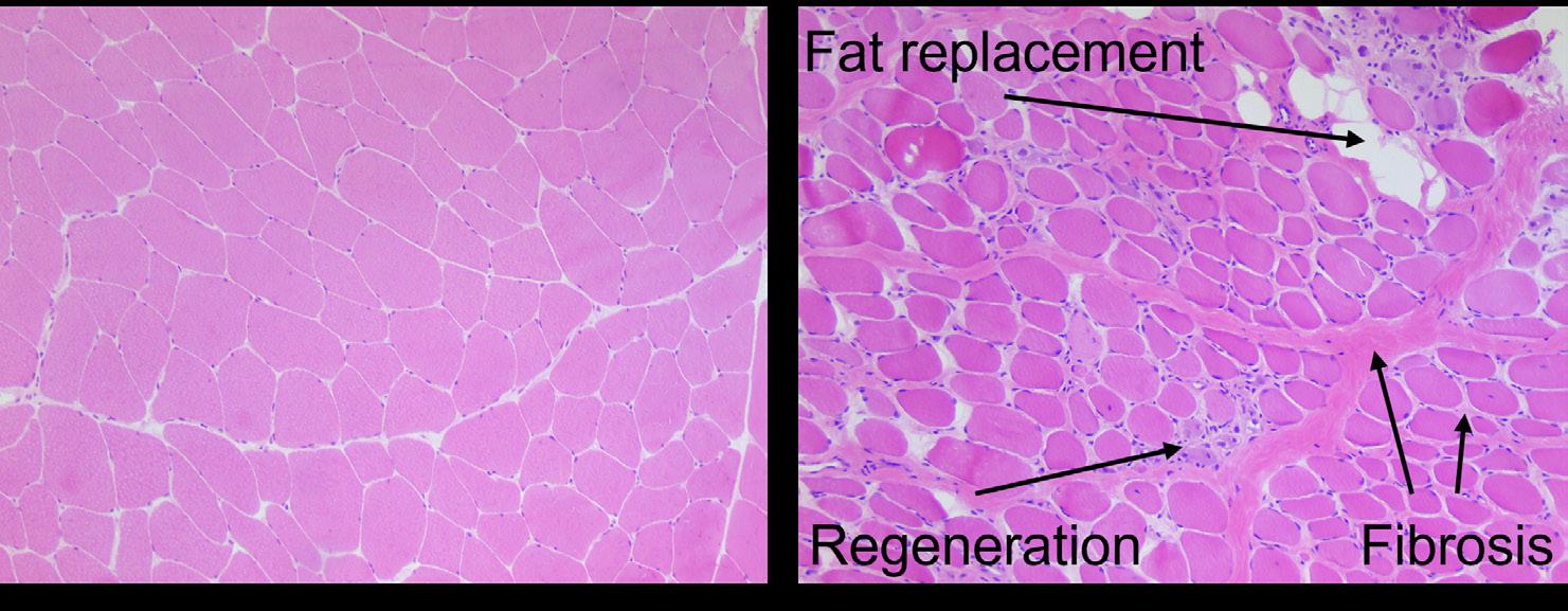
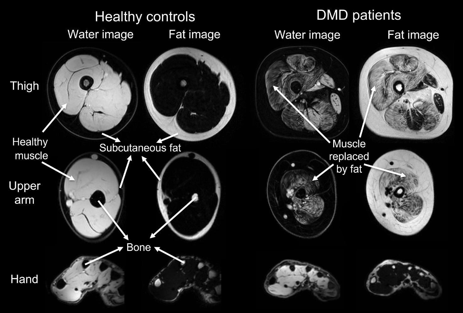
The full-length dystrophin protein is expressed in skeletal muscle, where it stabilizes the muscle fiber membrane and protects it from contraction induced damage.14 It also has a function as signaling complex in skeletal muscle by providing binding sites for signaling proteins such as nitric oxide synthase.14 In DMD, disruption of these functions is assumed to cause muscle fibers to be easily damaged, which leads to fiber degeneration and regeneration, inflammation and finally muscle wasting with irreversible replacement of muscle fibers with fat and fibrotic tissue (Figure 1).15
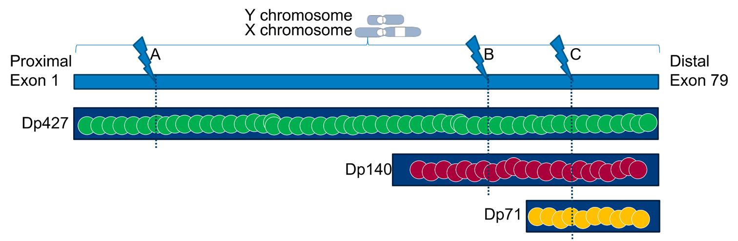
Dystrophin proteins with different lengths (i.e. isoforms) are encoded by the DMD gene. The full-length dystrophin protein is the primary isoform in skeletal muscle, but at least three dystrophin isoforms are also expressed in the brain: full-length Dp427, and the shorter Dp140 and Dp71 (Figure 2).16 The location of the mutation within the DMD gene influences the number of dystrophin isoforms that are lacking. In Figure 2, a mutation at location A (exon 1 until 44) will only lead to absence of Dp427. In case of a mutation at location B (exon 51 until 62), Dp140 will be lacking in addition to Dp427. Finally, a mutation at location C (exon 63 until 79) will lead to absence of all three dystrophin isoforms. DMD patients lacking the Dp140 isoform have been demonstrated to on average perform more poorly on neuropsychological tests and have a higher incidence of learning and behavioral disabilities.17, 18 These seem even more pronounced in patients lacking all three brain isoforms.19, 20
Image representing mutation locations in the DMD gene and their effect on the different dystrophin isoform expression. The full-length dystrophin Dp427 and the shorter Dp140 and Dp71 isoforms are present in the healthy brain. A mutation at location A, in exon 1 until 44, will only lead to absence of Dp427. Dp140 will be lacking in addition to Dp427 with a mutation at location B, in exon 51 until 62. A mutation at location C, which is in exon 63 until 79, will lead to absence of all three dystrophin isoforms. Modified with permission from Dr. N. Doorenweerd.
Clinical trials in DMD and the importance of outcome measures
The International standards of care for DMD have been published in 2010 and updated in 2018. Guidelines on the implementation of these standards of care in the Dutch healthcare system can be found on the website of the Duchenne Center Netherlands: www. duchenneexpertisecentrum.nl/passende-zorg/zorgverleners/duchenne-richtlijn/.4, 21, 22 Treatment with glucocorticoids is recommended and has been shown to delay loss of ambulation and upper limb disease progression, to reduce the need for scoliosis surgery, to improve pulmonary function, and to delay cardiomyopathy onset.5, 23 Life expectancy has shifted to the late twenties and thirties due to the combination of improved cardiac care, orthopedic interventions, the use of glucocorticoids and, most importantly, advancements in respiratory care such as air stacking, use of cough assist devices, and assisted ventilation.24, 25 Nonetheless, more severely affected patients can still die in the late teens or early twenties. A fully approved cure for DMD is currently lacking, but three dystrophin-restoring drugs have received conditional approval, two by the U.S. Food and Drug Administration (FDA) and another one by the European Medicines Agency (EMA).26, 27 Unfortunately, these drugs only seem to have a limited effect on disease progression.26
Over the past decade, many clinical trials in DMD have been conducted in primarily ambulant patients to study effectiveness of therapies aiming at dystrophin restoration or improvement of muscle quality, and many trials are still being conducted.26 Assessment of the safety and efficacy of a new therapy follows a standardized order. After preclinical studies, a new drug is studied in humans in four sequential clinical trial phases. In phase I, the optimal dosage is determined based on safety in healthy volunteers. In phase II, efficacy and side effects are assessed in a small group of patients. In DMD, phase I and II are often combined and called phase II, because of potential harm in healthy volunteers when RNA modifying drugs are used, such as antisense oligonucleotides designed to alter RNA splicing. Phase II is sometimes split in IIa and IIb, where phase IIa is specifically designed to assess the required dose and phase IIb to study efficacy in a small group of patients. Phase III is used to assess efficacy, effectiveness and safety in a larger patient population, that is predefined using a power calculation based on the effect in the phase II trials, compared to a placebo. In such randomized controlled trials (RCT), patients are randomized into the study drug arm or comparative arm, and both patients and study personnel, including clinicians and clinical evaluators, are blinded for the received treatment. It is the last phase before approval is requested from the regulators. Phase IV consists of post-marketing surveillance.
Phase III trials have a number of important characteristics. The main purpose is to demonstrate the effect of the intervention on one outcome measure, the primary endpoint. Secondary endpoints may contribute supportive information about the effectiveness of a drug, but they are less important for drug approval, because the sample size was usually not determined to find an effect on secondary endpoints. Approval of the regulatory agencies to use an outcome measure as primary endpoint depends on the natural history of the disease, the disease phase that is studied, the availability of outcome measures, and the expected effect and mechanism of action of the intervention.28
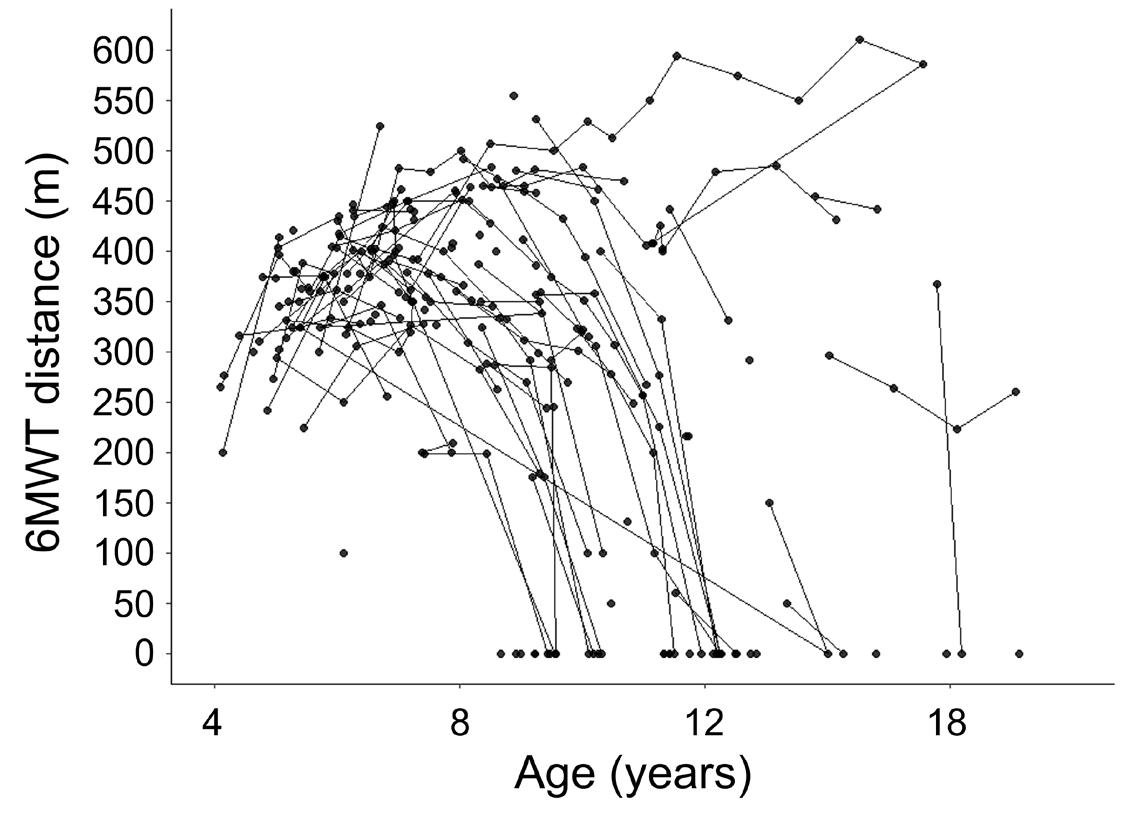
When the first phase IIb RCT in DMD was conducted using ataluren as stop codon readthrough in 2008, the six minute walking test (6MWT) was available from pulmonary and metabolic disease studies and was acceptable for regulators as primary endpoint in DMD.2931 However, no natural history data were available at that time, and it was not until the placebo arm of this trial was analyzed that it became clear that the total distance walked in the 6MWT by DMD patients first improved with age due to maturation, then stabilized and finally declined due to disease progression until loss of ambulation (Figure 3).32 Many natural history studies have since described that 6MWT distances vary largely between and within patients over time due the non-linear progression, different progression rates, and other factors such as interobserver variation and motivational issues (Figure 3).32-35 The ataluren RCT demonstrated the hurdles that arise in a maturing population of ambulant DMD patients with a heterogeneous disease course, an outcome measure that has a motivational component and with drugs that potentially limit the progression rather than improve muscle strength, and thus have a limited effect. This initiated research into the natural history and the development of new outcome measures.28 As a consequence, following trials narrowed inclusion criteria to select a more homogenous study population, which complicated study inclusion in this rare disease. Furthermore, different primary endpoints were selected that potentially had a linear progression and were more sensitive to change, such as the North Star Ambulatory Assessment (NSAA), the time to climb four stairs and the time to rise from the floor (examples of trials using these as primary endpoint: NCT04281485, NCT04632940, NCT02851797, NCT04587908 and NCT03439670). 28, 36, 37 The NSAA is an assessment of ambulatory motor performance that consists of 17 items and yields a maximum score of 34 points.37
In the past, the FDA and EMA have ruled differently in the conditional approval phase of new treatments for DMD. The FDA ruled in favor of treatments that demonstrated an increase of dystrophin levels, which has a causal relation with the symptoms in DMD and therefore were anticipated to have a potential clinical benefit upon continued treatment.38 The FDA conditionally approved eteplirsen based on an open label study that showed a minor increase in dystrophin levels of up to 0.9% after 180 weeks of treatment.26, 39 The EMA ruled in favor of treatments that demonstrated a clinically relevant effect for patients, and expected this to originate from the intended effect of a therapy. 40 The EMA therefore conditionally approved ataluren based on a subgroup analysis that showed a 6MWT difference of 68.2 meters and a favorable side-effect profile, although the study did not meet the primary endpoint for the whole group.26, 29 These differences in the FDA‘s and EMA‘s rulings thus led to conditional approval of one drug by the FDA that the EMA did not approve, and vice versa.26
There are several types of outcome measures: clinical outcome measures that require specific patient related tasks, such as the 6MWT or Performance of the Upper Limb, patient reported outcome measures, such as the DMD Upper Limb Patient Reported Outcome Measure (PROM), and biomarkers that for example reflect tissue characteristics, such as fat fraction measured using muscle magnetic resonance imaging (MRI), or dystrophin levels using muscle biopsies, or circulating muscle related micro RNA or proteins in blood or urine samples. Clinical relevance could be assumed for clinical outcome measures and patient reported outcome measures that are relevant for a specific disease and disease stage, although the amount of change that is clinically relevant is topic of debate. For biomarkers clinical relevance is less clear. One method to study clinical relevance is by using the biomarker to predict loss of an important disease milestone, such as the ability to ambulate or to bring a glass to the mouth. Another method is to determine the minimally clinically important difference, for instance via the Delphi method by using a panel of experts, or via an anchor-based approach where the biomarker is linked to an independent measure with clinical relevance to patients, such as a global rating of change.41, 42
All outcome measures should demonstrate sufficient reliability, construct validity, concurrent validity, longitudinal change and accessibility. A measure is reliable, when repeated tests lead to similar results. Construct validity is the extent to which the test measures what it is intended to measure. Concurrent validity is the extent to which the studied outcome measure correlates with an established outcome measure. An outcome measures should be sensitive to longitudinal change in DMD, even more so because therapies so far have been expected to reach a stabilizing effect in this progressive disease. The sensitiveness to change of the outcome measure and the expected effect of a therapy together determine the number of participants that is required per clinical trial. Due to its variability between patients and within patients over time, the 6MWT required relatively large sample sizes.33, 34 A more sensitive outcome measure or more effective drugs would obviously lead to a smaller required sample size and thus increase the likelihood of such a trial to be completed successfully.43 Accessibility of outcome measures is important in DMD, because trials are usually conducted worldwide over a period of years due to the rarity of the disease and use of stringent in- and exclusion criteria. Outcome measures should therefore preferably be easily operatable and accessible to use over a long period of time. It can be considered to improve existing outcome measures by changing the included items, however, the disadvantage is that new natural history data is required, such as with the improvement of the Performance of the Upper Limb from version 1.2 to 2.0.44, 45
Another important characteristic of phase III trials are the in- and exclusion criteria, which influence the generalizability of the results. Inclusion criteria often contain a statement that patients have to be able to follow study instructions, which could be harder for patients with learning and behavioral disabilities. This could result in inclusion of less patients with a distal mutation in exon 63-79, because of a higher prevalence of these symptoms in this population. Whether specific patient characteristics influence the likeliness to participate in studies could be studied by comparing baseline characteristics of participating patients to patients who were eligible but decided together with their parents to refrain from participation.
Outcome measures of the upper extremities
Outcome measures of lower extremity function, such as the 6MWT, can only be used in ambulant patients. Drugs that restore dystrophin or improve the muscle quality need sufficient muscle tissue to target and the progressive replacement of muscle by fat and fibrosis, currently considered to be irreversible, limits the amount of muscle that can be targeted. Therefore, drugs that are proven effective in ambulant patients need to be studied separately and with different outcome measures in non-ambulant patients before the EMA and FDA will approve use in these populations as well.38, 40 Most studies in non-ambulant patients have focused on the early non-ambulant phase. The available outcome measures often have a floor and ceiling effect showing little to no longitudinal change in patients who have limited function left or have little functional impairment. An example is the stable phase in the 6MWT and a maximum PUL score in many ambulant patients.34, 46 In addition, there is a lack of outcome measures that are suitable for the more advanced stages of the disease. For known outcome measures there is a need for longitudinal natural history data in non-ambulant patients. Examples are strength tests, the Performance of the Upper Limb (PUL) motor function measure and the PROM. To overcome issues of the described outcome measures, innovative outcome measures and their characteristics could be explored, such as Kinect and Leap Motion outcome measures and quantitative muscle MRI.
Upper extremity muscle strength tests
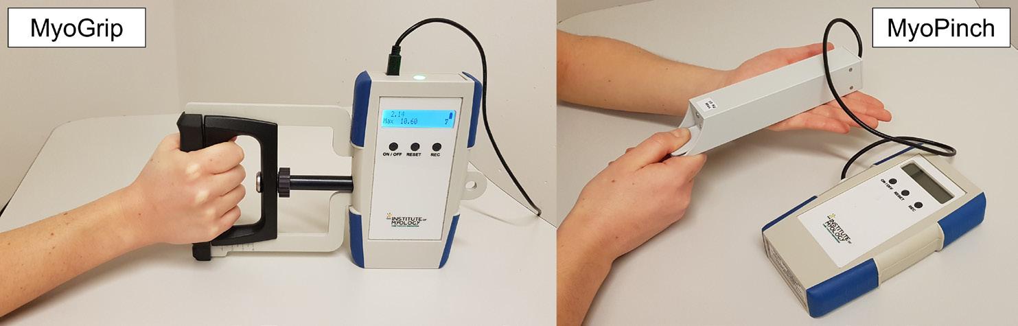
Muscle strength is considered an important outcome measure, because it can quantitatively assess the clinical effect of the underlying muscle pathology in DMD.40 However, it is not approved as primary endpoint, because FDA and EMA ruled that clinical relevance has to be established.40 Muscle strength can be measured reliably using a hand-held dynamometer,47 but this method cannot be used to assess distal muscle function in very weak patients. For this the MyoGrip and MyoPinch have been developed to assess isometric grip and pinch strength respectively (Figure 4).48 The smallest change that can be measured is 0.01 kg for the MyoGrip and 0.001 kg for the MyoPinch. Both devices have been shown to be reliable and to show differences between DMD patients and controls.48 Disadvantages are the need for a trained assessor, and vulnerable accessibility with a single manufacturer that is also needed for maintenance. Furthermore, the ability of MyoGrip and MyoPinch to predict important disease milestones has not yet been demonstrated.
Performance of the Upper Limb
The only approved outcome measure in non-ambulant patients is the Performance of the Upper Limb (PUL).44, 45 It has been used as primary endpoint in two recent clinical trials in non-ambulant patients (NCT03406780 and NCT04371666). The first widely used version of the PUL was version 1.2 consisting of 20 items with a maximum total score of 74 points. Using a Rasch analysis and input from clinicians, the scoring per item was simplified to options of 0, 1, or 2 points and some items were removed because of redundancy and some were separated or added.45 The resulting PUL 2.0 consists of 22 motor items that are assessed by an observer and it yields a maximum total score of 42 points.45 The items can be divided into three dimensions with a maximum score of 12 for the shoulder dimension, 17 for the elbow dimension and 13 for the distal wrist/hand dimension. During the course of this thesis, the first PUL longitudinal natural history data in DMD has been published. For a cohort of 177 DMD patients the annual change was found to be -1.5 points, where nonambulant patients lost on average 1.1 points more than ambulant patients.49 Limitations of the PUL are a ceiling effect in the upper and lower regions of the score and observerdependence. Its prediction of important disease milestones has not yet been demonstrated, nor is a minimal clinically important difference available.
Patient reported outcome measures for the upper extremity
For the upper extremity in DMD there is one patient reported outcome measure: the DMD Upper Limb Patient Reported Outcome Measure (PROM). The most recent version of the PROM questionnaire contains 32 daily-life activity items which are scored on a three-level scale (‘cannot do’; ‘with difficulty’; ‘easy’) with a maximum total score of 64 points.50 It describes self-reported meaningful upper extremity daily-life activities that could not otherwise be observed in a clinical or research setting (e.g. feeding, washing, and leisure activities). Internal consistency and test-retest reliability have been demonstrated. 50 Advantages are the fact that an assessor is not required and that data can be acquired offsite. Disadvantages are the subjective nature of the questionnaire and the current lack of change over time data and a minimal clinically important difference, reflecting relevant change for a patient. The FDA and EMA recommend the
use of Patient Reported Outcome
Measures as secondary outcome measure, because these can aid in determining clinical meaningfulness of objective findings of small magnitude, contribute to the assessment of benefit and risk, and assess the effect of a therapy on daily life activities.38, 40
Innovative outcome measures using Leap Motion and Microsoft Kinect
The Leap Motion controller and Microsoft Kinect v2 sensor are two innovative, low-cost marker-less motion capture systems that were developed by the gaming industry. Outcome measures of upper extremity motor function using these devices provide a continuous outcome parameter without a maximum score. This mitigates disadvantages of current outcome measures, such as a floor and ceiling effect. The Leap Motion controller uses infrared cameras to estimate the location of wrist and hand joints. This information can be used to calculate active ranges of motion (aROM) of these joints, and could have potential as outcome measure in advanced stages of DMD.51 The Kinect obtains depth data with an infrared laser transmitter and an infrared camera. Using this data, real-time 3D-coordinates of body points, including the head, shoulders, elbows and wrists, are provided to estimate human posture. So far, two methods have used Kinect to assess the reaching ability of the arms in DMD: the ‘reachable workspace envelope relative surface area’ (RSA) and the ‘Ability Captured Through Interactive Video Evaluation’ (ACTIVE).52, 53 Both measures could be used to objectively quantify upper extremity motor function without ceiling effect or observerdependence, and might therefore be more sensitive to disease progression. Potential drawbacks could be the lack of insight in constraints of the software and hardware due to intellectual property, and possible software updates and hardware discontinuation which could jeopardize their use in clinical trials.
Quantitative muscle MR methods
Quantitative muscle magnetic resonance (qMR) methods such as MRI and MR spectroscopy (MRS) can be used to assess muscle pathology in muscular dystrophies including DMD. qMR is considered to be a promising biomarker, because it has the potential to accurately assess different aspects of muscle wasting in this muscle wasting disease.54-59 MRI is non-invasive and safe, and most patients aged five years and older tolerate scanning up to one hour well without anesthesia.60-62 However, lying still for prolonged periods of time in a specific position can be strenuous for patients. qMR methods of the lower extremity have been shown to accurately describe fat replacement and tissue edema in DMD, and to have excellent testretest reproducibility within and across centers.55, 57-59, 63-66 At the start of the projects described in this thesis, muscle MRI of the upper extremities had only been studied in small cohorts of primarily ambulant DMD patients and mainly of the forearm muscles.58, 67, 68 Muscle MRI of the upper extremities could pose extra hurdles compared to the lower extremities, because of the smaller muscle mass and position of the arms at the side of the body and therefore not in the center of the MR scanner, which decreases the image quality and frequently causes artefacts. A position where the participants are lying on their side could be chosen to overcome the off-center position of the upper extremity, but this could be difficult for patients to maintain.
Different qMR techniques can be used to measure fat replacement and tissue edema. Muscle fat fraction (FF) is the most studied qMR parameter and can be assessed using water-fat MRI scans or MRS. MRS is the gold standard for FF determination with a high accuracy and reproducibility.65 A disadvantage of MRS is that results are usually obtained for one prespecified region of interest, resulting in information for only a specific part of a single muscle or muscle group. Therefore, information about spatial variation within or between muscles cannot be obtained. Tissue edema or inflammation can be studied via the T2 relaxation time of water in the muscle (T2water), which can be determined using multi-echo spin-echo (MSE) imaging or spectroscopy sequences.69-71
Fat-water imaging
Chemical shift based fat-water imaging is based on the difference in precession speed between protons in water and fat.65 In a technique originally shown by Dixon,72 images are acquired at different echo times, or phases, and from these images a purely water and fat image can be obtained (Figure 5). The FF in a specific area, typically a single muscle or muscle group, can be calculated by dividing the signal on the fat image by the combined signal of the water and fat image in that area.
qMR muscle fat fraction
In the lower extremities, muscle FF has been shown to increase with age and to correlate with motor function tests and strength of specific muscle groups.54, 57, 58, 73, 74 A study by Willcocks at al. illustrated that FF is more sensitive to disease progression than functional tests, by showing that FF increased significantly over 12 months, even in some boys in whom 6MWT distances increased due to growth.58 Also in forearm muscles FF has been shown to increase over six or 12 months, and in upper arm muscles FF correlated with the PUL and grip and pinch strength.67, 68, 75 In non-ambulant patients, limited data on qMR FF of upper arm muscles is available.75 No studies have examined very distal muscles in DMD, which are thought to be relatively spared in advanced disease stages. Furthermore, the extensively described correlations between muscle FF, strength, and function at the time of qMR do not prove causality.54, 56, 57, 68, 74, 76-79 Even unrelated biological parameters that consistently increase or decrease with age will inherently correlate with functional parameters in a progressive disease. Therefore, such correlations alone are not sufficient to show clinical relevance of muscle FF. For this, qMR FF should be able to predict loss of an important disease milestone, such as the ability to ambulate or bring a glass to the mouth. Such a predictive ability has not yet been demonstrated, and would help to qualify muscle qMR FF as outcome measure that can be used as primary or secondary endpoint in trials.
T2 relaxation time of water in muscle
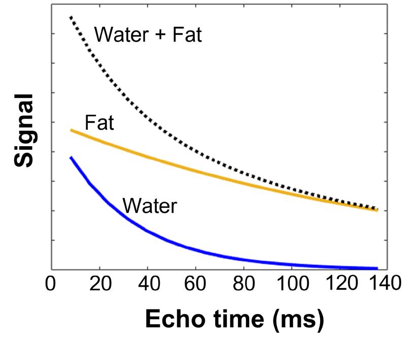
The T2 relaxation time is an MR parameter which is prolonged by increased water mobility, such as in inflammation, necrosis and fatty replacement.70 The T2 relaxation time of water in muscle (T2water) is relatively short. This parameter has been used to study tissue edema or inflammation, and thus disease activity, as these changes are thought to occur in muscles prior to fat replacement and fibrosis.69-71 T2water can be determined accurately using MRS, as water and fat signals can be easily discriminated using this method. However, this method is commonly applied using a single voxel approach due to time constraints, and hence only data are acquired from a pre-specified region of interest. T2water can also be determined with an imaging based method using an MSE sequence. In this way, spatial information is preserved and data can be obtained for a large field of view that covers different muscles or muscle groups. The T2 relaxation time of fat (T2fat) is much longer (~ 130-170 ms at 3 Tesla) than the T2water in muscle (25-30ms), and the increase in T2water due to disease activity (~ 1-4 ms).69-71 An MRI MSE sequence consist of multiple spin echoes from which the ‘global’ T2 relaxation time can be determined using a monoexponential fit of the signal decay as a function of the echo time (Figure 6).70 This global T2 consists of both the signal decay of water and fat. In DMD, due to progressive fat replacement, the signal of fat dominates the value of the global T2. Therefore, it is necessary to separate the signal decay into a water and fat component, to determine the T2water and T2fat (Figure 6).71
T2water has been studied as a qMR marker for disease activity,69, 70 and in young DMD patients, lower extremity muscles with limited fat replacement were shown to have an elevated T2water 69 Some data on T2water in forearm muscles is available, but T2water is more difficult to interpret in fat replaced muscles and therefore more difficult to study in advanced disease stages.68 No studies have examined T2water in very distal muscles, which might show interesting results in advanced disease stages in case of limited fat replacement.
Objectives of this thesis
DMD is a rare and fatal muscle wasting disorder. Beside chronic use of glucocorticoids there is currently no fully approved therapy available. Due to improved care and glucocorticoids, patients have a longer life-expectancy and therefore go longer through life in the nonambulant phase. The importance of outcome measures and natural history data was demonstrated when the 6MWT was used as primary endpoint in the first clinical trials in DMD. Outcome measures should demonstrate sufficient reliability, construct validity, concurrent validity, longitudinal change and accessibility. Clinical relevance of outcome measures should also be demonstrated, for instance via their relation with important disease milestones. Due to a progressive reduction in muscle tissue to be targeted by drugs, separate clinical trials need to be performed in non-ambulant patients, therefore specific outcome measures are required for this disease stage. The overall aim of this thesis was to identify outcome measures in DMD, specifically for non-ambulant patients, that detect an effect of a new therapy that is clinically relevant for patients in a relatively short period of time according to what is feasible in clinical trials. The use of such outcome measures could lead to a reduction in the sample sizes and potentially to a lower burden for patients.
Outline of this thesis
Chapter 2 describes reasons why DMD and BMD patients and parents declined participation in observational studies and discusses the presence of selection bias by comparing characteristics of participants and non-participants in these studies. These results can be used to optimize patient participation in studies on this rare disease and to stimulate representative observational research.
Chapter 3 describes the relation between quantitative MRI (qMRI) FF of a lower extremity muscle and loss of ambulation on top of the effect of age in ambulant patients with DMD. The aim was to predict the loss of this important milestone and thus establish the clinical relevance of muscle qMRI FF to support its use as an outcome measure in clinical trials.
Chapter 4 describes the relation between qMRI FF of an upper extremity muscle and loss of hand-to-mouth movement on top of the effect of age in non-ambulant patients with DMD. The aim was to establish clinical relevance by predicting the loss of this important upper extremity disease milestone and thus to support the use of muscle qMRI FF in clinical trials in non-ambulant patients.
Chapter 5 describes qMRI results of the thenar muscles and hand function over one year to establish the value of the thenar muscles for monitoring treatment effects in nonambulant DMD patients. Preservation of these muscles and measurable disease progression in these muscles would indicate that the thenar muscles are a valuable and quantifiable biomarker and target for systemic or local therapy in the later non-ambulant stages of the disease.
Chapter 6 illustrates the potential and constraints of using sensors from the gaming industry to develop upper extremity outcome measures for non-ambulant patients with DMD. The results of these novel outcome measures are compared to the currently used PUL and PROM outcome measures.
References
1. Mendell JR, Shilling C, Leslie ND, et al. Evidence-based path to newborn screening for Duchenne muscular dystrophy. Ann Neurol 2012;71:304-313. doi:10.1002/ana.23528
2. Moat SJ, Bradley DM, Salmon R, et al. Newborn bloodspot screening for Duchenne muscular dystrophy: 21 years experience in Wales (UK). Eur J Hum Genet 2013;21:1049-1053. doi:10.1038/ejhg.2012.301
3. Ciafaloni E, Fox DJ, Pandya S, et al. Delayed Diagnosis in Duchenne Muscular Dystrophy: Data from the Muscular Dystrophy Surveillance, Tracking, and Research Network (MD STARnet). J Pediatr-Us 2009;155:380-385. doi:10.1016/j.jpeds.2009.02.007
4. Birnkrant DJ, Bushby K, Bann CM, et al. Diagnosis and management of Duchenne muscular dystrophy, part 1: diagnosis, and neuromuscular, rehabilitation, endocrine, and gastrointestinal and nutritional management. Lancet Neurol 2018;17:251-267. doi:10.1016/S1474-4422(18)30024-3
5. McDonald CM, Henricson EK, Abresch RT, et al. Long-term effects of glucocorticoids on function, quality of life, and survival in patients with Duchenne muscular dystrophy: a prospective cohort study. Lancet 2018;391:451-461. doi:10.1016/S0140-6736(17)32160-8
6. van den Bergen JC, Ginjaar HB, van Essen AJ, et al. Forty-Five Years of Duchenne Muscular Dystrophy in The Netherlands. J Neuromuscul Dis 2014;1:99-109
7. Janssen M, Harlaar J, Koopman B, de Groot IJM. Dynamic arm study: quantitative description of upper extremity function and activity of boys and men with duchenne muscular dystrophy. J Neuroeng Rehabil 2017;14:45. doi:10.1186/s12984-017-0259-5
8. Hogrel JY, Decostre V, Ledoux I, et al. Normalized grip strength is a sensitive outcome measure through all stages of Duchenne muscular dystrophy. Journal of neurology 2020;267:2022-2028. doi:10.1007/ s00415-020-09800-9
9. Heutinck L, Kampen NV, Jansen M, Groot IJ. Physical Activity in Boys With Duchenne Muscular Dystrophy Is Lower and Less Demanding Compared to Healthy Boys. J Child Neurol 2017;32:450-457. doi:10.1177/0883073816685506
10. Banihani R, Smile S, Yoon G, et al. Cognitive and Neurobehavioral Profile in Boys With Duchenne Muscular Dystrophy. J Child Neurol 2015;30:1472-1482. doi:10.1177/0883073815570154
11. Monaco AP, Bertelson CJ, Liechti-Gallati S, et al. An explanation for the phenotypic differences between patients bearing partial deletions of the DMD locus. Genomics 1988;2:90-95
12. Bushby KM, Gardner-Medwin D. The clinical, genetic and dystrophin characteristics of Becker muscular dystrophy. I. Natural history. Journal of neurology 1993;240:98-104. doi:10.1007/BF00858725
13. Clemens PR, Niizawa G, Feng J, et al. The CINRG Becker Natural History Study: Baseline characteristics. Muscle Nerve 2020;62:369-376. doi:10.1002/mus.27011
14. Allen DG, Whitehead NP, Froehner SC. Absence of Dystrophin Disrupts Skeletal Muscle Signaling: Roles of Ca2+, Reactive Oxygen Species, and Nitric Oxide in the Development of Muscular Dystrophy. Physiol Rev 2016;96:253-305. doi:10.1152/physrev.00007.2015
15. Cros D, Harnden P, Pellissier JF, Serratrice G. Muscle hypertrophy in Duchenne muscular dystrophy. A pathological and morphometric study. Journal of neurology 1989;236:43-47. doi:10.1007/bf00314217
16. Doorenweerd N, Mahfouz A, van Putten M, et al. Timing and localization of human dystrophin isoform expression provide insights into the cognitive phenotype of Duchenne muscular dystrophy. Sci Rep 2017;7:12575. doi:10.1038/s41598-017-12981-5
17. Chamova T, Guergueltcheva V, Raycheva M, et al. ASSOCIATION BETWEEN LOSS OF Dp140 AND COGNITIVE IMPAIRMENT IN DUCHENNE AND BECKER DYSTROPHIES. Balk J Med Genet 2013;16:21-29. doi:10.2478/bjmg-2013-0014
18. Ricotti V, Mandy WPL, Scoto M, et al. Neurodevelopmental, emotional, and behavioural problems in Duchenne muscular dystrophy in relation to underlying dystrophin gene mutations. Developmental Medicine and Child Neurology 2016;58:77-84. doi:10.1111/dmcn.12922
19. Taylor PJ, Betts GA, Maroulis S, et al. Dystrophin gene mutation location and the risk of cognitive impairment in Duchenne muscular dystrophy. PLoS One 2010;5:e8803. doi:10.1371/journal.pone.0008803
20. Pane M, Lombardo ME, Alfieri P, et al. Attention deficit hyperactivity disorder and cognitive function in Duchenne muscular dystrophy: phenotype-genotype correlation. J Pediatr 2012;161:705-709 e701. doi:10.1016/j.jpeds.2012.03.020
21. Birnkrant DJ, Bushby K, Bann CM, et al. Diagnosis and management of Duchenne muscular dystrophy, part 2: respiratory, cardiac, bone health, and orthopaedic management. Lancet Neurol 2018;17:347-361. doi:10.1016/S1474-4422(18)30025-5
22. Birnkrant DJ, Bushby K, Bann CM, et al. Diagnosis and management of Duchenne muscular dystrophy, part 3: primary care, emergency management, psychosocial care, and transitions of care across the lifespan. Lancet Neurol 2018;17:445-455. doi:10.1016/S1474-4422(18)30026-7
23. Gloss D, Moxley RT, Ashwal S, Oskoui M. Practice guideline update summary: Corticosteroid treatment of Duchenne muscular dystrophy Report of the Guideline Development Subcommittee of the American Academy of Neurology. Neurology 2016;86:465-472. doi:10.1212/Wnl.0000000000002337
24. Landfeldt E, Thompson R, Sejersen T, et al. Life expectancy at birth in Duchenne muscular dystrophy: a systematic review and meta-analysis. Eur J Epidemiol 2020;35:643-653. doi:10.1007/s10654-020-00613-8
25. Bushby K, Finkel R, Birnkrant DJ, et al. Diagnosis and management of Duchenne muscular dystrophy, part 1: diagnosis, and pharmacological and psychosocial management. Lancet Neurol 2010;9:77-93. doi:10.1016/S1474-4422(09)70271-6
26. Verhaart IEC, Aartsma-Rus A. Therapeutic developments for Duchenne muscular dystrophy. Nature reviews Neurology 2019;15:373-386. doi:10.1038/s41582-019-0203-3
27. Roy B, Griggs R. Advances in Treatments in Muscular Dystrophies and Motor Neuron Disorders. Neurol Clin 2021;39:87-112. doi:10.1016/j.ncl.2020.09.005
28. Straub V, Mercuri E, Grp DOMS. Report on the workshop: Meaningful outcome measures for Duchenne muscular dystrophy, London, UK, 30-31 January 2017. Neuromuscular Disorders 2018;28:690-701. doi:10.1016/j.nmd.2018.05.013
29. Bushby K, Finkel R, Wong B, et al. Ataluren treatment of patients with nonsense mutation dystrophinopathy. Muscle Nerve 2014;50:477-487. doi:10.1002/mus.24332
30. Mendell JR, Goemans N, Lowes LP, et al. Longitudinal effect of eteplirsen versus historical control on ambulation in Duchenne muscular dystrophy. Ann Neurol 2016;79:257-271. doi:10.1002/ana.24555
31. McDonald CM, Henricson EK, Han JJ, et al. The 6-Minute Walk Test as a New Outcome Measure in Duchenne Muscular Dystrophy. Muscle & Nerve 2010;41:500-510. doi:10.1002/mus.21544
32. McDonald CM, Henricson EK, Abresch RT, et al. The 6-minute walk test and other endpoints in Duchenne muscular dystrophy: longitudinal natural history observations over 48 weeks from a multicenter study. Muscle Nerve 2013;48:343-356. doi:10.1002/mus.23902
33. Goemans N, Vanden Hauwe M, Signorovitch J, et al. Individualized Prediction of Changes in 6-Minute Walk Distance for Patients with Duchenne Muscular Dystrophy. PLoS One 2016;11:e0164684. doi:10.1371/ journal.pone.0164684
34. Mercuri E, Signorovitch JE, Swallow E, et al. Categorizing natural history trajectories of ambulatory function measured by the 6-minute walk distance in patients with Duchenne muscular dystrophy. Neuromuscul Disord 2016;26:576-583. doi:10.1016/j.nmd.2016.05.016
35. Arora H, Willcocks RJ, Lott DJ, et al. Longitudinal timed function tests in Duchenne muscular dystrophy: ImagingDMD cohort natural history. Muscle & Nerve 2018;58:631-638. doi:10.1002/mus.26161
36. McDonald CM, Henricson EK, Abresch RT, et al. The 6-minute walk test and other clinical endpoints in duchenne muscular dystrophy: reliability, concurrent validity, and minimal clinically important differences from a multicenter study. Muscle Nerve 2013;48:357-368. doi:10.1002/mus.23905
37. Mazzone E, Martinelli D, Berardinelli A, et al. North Star Ambulatory Assessment, 6-minute walk test and timed items in ambulant boys with Duchenne muscular dystrophy. Neuromuscul Disord 2010;20:712716. doi:10.1016/j.nmd.2010.06.014
38. CDER, CBER. Duchenne Muscular Dystrophy and Related Dystrophinopathies: Developing Drugs for Treatment. Guidance for Industry. https://www.fda.gov/media/92233/download. Accessed on May 1, 2019. US Food & Drug Administration (FDA) 2018
39. Charleston JS, Schnell FJ, Dworzak J, et al. Eteplirsen treatment for Duchenne muscular dystrophy: Exon skipping and dystrophin production. Neurology 2018;90:e2146-e2154. doi:10.1212/ WNL.0000000000005680
40. CHMP. Guideline on the clinical investigation of medicinal products for the treatment of Duchenne and Becker muscular dystrophy. In European Medicines Agency (EMA). Available at: http://www.ema.europa. eu/docs/en_GB/document_library/Scientific_guideline/2015/12/WC500199239.pdf. Accessed December 18, 2020.
41. Johnston BC, Ebrahim S, Carrasco-Labra A, et al. Minimally important difference estimates and methods: a protocol. Bmj Open 2015;5. doi:ARTN e007953 10.1136/bmjopen-2015-007953
42. King MT. A point of minimal important difference (MID): a critique of terminology and methods. Expert Rev Pharm Out 2011;11:171-184. doi:10.1586/Erp.11.9
43. Straub V, Balabanov P, Bushby K, et al. Stakeholder cooperation to overcome challenges in orphan medicine development: the example of Duchenne muscular dystrophy. Lancet Neurol 2016;15:882-890. doi:10.1016/S1474-4422(16)30035-7
44. Mayhew A, Mazzone ES, Eagle M, et al. Development of the Performance of the Upper Limb module for Duchenne muscular dystrophy. Dev Med Child Neurol 2013;55:1038-1045. doi:10.1111/dmcn.12213
45. Mayhew AG, Coratti G, Mazzone ES, et al. Performance of Upper Limb module for Duchenne muscular dystrophy. Dev Med Child Neurol 2019. doi:10.1111/dmcn.14361
46. Pane M., Mazzone E.S., Sivo S., et al. The 6 minute walk test and performance of upper limb in ambulant duchenne muscular dystrophy boys. PLoS Curr 2014. doi:10.1371/currents.md.a93d9904d57dcb08936f 2ea89bca6fe6
47. Mendell JR, Florence J. Manual muscle testing. Muscle Nerve 1990;13 Suppl:S16-20. doi:10.1002/ mus.880131307
48. Servais L, Deconinck N, Moraux A, et al. Innovative methods to assess upper limb strength and function in non-ambulant Duchenne patients. Neuromuscul Disord 2013;23:139-148. doi:10.1016/j. nmd.2012.10.022
49. Pane M, Coratti G, Brogna C, et al. Upper limb function in Duchenne muscular dystrophy: 24 month longitudinal data. PLoS One 2018;13:e0199223. doi:10.1371/journal.pone.0199223
50. Klingels K, Mayhew AG, Mazzone ES, et al. Development of a patient-reported outcome measure for upper limb function in Duchenne muscular dystrophy: DMD Upper Limb PROM. Dev Med Child Neurol 2017;59:224-231. doi:10.1111/dmcn.13277
51. Nizamis K, Rijken NHM, Mendes A, et al. A Novel Setup and Protocol to Measure the Range of Motion of the Wrist and the Hand. Sensors (Basel) 2018;18. doi:10.3390/s18103230
52. Han JJ, de Bie E, Nicorici A, et al. Reachable workspace and performance of upper limb (PUL) in duchenne muscular dystrophy. Muscle Nerve 2016;53:545-554. doi:10.1002/mus.24894
53. Lowes LP, Alfano LN, Crawfis R, et al. Reliability and validity of active-seated: An outcome in dystrophinopathy. Muscle Nerve 2015;52:356-362. doi:10.1002/mus.24557
54. Akima H, Lott D, Senesac C, et al. Relationships of thigh muscle contractile and non-contractile tissue with function, strength, and age in boys with Duchenne muscular dystrophy. Neuromuscul Disord 2012;22:1625. doi:10.1016/j.nmd.2011.06.750
55. Azzabou N, Loureiro de Sousa P, Caldas E, Carlier PG. Validation of a generic approach to muscle water T2 determination at 3T in fat-infiltrated skeletal muscle. J Magn Reson Imaging 2015;41:645-653. doi:10.1002/jmri.24613
56. Barnard AM, Willcocks RJ, Finanger EL, et al. Skeletal muscle magnetic resonance biomarkers correlate with function and sentinel events in Duchenne muscular dystrophy. PLoS One 2018;13:e0194283. doi:10.1371/journal.pone.0194283
57. Bonati U, Hafner P, Schadelin S, et al. Quantitative muscle MRI: A powerful surrogate outcome measure in Duchenne muscular dystrophy. Neuromuscul Disord 2015;25:679-685. doi:10.1016/j.nmd.2015.05.006
58. Willcocks RJ, Rooney WD, Triplett WT, et al. Multicenter prospective longitudinal study of magnetic resonance biomarkers in a large duchenne muscular dystrophy cohort. Ann Neurol 2016;79:535-547. doi:10.1002/ana.24599
59. Strijkers GJ, Araujo ECA, Azzabou N, et al. Exploration of New Contrasts, Targets, and MR Imaging and Spectroscopy Techniques for Neuromuscular Disease - A Workshop Report of Working Group 3 of the Biomedicine and Molecular Biosciences COST Action BM1304 MYO-MRI. Journal of Neuromuscular Diseases 2019;6:1-30. doi:10.3233/Jnd-180333
60. Chou IJ, Tench CR, Gowland P, et al. Subjective discomfort in children receiving 3 T MRI and experienced adults’ perspective on children’s tolerability of 7 T: a cross-sectional questionnaire survey. BMJ Open 2014;4:e006094. doi:10.1136/bmjopen-2014-006094
61. Mercuri E, Pichiecchio A, Counsell S, et al. A short protocol for muscle MRI in children with muscular dystrophies. Eur J Paediatr Neurol 2002;6:305-307
62. Tornqvist E, Mansson A, Hallstrom I. Children having magnetic resonance imaging: A preparatory storybook and audio/visual media are preferable to anesthesia or deep sedation. J Child Health Care 2015;19:359-369. doi:10.1177/1367493513518374
63. Forbes SC, Walter GA, Rooney WD, et al. Skeletal muscles of ambulant children with Duchenne muscular dystrophy: validation of multicenter study of evaluation with MR imaging and MR spectroscopy. Radiology 2013;269:198-207. doi:10.1148/radiol.13121948
64. Wokke BH, Bos C, Reijnierse M, et al. Comparison of dixon and T1-weighted MR methods to assess the degree of fat infiltration in duchenne muscular dystrophy patients. Journal of Magnetic Resonance Imaging 2013;38:619-624. doi:10.1002/jmri.23998
65. Burakiewicz J, Sinclair CDJ, Fischer D, et al. Quantifying fat replacement of muscle by quantitative MRI in muscular dystrophy. Journal of neurology 2017. doi:10.1007/s00415-017-8547-3
66. Hooijmans MT, Niks EH, Burakiewicz J, et al. Non-uniform muscle fat replacement along the proximodistal axis in Duchenne muscular dystrophy. Neuromuscul Disord 2017;27:458-464. doi:10.1016/j. nmd.2017.02.009
67. Ricotti V, Evans MR, Sinclair CD, et al. Upper Limb Evaluation in Duchenne Muscular Dystrophy: FatWater Quantification by MRI, Muscle Force and Function Define Endpoints for Clinical Trials. PLoS One 2016;11:e0162542. doi:10.1371/journal.pone.0162542
68. Hogrel JY, Wary C, Moraux A, et al. Longitudinal functional and NMR assessment of upper limbs in Duchenne muscular dystrophy. Neurology 2016;86:1022-1030. doi:10.1212/WNL.0000000000002464
69. Arpan I, Willcocks RJ, Forbes SC, et al. Examination of effects of corticosteroids on skeletal muscles of boys with DMD using MRI and MRS. Neurology 2014;83:974-980. doi:10.1212/WNL.0000000000000775
70. Carlier PG. Global T2 versus water T2 in NMR imaging of fatty infiltrated muscles: different methodology, different information and different implications. Neuromuscul Disord 2014;24:390-392. doi:10.1016/j. nmd.2014.02.009
71. Keene KR, Beenakker JM, Hooijmans MT, et al. T2 relaxation-time mapping in healthy and diseased skeletal muscle using extended phase graph algorithms. Magn Reson Med 2020;84:2656-2670. doi:10.1002/ mrm.28290
72. Dixon WT. Simple Proton Spectroscopic Imaging. Radiology 1984;153:189-194. doi:DOI 10.1148/ radiology.153.1.6089263
73. Godi C, Ambrosi A, Nicastro F, et al. Longitudinal MRI quantification of muscle degeneration in Duchenne muscular dystrophy. Ann Clin Transl Neurol 2016;3:607-622. doi:10.1002/acn3.319
74. Wokke BH, van den Bergen JC, Versluis MJ, et al. Quantitative MRI and strength measurements in the assessment of muscle quality in Duchenne muscular dystrophy. Neuromuscul Disord 2014;24:409-416. doi:10.1016/j.nmd.2014.01.015
75. Forbes SC, Arora H, Willcocks RJ, et al. Upper and Lower Extremities in Duchenne Muscular Dystrophy Evaluated with Quantitative MRI and Proton MR Spectroscopy in a Multicenter Cohort. Radiology 2020;295:616-625. doi:10.1148/radiol.2020192210
76. Fischmann A, Hafner P, Gloor M, et al. Quantitative MRI and loss of free ambulation in Duchenne muscular dystrophy. Journal of neurology 2013;260:969-974. doi:10.1007/s00415-012-6733-x
77. Wary C, Azzabou N, Giraudeau C, et al. Quantitative NMRI and NMRS identify augmented disease progression after loss of ambulation in forearms of boys with Duchenne muscular dystrophy. NMR Biomed 2015;28:1150-1162. doi:10.1002/nbm.3352
78. Willcocks RJ, Triplett WT, Forbes SC, et al. Magnetic resonance imaging of the proximal upper extremity musculature in boys with Duchenne muscular dystrophy. Journal of neurology 2017;264:64-71. doi:10.1007/s00415-016-8311-0
79. Gaeta M, Messina S, Mileto A, et al. Muscle fat-fraction and mapping in Duchenne muscular dystrophy: evaluation of disease distribution and correlation with clinical assessments. Preliminary experience. Skeletal Radiol 2012;41:955-961. doi:10.1007/s00256-011-1301-5
Chapter 2
Decision-making and selection bias in four observational studies on Duchenne and Becker muscular dystrophy
Published in: J Neuromuscul Dis Sept 2020; 7(4); 433-442; DOI:10.3233/JND-200541
Abstract
Background
Natural history data are essential for trial design in Duchenne (DMD) and Becker muscular dystrophy (BMD), but recruitment for observational studies can be challenging.
Objective
We reviewed reasons why patients or caregivers declined participation, and compared characteristics of participants and non-participants to assess possible selection bias in four observational studies, three on DMD and one on BMD.
Methods
Three pediatric DMD studies focused on cross-sectional cognitive function and brain MRI (DMDbrain, n=35 and DMDperfusion, n=12), and on longitudinal upper extremity function and muscle MRI (DMDarm, n=22). One adult BMD study assessed longitudinal functioning (n=36). Considerations for non-participation were retrospectively reviewed from screening logs. Age, travel-time, DMD gene mutations and age at loss of ambulation (DMDarm and BMD study only), of participants and non-participants were derived from the Dutch Dystrophinopathy Database and compared using nonparametric tests (p<0.05).
Results
The perceived burden of the protocol (38.2%), use of MRI (30.4%), and travel-time to the study site (19.1%) were the most frequently reported considerations for non-participation. Only few patients reported lack of personal gain (0.0-5.9%). Overall, participating patients were representative for the studied sub-populations, except for a younger age of DMDarm study participants and a complete lack of participants with a mutation beyond exon 63.
Conclusion
Optimizing patient involvement in protocol design, improving MRI experiences, and integrating research into clinics are important factors to decrease burden and facilitate participation. Nationwide registries are essential to compare participants and nonparticipants and ensure representative observational research. Specific effort is needed to include patients with distal mutations in cognitive studies.
Introduction
Duchenne muscular dystrophy (DMD) and Becker muscular dystrophy (BMD) are caused by mutations in the DMD gene.1 This leads to absence of dystrophin in DMD, and to a truncated and partly functional protein in BMD muscles. These neuromuscular diseases form a spectrum in which DMD patients lose ambulation around their early teens, while BMD patients have a milder but more variable disease course.2, 3 In both diseases, a higher prevalence of learning and behavioral disabilities has been reported.4-6 In DMD, this is associated with absence of different dystrophin isoforms in the brain.7, 8
Although the first drugs in DMD have now received regulatory approval, there is no cure yet.9 Currently, many studies worldwide are recruiting DMD patients simultaneously: 21 interventional clinical trials and 15 observational studies (ClinicalTrials.gov accessed on February 2nd 2020). BMD patients are being recruited for three interventional clinical trials and two observational studies worldwide (ClinicalTrials.gov accessed on February 2nd 2020). These clinical trials are challenging because of the rarity of the diseases and a variable rate of progression.9 which stresses the importance of detailed knowledge of the natural history.10 The possibility to use historical controls reduces the required number of participants per study,11 but even further highlights the need for high quality natural history data. In observational studies however, direct benefit to patients is lacking, while the added burden of research on top of the disease and clinical care could be perceived as high. Knowledge of factors that influence the decision-making process for participation can be used when designing study protocols in order to increase the participation rate and avoid selection bias. Such detailed and high quality natural history data would enable their use for placebo arms, and for determination of primary and secondary outcome measures in interventional trials. While considerations for not participating have been described for interventional trials,12-14 only one observational study reported on this topic.5
In the present study, we reviewed the decision-making considerations reported by eligible patients and compared patient characteristics of participants and non-participants in three DMD and one BMD observational studies conducted at our institute.
Methods
DMD and BMD patients were recruited in the following observational studies at the Leiden University Medical Center (LUMC): ‘Non-invasive assessment of brain involvement in DMD’ (DMDbrain; ABR number NL23184.058.09; onset of recruitment in 2010), ‘The background of the reduced cerebral blood flow in DMD’ (DMDperfusion; ABR number NL58182.058.16; onset of recruitment in 2017), ‘Upper extremity outcome measures in non-ambulant DMD patients’ (DMDarm; ABR number NL63133.058.17; onset of recruitment in 2018), and ‘The natural history study of BMD’ (BMD; ABR number NL50171.058.14; onset of recruitment in 2014). All studies are registered at ToetsingOnline (www.toetsingonline.nl). For recruitment, the Dutch
Dystrophinopathy Database (DDD) was used (‘Epidemiology, natural course and registration of dystrophinopathies in the Netherlands’; ABR number NL21411.058.08) 3. This nationwide registry, initiated in 2008, provided the opportunity for all Dutch DMD and BMD patients to list their names and contact details together with details on comorbidities, medication use, disease history and current functional status. The local ethics committee approved all studies and the registry in accordance with the ethical standards laid down in the 1964 Declaration of Helsinki and its later amendments. Written informed consent had been obtained from all patients and from legal representatives for patients under 16 years of age.
DMDbrain – Non-invasive assessment of brain involvement in DMD
Thirty-five DMD patients were recruited from the DDD and through the Duchenne Parent Project Netherlands (DPP NL) newsletter. Inclusion criteria were: male, genetically confirmed DMD patients ≥8 years old. Exclusion criteria were: MRI contra-indications such as scoliosis surgery, daytime artificial ventilation or the inability to lie supine for 45 minutes. Patients were included in the study from March 2010 until October 2012. The cross-sectional study design consisted of a single visit and included a one-hour neuropsychological assessment and two 30 minute MRI scans (at 3 Tesla and at 7 Tesla) of the brain. Results have previously been reported.15-18
DMDperfusion – The background of the reduced cerebral blood flow in DMD
Thirteen DMD patients were recruited from the DDD, the LUMC outpatient clinic, and through a poster at the DPP NL annual conference. Inclusion criteria were: ambulant male, genetically confirmed DMD patients ≥10 years old. Exclusion criteria were MRI contraindications, a medical history of cardiovascular disease, diabetes mellitus, neurological disease (other than DMD), recurrent syncope, and joint contractures preventing the use of the tilting table. Patients were included in the study beginning January 2017 and recruitment is ongoing. The cross-sectional protocol includes a one-and-a half hour tilting table experiment with transcranial doppler and blood pressure measurements, 30 minute (taskbased) MRI of the brain and cerebral vasculature, brief neuropsychological assessment (20 minutes), and cardiac ultrasound if this was not available from a recent clinical care visit.
DMDarm – Upper extremity outcome measures in non-ambulant DMD patients
Twenty-two DMD patients were recruited from the DDD, via Dutch neurologists and rehabilitation specialists, and through the Spierziekten Nederland (SN) website, the DPP NL website and Facebook page, and a poster at the DPP NL and SN annual conferences. Inclusion criteria were male, non-ambulant genetically confirmed DMD patients ≥8 years old. Exclusion criteria were: MRI contra-indications, exposure to an investigational drug ≤6 months prior to participation and recent (≤6 months) upper extremity surgery or trauma. Patients were included in the study from April 2018 until June 2019. This ongoing longitudinal study consist of three half-day visits at 0-12-18 months and the protocol includes functional upper extremity outcome measures and a 45 minute MRI scan of the upper extremity.
BMD – The natural history study of BMD
Thirty-six BMD patients were recruited from the DDD and the LUMC outpatient clinic. Inclusion criteria were male BMD patients ≥18 years old. BMD was defined as follows: an in-frame mutation in the dystrophin gene, or a reduced amount of dystrophin in a muscle biopsy, or an out-of-frame mutation with a mild disease course (>16 years old at loss of ambulation). Patients were included in the study from November 2014 until June 2016. The longitudinal study required four half-day to full-day visits at 0-12-24-36 months including functional tests, cardiac ultrasound and pulmonary function tests, and a single neuropsychological assessment. Optionally, patients could also participate in the following sub studies: 1) yearly blood sample collection for biomarker studies, 2) muscle biopsies at one time-point, and 3) lower extremity muscle MRI at two time-points. Last follow-up visit took place in August 2019.
Data collection Review of considerations for non-participation
All considerations for non-participation were obtained during the telephone calls used for the inclusion. For the DMDbrain study, the study information letter was sent first, and potential participants or their legal representatives were called within a few weeks to discuss inclusion. If patients or their legal representatives decided not to participate, they were not actively asked for reasons for non-participation as this could be perceived as pressure to participate. When they volunteered a reason, this was recorded. For the other three studies, the decision not to participate could either be made at the first telephone call, before the study information letter was sent, or at the second telephone call after reading the study information letter. At the time of these studies, more thorough implementation of Good Clinical Practice (GCP) guidelines led to more in depth logging of the screening and enrollment process. Therefore, information on considerations for non-participation in these three studies was actively requested, although patients were always allowed to not answer this question.
All considerations for non-participation that had been recorded in the screening and enrollment logs of all studies were gathered retrospectively by one observer (KJN) and checked by a second observer (ND). Patients or parents could provide one or more considerations, and these considerations were divided in the following groups: ‘Burden of protocol’, ‘Travel-time’, ‘Burden of clinical care’, ‘Other research’, ‘No advantage’, ‘Not interested’, ‘MRI’. Definitions and examples of these considerations can be found in Table 1.
Assessment of patient characteristics
Age for both participants and non-participants was defined as the age at which study information was received. For the DMDbrain study this exact date was unavailable for 30 subjects, resulting in a maximal uncertainty of nine months. Travel-time to the LUMC was derived with registered postal codes from the DDD, using ‘www.google.nl/maps’ and setting the date and time at a Monday in June 2019 outside rush hour.


