5 minute read
THE FUTURE OF CLINICAL PATHOLOGY


The future of digital pathology
As we continue to battle the COVID-19 pandemic, cancer doesn’t stop, and any delay in the diagnosis and treatment in oncology care can pose a high risk to patients.
The rapid adoption of digital pathology services has been critical in ensuring the continuation of clinical services during the pandemic, with pathologists able to conduct primary diagnoses from home while also protecting themselves and those around them.
Throughout the pandemic, the pathology department experienced a significant transformation at a scale not seen before in the field. In fact, digital pathology — the acquisition, management, sharing and interpretation of pathology information in a digital environment — has ‘come of age’ over the last two years, with research from Signify indicating the market saw 40.9% growth year over year in 2020.
Health providers and CMIOs are increasingly focusing on pathology within their wider digitalisation strategies, enabling a fully digital care solution to speed up the processing of viewing slides to help enhance decision-making. While challenges lie ahead, the power of virtualisation and the ability to connect with other teams, coupled with advances in AI, mean digital pathology is key to a new paradigm of diagnostic precision.
The power of virtualisation and care orchestration One of the main challenges pathology departments face is an increasing shortage of pathologists. In addition, pathologists are spread across multiple locations while trying to be subspecialised to provide the right expertise for difficult cases. This creates a complex workflow, where slides must be distributed optimally to the pathologists across the system, balancing workloads, but also targeting the right cases to the right experts. Complicating matters, once acquired digitally, pathology data is growing exponentially, housed in disparate systems and scattered across various departments. This lack of a fully integrated, interoperable and secure set of harmonised systems keeps data, clinicians and workflows siloed and inefficient.
Enterprise-wide digital pathology solutions are able to tackle this issue head on with technology designed to accommodate current histopathology needs for routine use in high-volume labs and integrated pathology networks. Through virtualisation and better care orchestration, cases can be routed anywhere within the network to be read, scaling access to specialists, optimising workloads and decreasing the rate of interpretation errors conducted by non-subspecialised pathologists.
Virtual networks also enable pathology departments to moderate the impact of increased caseloads as a result of the pandemic by enabling efficient diagnoses and facilitating the speedier transfer of complex cases for second opinions. Connections to other teams also provide the opportunity for pathologists to collaborate with multiple professionals, helping to improve knowledge transfer and learning opportunities. Enabling AI in pathology for deeper insights Digital pathology also opens the door for artificial intelligence (AI) and automated tools for reading slides to help empower clinicians to deliver clear care pathways with predictable outcomes for every patient.
AI-powered workflows have the potential to provide a continuous pathway, where critical patient data is made visible to both pathologists and oncologists more rapidly, helping improve the clinician experience and enhance patient care. This will be particularly important in the years ahead, as the industry balances workforce shortages with the need to meet the increasing demand for pathology services and the ongoing impact of COVID-19.
The key to a new model of diagnostic precision is bringing together multiple diagnostic insights within the healthcare continuum — like radiology, pathology and genomics — at critical states along a patient’s journey. By providing pathologists with the interoperability and connectivity to share highquality images, utilise new technologies enabled by digitisation (such as AI) and expand diagnostic insights across networks, they will become key stakeholders in the data-driven healthcare systems of the future.
Kees Wesdorp is the Chief Business Leader of Precision Diagnosis at Philips. Reposted with permission from HealthCare Business News.
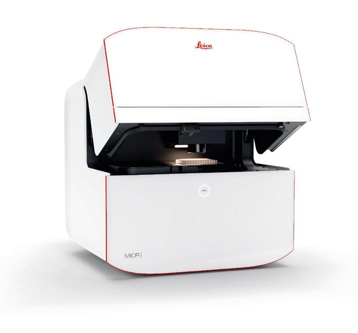
Imaging platform
Leica Microsystems has launched Mica — a type of wholly integrated imaging solution that leverages machine learning software, automation tools and fluorescence unmixing techniques to automate the imaging workflow for researchers, regardless of their microscopy experience levels. The product has been built for those who wish to utilise complex imaging in their research, while focusing more on their biology than the specialism of microscopy.
All researchers, regardless of expertise, can now work in a single digital imaging platform, moving from set-up to beautifully visualised results. The product intelligently automates sample-finding, parameter-setting and focus constancy, replacing manual set-up with just one push of a button. It is designed to eliminate over 85% of the tedious set-up steps in the conventional imaging workflow.
Users can visualise four labels simultaneously in widefield using Leica’s FluoSync technology, claimed to offer four times more data with 100% correlation compared to traditional fluorescence imaging methods. They can then switch seamlessly to confocal without moving the sample, to explore unexpected paths with no constraints.
The platform also fully integrates everything a researcher needs for radically simplified workflows, using automation and AI to enable deep understanding and a fast track to publication. For example, to perform a complex experiment such as a fluorescence multi-well plate assay, the current workflow can be simplified from 24 steps using a conventional microscope to just eight steps.
Leica Microsystems Pty Ltd www.leica-microsystems.com
Anti-daratumumab antibodies
Bio-Rad Laboratories has launched a range of antidaratumumab antibodies that are specific for daratumumab (Darzalex) and inhibit the binding of the drug to its target, CD38. The highly specific and high-affinity recombinant antibodies are suitable for bioanalysis and drug monitoring of daratumumab and its biosimilars.
Daratumumab is an anticancer drug that binds to the CD38 protein overexpressed in multiple myeloma cells, leading to immune-mediated apoptosis of the tumour cell. The range of anti-daratumumab antibodies comprises three fully human IgG1 inhibitory antibodies with varying levels of affinity. The antibodies are suitable for use as surrogate positive controls in anti-drug antibody assays, and for the development of pharmacokinetic bridging ELISAs to measure free drug.
Bio-Rad offers a portfolio of recombinant, monoclonal, nonanimal-derived anti-idiotypic antibodies and drug–target complex binders for the development of highly selective and sensitive assays. These critical reagents enable researchers to develop robust methods in a short timescale and produce translatable and reproducible results. The antibodies are generated using the Human Combinatorial Antibody Libraries (HuCAL) and CysDisplay, a proprietary method of phage display, along with guided selection methods to obtain highly targeted reagents.
The company’s recombinant production methods result in batch-to-batch consistency, so the anti-daratumumab antibodies should deliver reproducible results over the life cycle of the user’s bioanalytical assays. The antibodies are approved for in vitro research purposes and for commercial applications providing in vitro testing services to support preclinical and clinical drug development.
Bio-Rad Laboratories Pty Ltd www.bio-rad.com