March - April 2023

MasterSeries 60 Minutes Interactive


Getting the Best Patient Outcomes in Chronic Venous Disease; From Micro to Macro


Introduction

Wounds progress from hemostasis to inflammatory, proliferative, and remodelling phases, but wounds are generally considered hard-to-heal if they are stalled in the inflammation phase and have not entered the proliferation phase after 4 weeks of standard of care therapy.
Venous Leg Ulcers (VLUs) account for up to 80% of the 2.5 million leg ulcer cases per year, with an increased incidence in an ever-increasing elderly population; aside from the associated reduced quality of life, the costs involved with this are substantial, involving a high frequency of hospitalizations, wound clinic visits, home healthcare, decreased work capacity, and job loss or time lost from work; in the United States, these costs amount to nearly $15 billion annually.
MasterSeries: Getting the Best Patient Outcomes in Chronic Venous Disease; From Micro to Macro includes a panel of global experts including Dr Abigail Chaffin, Associate Professor of Surgery and Medical Director of the MedCentris Wound Healing Institute at Tulane University; editorial board


member Dr M. Mark Melin, Medical Director of the M Health Wound Healing Institute; Dr Lee Ruotsi, Medical Director at the Saratoga Hospital Centre for Wound Healing and Hyperbaric Medicine; Dr Peter Gloviczki, Editor-in-chief of the Journal of Vascular Surgery and Professor of Surgery (Emeritus) at the Mayo Clinic Rochester, Minnesota, and Dr Monika Gloviczki, scientist and artist at Vascular Science and Art.
Associate Professor of Surgery and Medical Director, MedCentris Wound Healing Institute at Tulane University New Orleans LA, United States


Reconstructive Considerations

In the 4 weeks of standard of care, there should be a 50% reduction in the wound size. If not, it may be necessary to signal for a change or additional therapies or advanced extracellular matrices (ECM). This is important, because generally wound area reduction after 4 weeks is predictive of complete wound healing by 24 weeks. Local and national guidelines should be followed, with incorporation of evidence-based modalities that lead to the highest quality outcomes with the most appropriate resource utilization in an integrated approach.
The goals are to alleviate pain, rapidly heal the ulcer

and to prevent long term sequelae.
General treatment algorithms for VLUs include commencing with standard compression, leg elevation, and then a thorough wound bed preparation with a sharp excisional debridement, a high consideration for bacterial control with appropriate wound cleansers (it is possible to use a pure hypochlorous acid preserved wound cleanser); early use of ECMs when appropriate for coverage of vital structures, dermal regeneration and accelerating primary healing of small VLUs (preferred option would be to use an ovine forestomach matrix graft). Split Thickness Skin Grafts (STSG) should be considered for definitive coverage of larger VLUs.
Pure Hypochlorous Acid (pHA) Preserved Cleanser
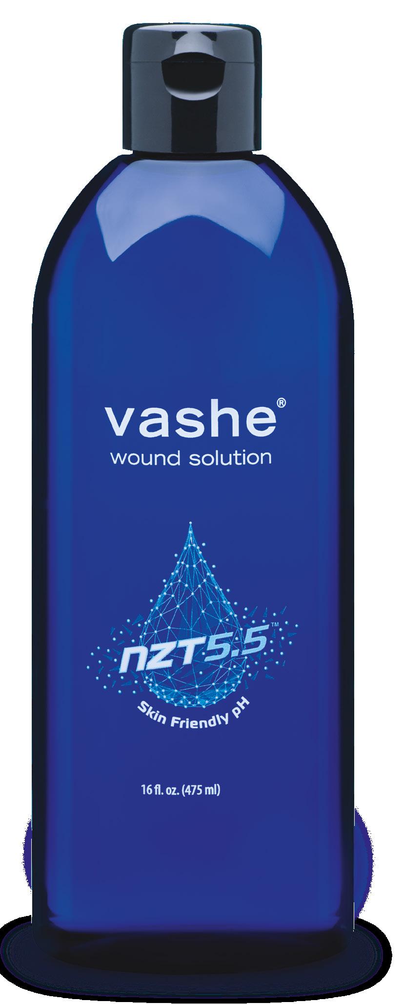
This is a pure Hypochlorous Acid (pHA) preserved cleanser (Vashe®, Urgo Medical North America, Fort Worth, TX), manufactured at 300 ppm (parts per million); it has a pH range of 3.5 - 5.5, which is conducive to healing.2 It is not cytotoxic,3 unlike standard sodium hypochlorite solutions, which are considered toxic even at low concentrations. This product is consumed by organic matter in the wound and dissipates in seconds, so it is safe to use with ECMs in the same setting.4 For wounds that have microbial adherent aggregates, 5-8 minutes of wound contact with the solution should be considered,5 in combination with sharp debridement. This mechanical disruption of germs and associated necrotic debris is a rapid process, and does not require long exposure like other some other modalities may.
Some evidence and very recent consensus documents and guidelines suggest discontinuation of cytotoxic cleansing agents such as Dakin’s or solutions that contain cytotoxic agents such as chlorhexidine gluconate. In 2022 consensus guidelines were published by the International Wound Infection Institute,6 which included discussions from global experts on the use of hypochlorous acid versus sodium hypochlorite. A key section of this guideline discusses the issue of margin of safety; this is the therapeutic index.7 Hypochlorous acid as a
preservative has a higher therapeutic index compared to the hypochlorite present in Dakin’s, and also some cleansers that combine variable proportions of elements of hypochlorous acid (harmless) and hypochlorite (toxic). The pH of 3.5 to 5.5 of pHA is well below the pKa of HOCl (7.5), with the added attribute that the pHA preserved cleanser has a greater efficacy with a lower toxicity to the tissue.
The hypochlorous acid molecule, a naturally evolving substance inside the human tissue, forms part of the cellular barrier immunity. Humans are designed to live with hypochlorous acid in their tissue, which explains the lack of cytotoxicity of the pHA based cleanser.
A study in an academic centre looking at sulfamylon or mafenide and bacterial adherent aggregates reports that the mafenide solution had a flat curve; very little effects occurred on the bacterial adherent aggregates, as compared to the pHA based cleanser solution. Antibiotic Stewardship is a key topic to incorporate into clinical practice and It is vital not to overuse antibiotics to prevent the ever evolving challenge of drug resistance. Mafenide was also unable to remove Candida colonies, while the pHA preserved cleanser did so easily; which also illustrates that while the pHA preserved cleanser is able to kill planktonic germs in seconds in a situation where microbial aggregates adherent to tissue is involved, it may be appropriate to soak the wound for 5 - 8 minutes.
Ovine (Sheep) Forestomach Matrix
An effective advanced ECM for use in VLUs is ovine forestomach matrix (OFM) (EndoformTM Antimicrobial; EndoformTM Natural; Myriad MatrixTM , Aroa Biosurgery, Auckland, New Zealand). After years of research, the forestomach of sheep in New Zealand was identified as an ideal starting tissue to construct advanced ECM technologies for soft tissue repair. This source tissue from juvenile sheep (less than 12 months of age) is highly abundant in New Zealand, and the animals are disease free. The forestomach, even from these young animals, is very large, meaning large sheets of ECM can be easily manufactured. The
“Humans are designed to live with hypochlorous acid in their tissue, which explains the lack of cytotoxicity of the pure hypochlorous acid (pHA) based cleanser.”
forestomach is a highly vascular tissue, designed for digestion that is integral for rapid nutrient absorption. This also means that residual vascular channels in the graft help it to revascularize more quickly, and then this tissue undergoes more rapid remodelling, where soft tissues are continuously renewed in the body; it has a high concentration of secondary macromolecules, which are known to be important for healing.12
A study of ovine forestomach matrix usage in a military veteran hospital in 201713 shows that in nine months, cellular or tissue-based graft unit usage decreased by 60%; expenditures on these grafts decreased by 66%, with a higher and faster rate of healing.
Chronic wounds tend to be deficient in a quality ECM; this is comprised of a complex process of cells, blood vessels, and macromolecules.10 The ECM is important for structural and biochemical support. Chronic wounds often have deficient ECM, senescent fibroblasts and increased matrix metalloproteinases (MMPs). Enhancing and optimising the ECM with a replacement ECM to heal these ulcers more effectively should be considered. To that end, it is important to consider what features make an ideal replacement? An ideal advanced ECM affords a rapid 100% or near 100% wound closure, long term tissue durability, cost effectiveness, and an ease of storage and transport. The outcomes that are measured by multiple studies include the percentage of closed wounds, the time to closure, wound recurrence or infections, need for amputation or hospitalization, and a time to return to baseline Activities of Daily Living (ADLs), pain reduction, odour and exudate reduction, and also other adverse effects.
Patients that have failed healing of large venous ulcers with standard of care in conjunction with a granulated wound base, with no bone or tendon exposure and a high quality of skin coverage is desired, a STSG may be considered.
Utilizing Synergistic Effects of Both Modalities

An ideal operative algorithm for STSGs could be considered as the following: a combination of performing a thorough intra-operative ultrasonic debridement with pure hypochlorous acid solutions; sharp debridement with a curette; and ovine forestomach matrix coverage. The graft can then be stapled or sutured in place with a thrombin sealant. A standard dressing protocol can be used which can include: a non-adherent silver silicone dressing with Negative Pressure Wound Therapy (NPWT); appropriate 4 or 5 layer compression wrap dressings, and then immobilization at the ankle or knee as indicated. The most complex patients are often admitted, put on bed rest, and some require a stay for optimal take of the graft; and compression and post-operative patient education.
The Typical Venous Leg Ulcer Checklist
It is important to run a typical VLU checklist like an airplane pilot’s pre-flight checks to maximize outcomes and achieve consistency in terms of treatment. To begin, a vital check is to make sure that there is adequate blood flow; in current demographics, about 20-25% of patients have some component of either diabetes or a metabolic syndrome which can compromise both microvascular as well as macrovascular arterial flow. Investigation with venous competency ultrasound is critical in identifying both saphenous and non-saphenous reflux and perforator status. The EVRA study and the ESCAR study suggest that axial and non-axial venous reflux should be treated early, managing the venous hypertension as a basis for wound healing. To attempt healing without managing the venous
“Decellularized extracellular matrix–based biomaterials are of great clinical utility in soft tissue repair applications due to their regenerative properties. ”Global expert Dr M. Mark Melin Medical Director of the M Health Wound Healing Institute Mineapolis MN, United States
hypertension is not found to be effective practice.
A nutrition evaluation typically falls way down on the checklist; it is established that the micronutrient components to maximize nitric oxide production which improves lymphatic function, micro arterial vasodilation, venous tone and immune function are all incredibly beneficial to these patients who often times may be micronutrient deficient. It is vital to focus on the B vitamins (B12, B6 folate), and high dose vitamin C to help with decreasing reactive oxygen species and antioxidants; a caveat on vitamin C is that if the patient has a history of renal stones, it may be necessary to be careful and discuss with the primary care physician. Vitamin D can also have an impact, not just on bone health, but on microvascular perfusion. Arginine certainly contributes to nitric oxide production and micronized purified flavonoid fractions (MPFFs). A new way to look at albumin is not necessarily as a marker of overall nutritional health; it is actually a component of the endothelial glycocalyx, it helps as a permeability layer, so when the albumin is low increased levels of microvascular hyperpermeability may be seen, which can influence subcutaneous edema, potentially compromising microvascular arterial perfusion, as well as immune function.
Certainly, looking at the medication list is important, because medications like amlodipine or other calcium channel blockers can contribute directly to lower extremity edema; typically amlodipine needs to be stopped to maximize edema control. Oncology patients undertaking chemotherapy, or who are on hydroxyurea, are certainly at a high risk of inhibited long term healing.
Good hyperglycaemic control is important; again, hyperglycaemia can shed the very fragile layer of the endothelial glycocalyx, which can result in more edema.
It is prudent to consider biopsy; concerning atypical vs typical ulcerations, if an atypical is missed such as a malignancy, or pyoderma gangrenosum, and it is treated as a typical venous leg ulcer, there will not
be a good outcome.
Evidence shows that the wound pH should be controlled at a mildly acidic, low pH range of between 3 to 6. It is established through peer reviewed, validated data that decreasing wound pH can help with decreasing pathogen proliferation, improving angiogenesis, and improving accelerated epithelialization, when with other types of adjunctive modalities.
Another important consideration is compression, which is critical; it is important to get certified lymphedema therapists involved and begin utilizing inelastic Velcro; this is easy to get on and off over the top of dressings, and it is important not to forget about both the foot component and the calf component.
Cost effective wound bed modulation, as shown with ovine forestomach, should be opted for. Also, smoking has to be ceased, including vaping.
For bacteria proliferation management, a pHA preserved cleanser is recommended; it has a stable shelf life of approximately 18 months, a pH range over its shelf life at 3.5 to 5.5, and it is noncytotoxic for wound and peri-wound environments; it decreases inhibitory wound bed pathogens; it can be used in conjunction with NPWT after surgical debridement to maximize lowering of the wound pH, and avoid recurrence of formation of adherent germ aggregations; it is applicable to all wound sites. It has been used in trauma, pyoderma wounds, and diabetic foot ulcerations. In terms of the extracellular matrix, ovine forestomach matrix should be used in the operating room, and Endoform™ Antimicrobial or Endoform™ Natural in the clinic. Myriad Morcells™ is another option that is available; it is a structured retention of architecture from the forestomach of young sheep, typically less than 12 months old. The stomach of sheep has to heal very quickly to maintain the health of the animal, so the ovine forestomach matrix has a diverse array of collagens including 1, 2, 3, and 5; as well as an abundance of glycoproteins and proteoglycans. All of these are preserved through a
“It is prudent to consider biopsy; concerning atypical vs typical ulcerations, if an atypical is missed such as a malignancy, or pyoderma gangrenosum, and it is treated as a typical venous leg ulcer, there will not be a good outcome.”
very specific process and retention of vascular channels, and it is cost effective.
It is an important consideration to try to focus on decreasing interstitial edema, and maximizing lymphatic functionality, also resulting in immune function enhancement, as by decreasing subcutaneous edema we can maximize microvascular arterial perfusion at the 5-micron level. To consider peri-wound lymphatic stasis, it is occurring in almost every wound; the epiboly we see in wounds is actually built-up lymphatic stasis, and when lymphatics are static, the immune system does not function well. By maximizing peri-wound lymphatic stasis resolution, overall wound healing can be improved.11,14,15
To conclude, the steps should include the checklist for VLUs with special attention to avoiding missing the identification of Peripheral Arterial Disease (PAD). It has been observed a growing demographic with diabetes and metabolic syndrome in the population. Another focus point is avoiding undertreating lymphedema; recognising malnutrition and treating it appropriately, not just the protein component but the micronutrients component. In terms of investigations venous ultrasounds must be done early. Vigilance with medications contributing to lower extremity edema and modify them. Help patients to wean themselves off tobacco.
and skin changes such as hemosiderosis (extravasated red blood cells in the breakdown of haemoglobin). Chronic inflammation and fibrosis of skin can often be mistaken for cellulitis. The morphology of VLUs tends to be irregularly shaped, full thickness ulcerations with moderate to large exudate, with typically no exposed underlying bone, tendon, or other structures.
General treatment guidelines include vascular workup; ensuring an adequate arterial workup, including ankle brachial indices and toe brachial indices to support that gold standard of multi-layer compression. It is important to remember that both clinically applied compression and the post-closure garments that the patients wear at home have to be tailored to the patient’s life status. Utilizing arteriograms, Computed Tomography Angiography (CTA) and Magnetic Resonance Arteriography (MRA).
Lymphedema is a common presentation to outpatient wound centers commonly presenting with open wounds with a multitude of challenges. Most outpatient wound centres are poorly equipped to manage lymphedema, and many lymphedema providers and clinics are unwilling to accept patients with open wounds. Therefore, this brings about a dilemma that these lymphoedematous wounds need to be closed before the lymphedema can actually be managed appropriately, with the skill and technique that can really only be provided by lymphedema therapists.
Arterial Insufficiency Ulceration
The Differential Diagnosis of Lower Extremity Ulcerations

VLUs begin distally with the feet and ankles, moving proximally as venous hypertension develops. Over time varicosities develop, and this is worse after prolonged standing or activity and the venous hypertension symptoms such as aching, discomfort
In this condition the clinical features tend to be punched out, with well-defined edges almost as though the wound was created with a drill-bit. They tend to occur over distal bony prominences; in appearance, the wound beds tend to have an unhealthy and pale granulation tissue. They tend to be fairly dry, with minimal exudate, black to brown eschars, and the surrounding skin tends to be ruborous and hairless. In terms of management of arterial insufficiency ulceration, it is vital to make the diagnosis and then refer to an appropriate vascular surgeon. It is important that there is an adequate arterial
“The critical catalyst for Buerger’s disease is cigarette smoking; in a patient who is not a cigarette smoker, a diagnosis of this disease cannot be made.”Global expert Dr Lee Ruotsi Medical Director at the Saratoga Hospital Center for Wound Healing and Hyperbaric Medicine Saratoga NY, United States
supply. Priorities should be completing the VLU checklist; thinking out of the box and ensuring that you make that diagnosis of arterial insufficiency in a timely fashion. Don’t always rely on your fingers; remember to use a hand-held doppler, MRA, and CTA.
Debridement should be performed cautiously, especially in the ischemic limb; and on the advice from the National Pressure Injury advisory panel: it is important not to debride dry stable heal eschar; leave it alone allowing it to demarcate.
Thromboangiitis Obliterans
Also known as Buerger’s disease, this is an arterial clotting in the hands and feet, with the resultant ulceration and gangrene. It is a vaso-occlusive, inflammatory vasculopathy, where inflammatory thrombi occlude vessels but spare the vessel walls. The critical catalyst for Buerger’s disease is cigarette smoking; in a patient who is not a cigarette smoker, a diagnosis of this disease cannot be made.
Vasculitis
Vasculitis can occur in any organ system, however interestingly the skin is the most common organ system affected. The bilateral lower legs are the most common location. The typical description is a painful palpable purpura. Biopsy is done for diagnosis; doing a biopsy on the most immature of the areas will lead to the highest return in diagnosis. Leukocytoclastic of inflammatory vasculitis is the most common form of cutaneous vasculitis.
Scleroderma
Scleroderma, most commonly the C.R.E.S.T. syndrome, and also sometimes known as progressive system sclerosis, is an autoimmune disorder of unknown etiology ulcers, usually over the digits, pre-tibial area, and bony prominences. One of the most troubling features of this disease process if the subcutaneous calcification, known as calcinosis cutis. There are yellow areas around the periphery of these wounds, so sometimes those areas are able to be distracted or extracted, almost like dental extractions,
but getting these wounds to epithelialize is very difficult.
These are painful ulcers of varying depth and size, with purulent wound beds and a blue-black wound edge. PG is most commonly associated with underlying autoimmune and occasionally malignant disease. The most common association is with inflammatory bowel disease such as ulcerative colitis or Crohn’s disease. System corticosteroids and other disease modifying agents such as cyclosporin are often used in treatment. A very important concept with pyoderma gangrenosum is that of pathergy where, if the ulcers are sharply debrided, they grow large.
This is an unusual form of squamous cell, or occasionally basal cell carcinoma. Marjolin’s ulcers were originally described by French surgeon JeanNicholas Marjolin in the late 1700’s, as a squamous cell cancer arising in an area of prior skin trauma. In the United States, these are colloquially called scar carcinomas or burn carcinomas, and are now known to be roughly 80% squamous cell and 20% basal cell. A 73-year-old lady presented to the clinic with a large VLU in an area where she had prior venous leg ulceration. It appeared as a fairly typical looking venous ulcer. She was quite mobility impaired, so home nursing care was arranged and she was brought back in 4 weeks for a follow-up. When she returned she had an odd looking area in the superior aspect of the wound; this was biopsied and it turned out to be a poorly differentiated squamous cell carcinoma. This patient’s prior area of skin damage ended up being her recurrent VLU; biopsy early and biopsy often; there are very few wounds that can be negatively affected with a 5mm punch biopsy.
Pyoderma Gangrenosum Marjolin’s Ulcer“Biopsy early and biopsy often; there are very few wounds that can be negatively affected with a 5mm punch biopsy.”
Diabetic Foot Ulcer
5 year mortality of neuropathic Diabetic Foot Ulcer (DFU) is almost 50%. Vascular assessment is of critical importance. Remember the tri-neuropathy; we always think of the sensory neuropathy, but remember that we have the motor neuropathy that leads to the bony deformities, and then the autonomic neuropathy that leads to the dry foot, leading to fissuring and cracking. Infection and osteomyelitis are common.
Treat reflux early; vascular specialists should be consulted as soon as possible because we want to treat the underlying incompetence (whether it is superficial or perforated) early.32
Treat iliofemoral venous obstruction; if the patient has associated deep venous obstruction, we certainly need to do this. This can be done very effectively with relatively low risk but good clinical success.
Interventions for Treatment of Chronic Venous Insufficiency
Chronic venous disease is a huge spectrum of medical conditions. This is one of the most prevalent medical conditions in the world. With the increasing age of the population, there is likely to be a higher prevalence of venous ulcerations, and the associated economic cost of this is well understood. Although it affects elderly patients more frequently, there is a significant economic burden because of loss of working days in people who are still working, and it is not infrequent to see these patients having to retire early, with a reduced quality of life.1,26
There are guidelines on how to treat patients. These guidelines need upgrades frequently, not only because there is a large number of new venous ulcer treatments that are not interventional, but because there have been tremendous changes in vascular reconstruction because of the endovascular revolution.27,28,29,30

Strategy for VLU
In establishing the diagnosis of venous ulcers, duplex scanning is extremely important to establish the status of venous incompetence, and excluding the underlying arterial disease is important, as is performing the VLU checklist, looking at diabetes vasculitis and other rare ulcerations. Compression treatment is of course a usual part of immediate treatment, and treatment of infection is extremely important. Cleanse and debride the ulcer and apply dressing. Maintain a moist surface, absorb and excess fluid, and protect the surrounding skin.
For persistent/ recurrent ulcers, treat residual reflux or obstruction. If stenting is not possible, the Palma procedure or surgical bypass should be performed. For nonhealing wound ulcers, duplex scanning should be repeated to look for new or recurrent reflux; the effectiveness here of skin grafting and bilayer cellular therapy is well evidenced. To summarize, the best strategy to treat venous ulcers is to confirm the right diagnosis, optimize the local care, treat the infection, initiate compression therapy, and treat the underlying reflux and obstruction early to prevent/ treat, if there is ulcer recurrence.
Scientific Societies' Guidelines
The last guidelines of the American Venous Forum and Society for Vascular Surgery suggest nutrition assessment in any patient with VLU and evidence of malnutrition; nutritional supplementation should be provided if malnutrition is identified. For long standing or large venous ulcers, the 2 societies recommended adjunctive therapy with pentoxifylline or Micronized Purified Flavonoid Fraction (MPFF).
The guidelines of the Wound Healing Society attributed recommendations of Level I for pentoxifylline and MPFF, in conjunction with compression therapy to improve healing of venous ulcers.
Just recently the ESVS published Clinical Guidelines on the management of Chronic Venous Disease and included adjuvant pharmacotherapy to the algorithm of treatment of patients with active venous ulcers. In the recommended pharmacologic treatments, based on the level A evidence, the guidelines listed MPFF, hydroxyethylrutosides, pentoxifylline or sulodexide, combined with compression and local wound care. All the guidelines were using the same definition of scientific evidence, levels A, B, and C.


