28 minute read
Characterizing geometrical accuracy in clinically optimized 7T and 3T MR images for high-precision radiation treatment of brain tumours
Chapter 5 - Characterizing geometrical accuracy in clinically optimized 7T and 3T MR images for high-precision radiation treatment of brain tumours
Jurgen Peerlings, Inge Compter, Fiere Janssen, Christopher J. Wiggins, Alida A. Postma, Felix M. Mottaghy, Philippe Lambin, Aswin L. Hoffmann
Advertisement
Physics and Imaging in Radiation Oncology. 2019;9:35-42.
Abstract
Background and Purpose
To assess whether the spatial accuracy in 7-Tesla (7T) MR images, previously optimised for anatomical imaging, is sufficient for clinically-acceptable radiation treatment planning(RTP) in neuro-oncology in respect to standard clinical imaging-modalities. Materials and Methods
System- and phantom-related geometrical distortion(GD) was quantified on clinically-relevant MR-sequences at 7T and 3T, and on CT-images using a dedicated anthropomorphic head-phantom incorporating a 3D grid-structure, creating 436 points-of-interest. Global GD was assessed by mean absolute deviation (MADglobal). Local GD relative to the magnetic isocentre was assessed by MADlocal. Using 3D displacement-vectors of individual points-of-interest, GD maps were created. For clinically acceptable radiotherapy, 7T-images need to meet the criteria for accurate dose delivery (GD<1 mm) and present comparable GD as tolerated in clinicallystandard 3T-MR/CT-based RTP.
Results
MADglobal in 7T- and 3T-images ranged from 0.3−2.2 mm and 0.2−0.8 mm, respectively. MADlocal increased with increasing distance from the isocentre, showed an anisotropic distribution, and was significantly larger in 7T MR-sequences (MADlocal=0.2-1.2 mm) than in 3T (MADlocal=0.1-0.7 mm)(p<0.05). Significant differences in GD were detected between 7T-images (p<0.001). However, maximum MADlocal remained ≤1mm within 68.7 mm diameter spherical volume. No significant differences in GD were found between 7T- and 3T-protocols near the isocentre.
Conclusions
System- and phantom-related GD remained ≤1 mm in central brain regions, suggesting that 7T MR-images could be implemented in radiotherapy with clinicallyacceptable spatial accuracy and equally tolerated GD as in 3T-MR/CT-based RTP. For peripheral regions, GD should be incorporated in safety margins for treatment uncertainties. Moreover, the effects of sequence-related factors on GD needs further investigation to obtain RTP-specific MR protocols.
Introduction
In radiation treatment planning (RTP) of brain tumours, magnetic resonance imaging (MRI) at 1.5 or 3 Tesla (T) is currently being used as the standard anatomical imaging modality owing to its superior soft-tissue contrast compared to computed tomography (CT)1. Generally, after co-registration of MR and CT images, the former is used for target volume definition and the latter is used for dose calculation, as tissue electron density information is missing in MR-images. However, the current clinical MR-techniques are limited in depicting detailed neurologic malformations such as intracerebral tumour spread2. With the prospect of clinically-certified ultrahigh field (UHF-)MRI systems (≥7T), images with higher signal-to-noise ratio (SNR), higher spatial resolution, and novel contrast mechanisms such as quantitative susceptibility-weighted images will become available, enabling the visualisation of small lesions, basal ganglia, and tumour angiogenesis3-5. The improved ability in detecting microvasculature (diameter ~100μm) could play a decisive role in staging primary brain tumours such as glioblastoma (GBM), as tumour angiogenesis is directly associated with tumour grade, and could aid in determining relevant location of dedifferentiated cells for image-guided biopsies6,7. In addition, 7T-MRI could aid target volume definition in RTP by visualising microvasculature outside the contrastenhanced tumour and infiltration of GBM cells along white matter tracts7-9. During follow-up scans, 7T-MRI has been shown to reveal radiation-induced microbleeds, indicating potential neurocognitive decline and the need to adapt RTP10,11 .
However, concerns regarding geometrical distortion (GD) with increasing static magnetic field strength (B0) have compromised the integration of UHF-MRI into radiation treatment planning. System-related GD are mainly induced by imperfections in hardware (B0-inhomogeneity, gradient field nonlinearity, and eddy currents), applied radiofrequency (RF) pulse and parameter settings (sequence-dependent GD), while phantom-related GD (chemical shift and susceptibility differences) are related to the object’s shape placed in the MR-system12,13. To be applicable for highprecision RTP, the spatial accuracy of anatomical images needs to be within 2 mm for non-stereotactic radiotherapy and within 1 mm for stereotactic radiotherapy, as is the case for 3T-MRI13-18. In fact, profound GD may have significant dosimetric impact in radiotherapy as the precision of beam targeting and dose calculations could be affected19,20. For high-precision dose delivery techniques that apply steep 3D dose gradients (intracranial stereotactic radiotherapy or radiosurgery), spatial inaccuracies of >1 mm could result in clinically-unacceptable dose variations of more than 15% that could lead to underdosage of target volume and/or overdosage of surrounding tissue20,21. Ultimately, inaccuracy of beam targeting and dose calculation could lead to a different clinical outcome and local tumour control13,18. For instance, techniques that apply extremely steep dose gradients, such as stereotactic radiotherapy and radiosurgery, are susceptible to significant dosimetric effects (5-32% underdosage) caused by even small GD of 1.3 mm21,22. The question therefore arises whether our
clinical 7T-MRI protocol, optimised for anatomical imaging, can produce images with acceptable spatial accuracy needed for reliable high-precision radiation treatment of brain tumours. Previously several investigators have evaluated geometrical distortion on either CT, 3T- or 7T-MRI, demonstrating a maximum GD of up to several mm in large cylindrical or cubical phantoms14,23-27 .
This study aims to investigate the clinical applicability of anatomical 7T-MRI in comparison to 3T-MRI and CT for reliable high-precision radiotherapy by evaluating the magnitude and location of hardware-related GD. We hypothesise: 1) acceptable submillimetre spatial accuracy could be achieved in our 7T-MRI protocol and no significant differences in GD are present between different 7T-sequences; 2) the tolerated level of GD in 7T-MRI is not significantly different from system-related GD in a predefined 3T-MRI protocol, clinically used for RTP. To test these hypotheses on both global level (GD throughout the entire volume-of-interest (VOI)) and on local level (GD relative to the distance from the magnetic isocentre), we performed quantitative image analyses using a dedicated anthropomorphic head-phantom in a clinically-realistic set-up. Hence, different sequence parameter settings and distortion correction methods were not explored in this study.
Materials and methods
Phantom
A dedicated anthropomorphic head-phantom (CIRS Model 603A, Computerized Imaging Reference Systems Inc., USA) was used to evaluate system-related GD in 7T-MRI, 3T-MRI, and CT-images (Fig. S.1). The anatomical shape demanded similar procedures used for clinically imaging patients (FOV setting, shimming, robust positioning inside the scanner and head-coil). Furthermore, the centre of the phantom was aligned with the magnetic isocentre.
The phantom was composed of a plastic-based bone substitute and soft-tissue equivalent fluid consisting of a water-based polyacrylamide with added NiCl2 (Zerdine®, CIRS). Inside the phantom, 3-mm thick nylon rods formed a 3D grid with 15 mm spacing (±0.05 mm manufacturing tolerance), defining a VOI of 15×12×13.5 cm3 (anterior-posterior(AP) × left-right(LR) × superior-inferior(SI)). At grid-intersections, 437 measurable points-of-interest (POIs) were identified. Nylon shows similar magnetic susceptibility properties to water (χ <3 ppm) and was assumed not to induce such susceptibility artefacts in spin-echo or gradient-echo sequences 24,28 .
Image acquisition
MR images were acquired on a 7T-MR system (Magnetom 7T, Siemens, DEU) and on a 3T-MR system (Achieva 3T, Philips, NL) using 32-channel head coils from Nova Medical inc. (1Tx/32Rx, Wilmington, USA) and Invivo Corp. (Florida, USA), respectively. The 7T-MR system was equipped with Siemens’ SC72-gradient system (80 mT/m peak gradient amplitude, 200 T/m/s maximum slew rate). The 3T-MR system was fitted with Philips’ Dual Quasar gradient system (80 mT/m peak gradient amplitude, 100 mT/m/s maximum slew rate). All MR sequences were previously optimised to acquire anatomical images of high image-quality within an acceptable scan duration, and were not altered for this study[9]. The selected 7T-MR sequences included magnetization-prepared rapid gradient-echo (MP2RAGE), T2-sampling perfection with application optimized contrasts using different flip angle evolution (T2-SPACE), T2-SPACE with fluid attenuation inversion recovery (T2-SPACE FLAIR), and multiecho gradient-echo (T1-GRE)9. Sequences with equivalent pulse schemes were selected from a predefined clinical 3T-MR protocol used in RTP and included turbo field echo (3D TFE), volume isotropic turbo spin-echo (T2-VISTA) with and without FLAIR, and fast field gradient echo (T1-FFE), respectively (Table S.1). Vendor-installed 3D distortion correction methods were applied to restore gradient non-linearity and 3D volume-based second order shimming procedures were applied to improve B0 homogeneity14,29,30 .
CT images were acquired (SOMATOM Sensation 10, Siemens, Germany) with a slice thickness of 1 mm, 306 slices, 50×50 cm2 FOV, 140 kV, and 400 mAs.
Methods of analyses
The known 3D coordinates of all POIs were defi ned as reference dataset and manually reconstructed in MRI and CT images using Eclipse treatment planning system (Varian Medical Systems, USA). The X-, Y-, and Z-axes defi ned the LR-, SI-, and AP-directions in both datasets, respectively.
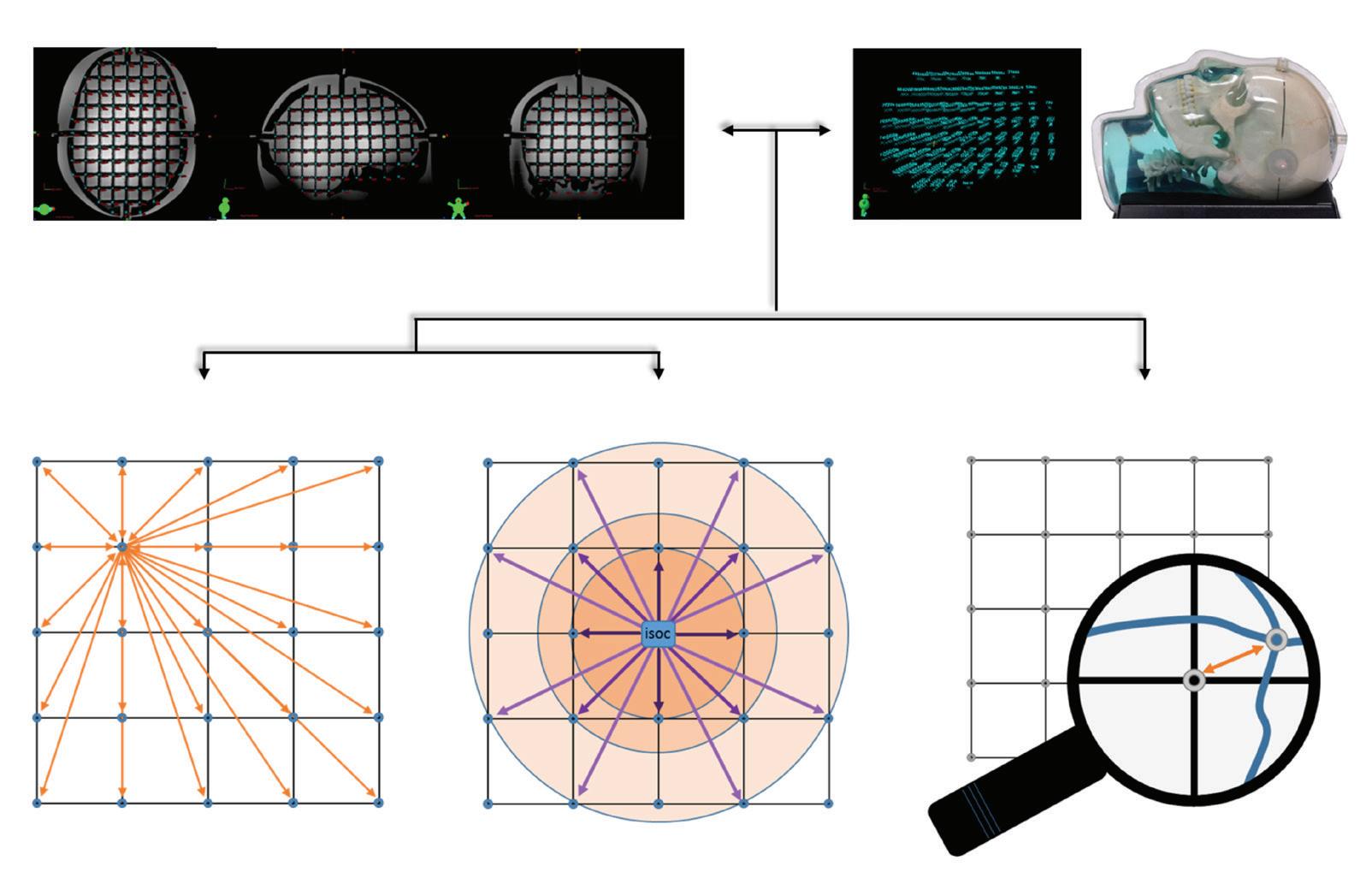
Three methods were used to assess GD at diff erent levels of sophistication using in-house developed MATLAB software (R2014b, MathWorks Inc, USA) (Fig.1). Firstly, mean absolute deviation (MAD) was computed to assess GD on a global level (MADglobal) throughout the entire VOI, independently of location within the phantom. Secondly, MAD was calculated to assess GD on a local level (MADlocal) relative to the distance from the magnetic isocentre. MAD was calculated according to:
(1)
where i = 1…M is an index into set Δ={δ1,δ2,…,δM } of M unique distances, Di (j) is the measured Euclidean distance for the j-th unique pair of POIs having an Euclidean distance in the reference dataset, and represents the number of unique pairs of POIs. For MADglobal, Δ contained all unique distances that could be observed within the entire phantom. For MADlocal, Δ contained only unique distances that originate from the POI nearest the magnetic isocentre. The mean MAD was averaged over all M unique distances in Δ
The third method of analyses pin-pointed GD by calculating 3D displacement of each individual POI by computing the Euclidean distance directly between the POIcoordinates in the image (Xm, Ym, Zm) and the coordinates in the reference datasets (Xref, Yref, Zref):
(2)
The resulting 3D displacement vector (Dxyz) indicated relative GD of each individual POI, independently of all other POIs.
The three methods of analyses provided complementary information on the geometrical quality of the images. The MADglobal quantifi ed the level of GD throughout the entire VOI, thereby ignoring information on the absolute and relative locations of the POIs. The MADlocal retained spatial information by quantifying the GD with respect to the diameter of a spherical volume (DSV) at the magnetic isocentre.

To quantify spatial integrity of an entire image, 95% confidence interval (|CI95|) was calculated from MAD and standard deviation (SD) as:
(3)
Based on this interval, a maximum acceptability-level of 1 mm was defined. The level of GD was considered acceptable when MADlocal ≤1 mm (at a specific DSV) and |CI95| ≤1 mm.

1a. Measured dataset: image-based 1b. Reference data: phantom-based
2a. Global MAD* 2. Mean Absolute Deviation (MAD)
2b. Local MAD 3. Relative deviation
3a. Displacement vector
Fig. 1: Overview of 3D data acquisition and analyses. XYZ-coordinates are determined on CT and MR images (1a.), and based on product characteristics of the CIRS’ phantom model 603A (1b.). Global MAD (2a.) is based on distances with 2 variable grid-intersection points (*example given for 1 intersection but applies for all points). Local MAD (2b.) is based on distances between magnetic field isocentre and 1 variable grid-intersection points. Displacements vectors (3a.) are determined between the measured and reference coordinates of each individual data-point and indicate the relative 3D geometrical distortion

Statistical Analyses
To assess differences in sequence-dependent GD, MADglobal and MADlocal were analysed between 7T-MRI sequences and between 3T-MRI sequences using a Kruskal-Wallis test. To assess differences in tolerated GD-levels, MADglobal and MADlocal were analysed between equivalent sequence pulse schemes in 7T- and 3T-MRI using a multiple student t-test with Bonferonni-Sidak correction. For MADlocal, DSV at which statistical significance in MADlocal between sequences occurred, was identified by a separate Kruskal-Wallis test for each set of unique distances. All statistical analyses were performed using GraphPad Prism v6.01 (GraphPad, USA). P-values <0.05 were considered to indicate statistical significance.
Results
Global GD
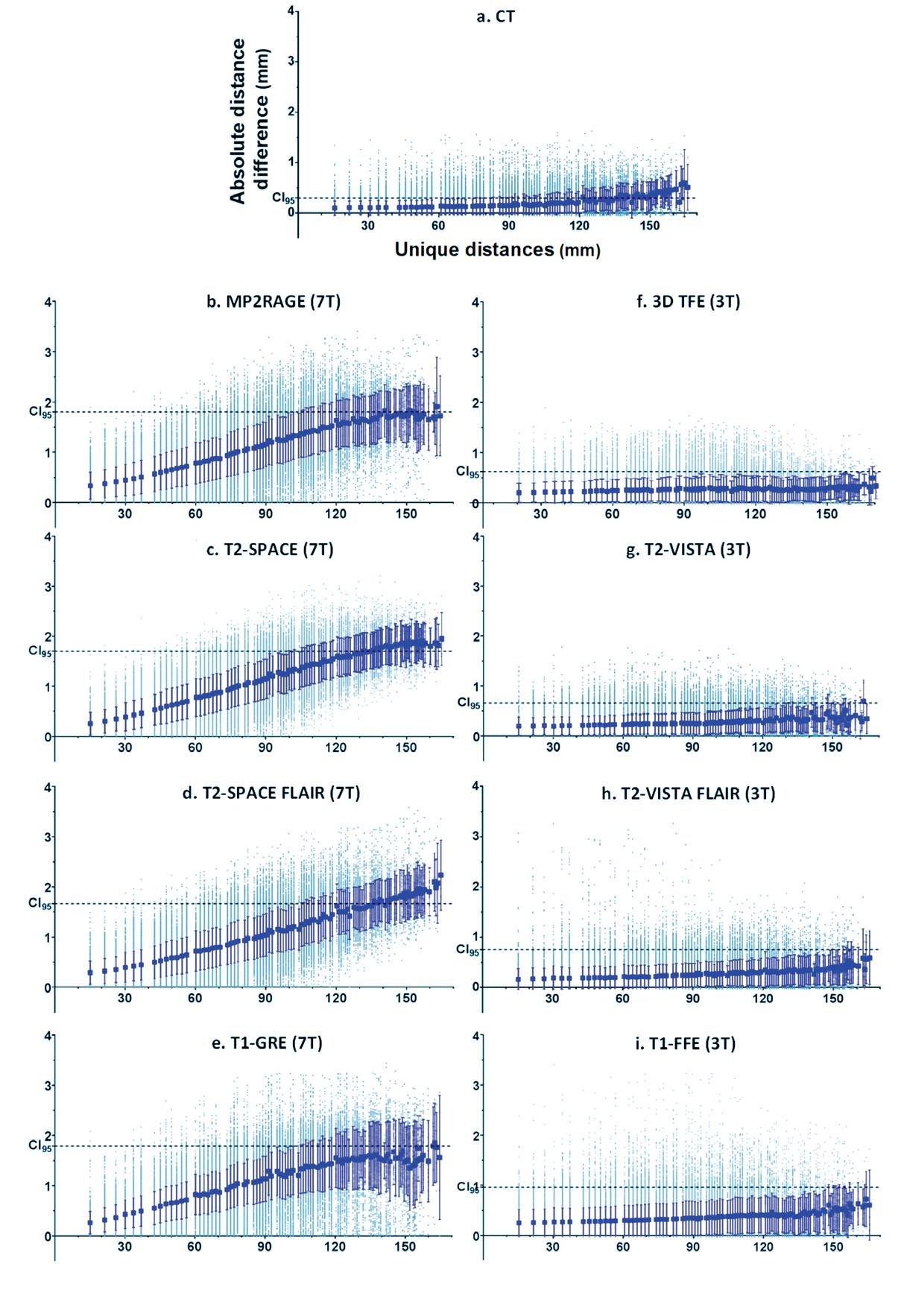
In total, 95.266 pairs of distances were identified and binned into 100 unique distances, ranging from 15 mm to 164.3 mm. In CT images, MADglobal ranged from 0.1−0.6 mm with mean MADglobal of 0.3±0.1 mm. For MR images, MADglobal ranged from 0.3−2.2 mm and 0.2−0.8 mm in 7T- and 3T-MRI, respectively (Table 1, Fig.2). Based on |CI95|, only 3T-MR sequences presented GD <1 mm throughout the entire image. No statistical significance in MADglobal were found between 7T-MR images (p=0.38) or 3T-MR images (p=0.13). However, significant differences in MADglobal were detected between all equivalent sequences on 7T and 3T MR-system(p<0.001). (Fig. 3.).
Table 1: Global geometrical deformation as measured on 3T and 7T MRI (*** = P < 0.001)
MADGlobal (in mm) MADGlobal (in mm)
B0 Sequence Range Mean ± sd |CI95| B0 Sequence Range Mean ± sd |CI95| P value 7T MP2RAGE 0.3–1.9 1.0 ± 0.3 1.7 3T 3D TFE 0.22–0.51 0.27 ± 0.23 0.7 ***
SPACE 0.3–2.0 1.0 ± 0.4 1.7 T2-VISTA 0.20–0.70 0.26 ± 0.20 0.7 ***
SPACEFLAIR 0.3–2.2 0.9 ± 0.3 1.6 T2-VISTA FLAIR 0.19–0.62 0.29 ± 0.23 0.7 ***
GRE 0.3–1.9 1.0 ± 0.4 1.8 T1-FFE 0.3–0.7 0.4 ± 0.3 1.0 ***
Local GD
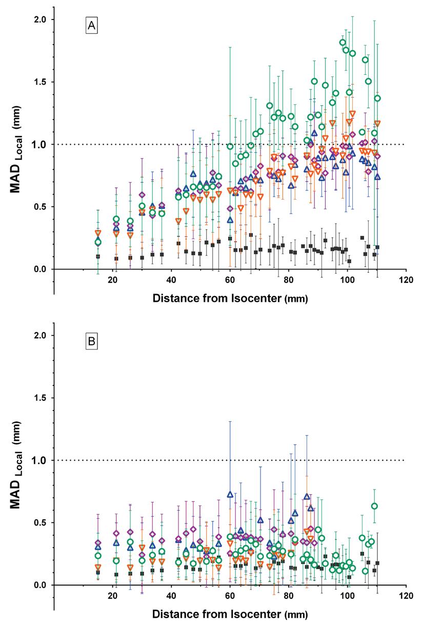
In CT images, MADlocal ranged from 0.1-0.3 mm with mean MADlocal of 0.2±0.04 mm( Fig.4). All MR images acquired at 7T presented a steady increase in MADlocal with increasing distance from the magnetic isocentre. MADlocal ranged from 0.2−1.8 mm for 7T-MRI and from 0.1−0.7 mm for 3T-MRI. Each 7T-MR sequence remained below the clinical tolerance-level of 1 mm up until a defined DSV (Table 2). For T1-GRE, MADlocal exceeded 1 mm only at DSV of 87.5-88.7 mm.
For all 3T MR sequences, MADlocal remained below 1mm throughout the whole VOI. MADlocal of MP2RAGE was statistically greater than all other 7T MR sequences (P < 0.001, Fig. S.2). No significant difference in MADlocal was found between T2-SPACE, T2- SPACE FLAIR (P=0.064), and T1-GRE (P=0.093), respectively. Within the 3T dataset, MADlocal of 3D TFE was significantly different from T2-VISTA (P < 0.001), T2-VISTA FLAIR (P=0.046), and T1-FFE (P < 0.001). T2-VISTA FLAIR presented significantly different MADlocal from T1-FFE (P < 0.001). MADlocal was not significantly different between T1-FFE and T2-VISTA (P=0.51), and the T2-VISTA and T2-VISTA FLAIR sequences (P=0.061). However, all sequences met the criteria for GD and seemed to have found an equilibrium with requirements for anatomical imaging.
Statistical analyses of MADlocal between equivalent 7T and 3T-sequences indicated significant differences for all sequences (p<0.001). However, no significant difference in MADlocal was found between equivalent sequences when the DSV was considered (Table 2).
Table 2: Local geometrical deformation as measured on 3T and 7T MRI (***=P < 0.001, §=DSV at which statistical significance between sequences occurred)
MADLocal (in mm)
B0 Sequence 7T MP2RAGE
SPACE
Range Mean ± sd |CI95| <1mm at DSV 0.22-1.81 0.85 ±0.30 1.45 68.6
0.21-1.08 0.68 ±0.28 1.23 101.7
SPACEFLAIR 0.27-1.24 0.61 ±0.28 1.17
GRE 0.24-1.09 0.68 ±0.27 1.23 92.5
87.5-88.7
MADLocal (in mm) P value B0 Sequence Range Mean ± sd |CI95| <1mm at DSV Sign. P<0.05 at DSV 3T 3D TFE 0.11-0.63 0.24 ±0.19 0.62 all *** 42.4 T2-VISTA 0.23-0.45 0.38 ±0.26 0.90 all *** 68.7 T2- VISTA FLAIR 0.14-0.43 0.21 ±0.17 0.55 all *** 47.4 T1-FFE 0.21-0.73 0.35 ±0.28 0.91 all *** 45-56.1
3D displacement
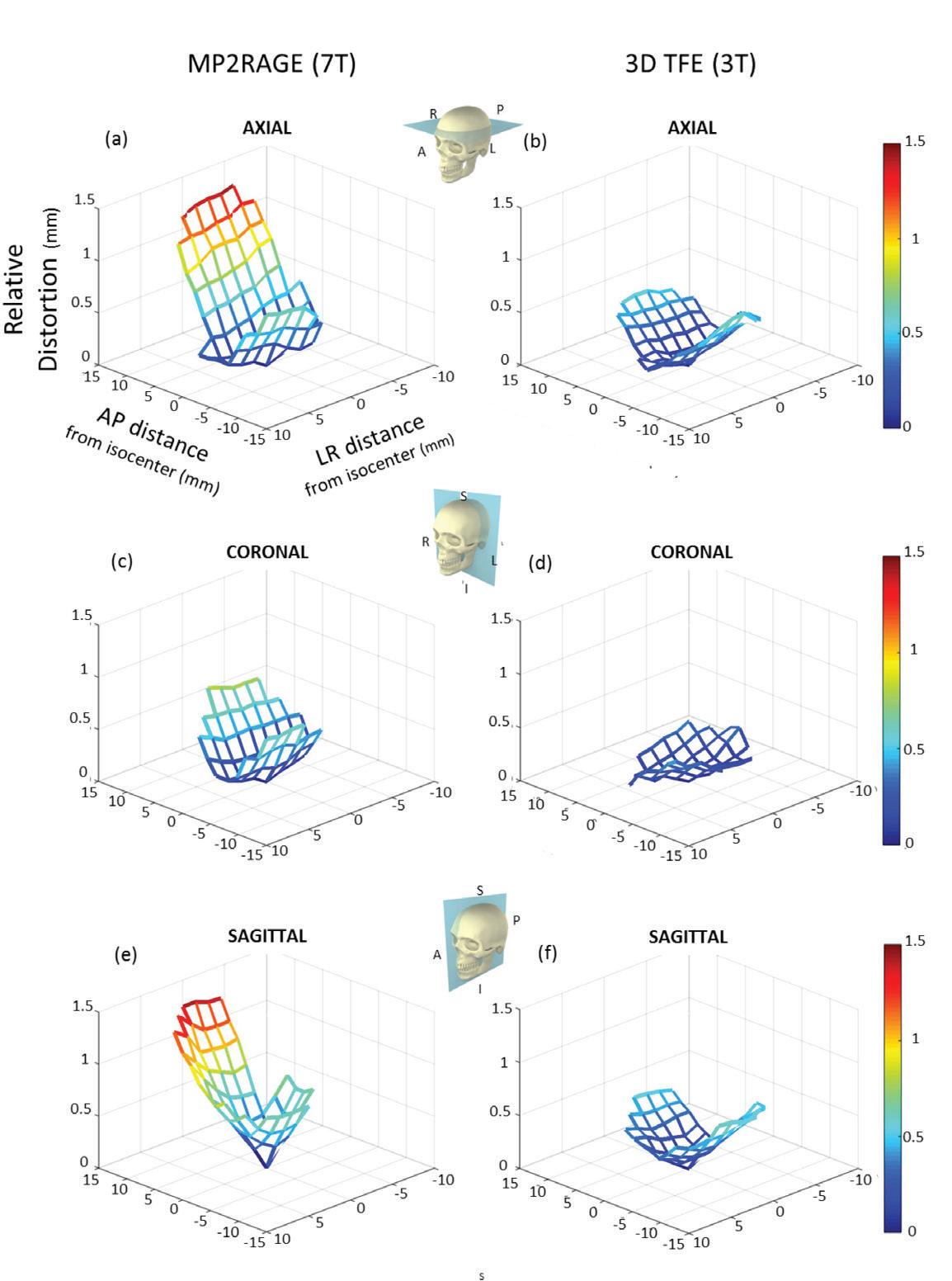
The magnitude of each displacement vector is presented in a 2D mesh-plot in axial (XZ), coronal (XY), and sagittal (YZ) planes, intersecting the magnetic isocentre. The worst GD was presented in MP2RAGE and 3D TFE (Fig.4). Additionally, anisotropic distributions of GD were observed along the phase-encoding direction and frequency-encoding direction. Particularly the anterior-superior section was affected by spatial deformation and maximal GD was observed in the right-superior section of the anterior region (frontal lobe). Distortion maps of all sequences are presented in Figures S.3-5.
Absolute distance difference (mm)
Absolute distance difference (mm)

Absolute distance difference (mm)
Absolute distance difference (mm)
Unique distances (mm)
Unique distances (mm)
Unique distances (mm)
Unique distances (mm) Unique distances (mm)
Unique distances (mm)
Unique distances (mm)
Unique distances (mm)
Fig. 2: Absolute differences in Euclidian distance between the measured and reference dataset (grey dot) relative to the unique distances found in the reference dataset, observed within CT (a), MP2RAGE (b), T2-SPACE (c), T2-SPACE FLAIR (d), T1-GRE (e), 3D TFE (f), T2-VISTA (g), T2-VISTA FLAIR (h), T1-FFE (i). The overall geometric distortion was quantified by MADglobal (±SD) (blue square). The 95 % confidence interval (CI95) is shown as the dotted horizontal line

Fig. 3: MADLocal values (±SD) relative to the distance from the magnetic isocentre at 7T (a) and 3T (b) MRI, both relative to CT (black square). Presented 7T MR sequence include MP2RAGE (green circle), T2-SPACE (purple diamond), T2-SPACE FLAIR (orange downward triangle), and T1-GRE (blue upward triangle). The same colour- and shape-code was used for the equivalent 3T sequences, 3D TFE, T2-VISTA, T2-VISTA FLAIR, and T1-FFE, respectively. The dotted horizontal line represents the 1 mm-acceptability level required for spatially reliable RTP

Fig. 4: Distortion maps of MP2RAGE on 7T MRI (a) and 3D TFE on 3T MRI (b) measured in the axial plane nearest the magnetic isocentre (Y=0), in the coronal plane (Z=0), and in the sagittal plane (X=0)
Discussion
This study reports both system- and phantom-related GD on clinically-relevant MRsequences at 7T-MRI for neurological imaging and allows direct comparison to GD on both 3T-MRI and CT-images. System-related GD was present in all 3T- and 7T-MRI and increased with increasing distance from the magnetic isocentre, even though vendor-provided correction methods and 2nd order 3D shimming were applied. However, GD of ≤1 mm could be assured within a sequence-dependent DSV near the magnetic isocentre.
While UHF-MRI promises clinical gain in terms of spatial resolution, SNR, contrast, and delineation of pathology, implementation of 7T-MRI in RTP requires mmscale spatial accuracy for reliable, high-precision radiotherapy. This geometrical requirement was met by all 3T-MR sequences throughout the entire VOI while the magnitude of GD in 7T-MRI depended on the distance from the magnetic isocentre and applied sequence. GD ≤1 mm was apparent in 7T-MRI within a sequence-specific DSV. Moreover, no significant differences in system-related GD were found between 3T- and 7T-images near the magnetic isocentre. This implies that these 7T-MRI with clinically-acceptable/tolerable levels of spatial uncertainty could be applied for highprecision RTP in central brain regions, under the assumption that tissue-related GD is negligible. In addition, it has previously been shown that GD in 7T-MRI did not affect targeting of basal ganglia and subthalamic nuclei in deep-brain stimulation23,31 .
Some sequences are more prone to GD than others as gradient pulse scheme and parameter settings affect B0-homogeneity and eddy currents25,29,32. Our MP2RAGE sequence, which acquires 2 gradient-echo images at different inversion times, presented the largest GD at 7T (and smallest DSV where GD <1 mm)33. Nevertheless, even in this worst-case scenario, system-related GD at 7T was below the clinically acceptable limit of ≤1mm within 68.7 mm DSV. Presumably, GD in MP2RAGE could be related to the combination of different sequence-parameter settings (receiver bandwidth (rBW), FOV, and matrix size)13,14,25,29,31,32,34. The effect of varying rBW on GD and SNR has extensively been studied by Walker et al. (2014) in 4 different MR-systems31. For instance, increasing rBW would reduce GD, chemical shift, and susceptibility artefacts but would also reduce SNR. Limiting FOV-settings (i.e., fewer acquisition lines in k-space) could subside the impact of time-dependent off-resonance effects, reducing GD34,35. However, this could also influence spatial resolution, SNR, and induce potential fold-over artefacts.
Residual GD in phase-encoding direction is expected to be related to gradient nonuniform and could be adequately reduced by vendor-installed distortion correction methods14,18,25,26. The technique behind this correction is similar for all main MRmanufacturers and achieve comparable and reproducible results for 3T- and 7T-MRI4,14,15,18,31. Nonetheless, some residual system-dependent GD was detected in
this study. It is therefore important to note that vendor-provided distortion correction is not flawless in correcting for B0-inhomogeneity and eddy currents, and gradient coils are designed with a tolerated performance variation24,36. For example, general gradient errors of body gradient coils should be >2% of the gradient strength over a 40 cm DSV37,38 .
Our findings of GD corresponded well with literature. At 3T, Stanescu et al. (2010) reported maximal overall GD of 0.6 mm in 3D TFE sequences and Schmidt et al. (2016) have shown global GD of less than 0.5 mm in an IR-sequence (rBW= 890 Hz/pixel mm, resolution 1 mm³)14,39. However, some studies report GD of several centimetres25-27 . These findings, however, occurred at the edge of the scanner’s FOV on a large cubical phantom that was placed in a body-coil. In contrast to this, we used a dedicated anthropomorphic head phantom to mimic a clinically realistic scan-procedure with standard RF-coils, shimming, distortion correction, and FOV settings (≤256 mm).
Similar to our approach and FOV setting, Dammann et al. (2011) investigated hardware-related GD in 7T-MRI using a cylindrical phantom (190 POIs) and presented local GD smaller than 1 mm at <80 mm DSV, except in T2-VISTA (rBW= 350 Hz/pixel 625 Hz/mm, resolution 0.56 mm² ) that showed maximal local GD of 1.6 mm26 . Presumably, this sequence was affected by B1-inhomogeneities and measurements at the phantom’s edge which could explain our finding of 1.2 mm local GD at 80 mm DSV. Differences in rBW-setting could explain GD differences found in MPRAGE (rBW= 600 Hz/mm, resolution 1 mm³) and MP2RAGE (rBW= 354 Hz/mm, 0.7 mm³). Recently, Lau et al. (2017) demonstrated differences in local GD between 3T and 7T larger than 1 mm at 80 mm DSV for T1-MPRAGE (rBW= 244 Hz/mm, 0.8 mm³)4. In our study, MP2RAGE showed differences in MADlocal between 3T and 7T to be below 1 mm up for a ≤87.5 mm DSV.
Distortion maps of MP2RAGE indicated an anisotropic distribution of GD along the phase-encoding (AP) and frequency-encoding (SI) direction (Fig.4). In a comparable 7T phantom study by Cho et al. (2010), coronal distortion maps showed 0.4 mm GD along the frequency-encoding direction and 0.8 mm along the phase-encoding direction22 .
We observed GD in CT-images with MADglobal ranging from 0.1−0.6 mm and MADlocal ranging from 0.1-0.3 mm. These findings were unanticipated as it is generally recognized that CT-images are distortion-free. Other studies have merged MRI with CT, ignoring this innate GD and measured fundamental errors. Artefacts in CT-images could originate from incorrectly reported scanner table-speed or shearing distortion (incorrectly reported gantry-tilt or table-bending). In this study, GD in CT-images could represent partial volume effects and variations in manual reconstruction leading to slightly different xyz-coordinates measurements of POIs (experimental inaccuracies).
In this study, we limited ourselves to evaluating system-related GD and not tissue-related GD. Relative to system-related GD, tissue-induced GD is rather small in 3T-sequences that are currently being used for RTP of brain tumours, but it is nevertheless not negligible16. Simulations on 3T-MRI suggest that susceptibility-induced GD were on average 0.6 mm at cranial air-cavities40. This has recently been confirmed by Schmidt et al. (2016), who specified susceptibility-related GD of 0.4 mm in the naso-oropharyngeal cavities and around the internal ear canal using a T1-weighted sequence (rBW= 890 Hz/mm, resolution 1 mm³)14. It remains to be determined what the impact of tissuerelated GD is for 7T-MRI. Susceptibility artefacts are proportional to the magnetic field strength and TE but could be reduced by increasing rBW12. Regardless, patientspecific correction methods are required16,37,40. Recently, Rai et al. (2018) developed an 3D printed human skull with sinuses and mastoid air cells, water-based brain tissue and eyes and surrogate cortical bone41. Such realistic anatomical skulls could be used to further assess tissue-induced GD in simulated air cavities, bony anatomy, and soft tissue. Since system-related GD could be further reduced by appropriate setting of MR-sequence parameters, research has been performed to systematically assess the impact of e.g., rBW, FOV, matrix size, scanning direction, and various (post-)processing correction algorithms16,18,27,31,36. However, 7T-MR protocols need to be optimised specifically for each clinical application and the trade-off between anatomical imagequality (spatial resolution, SNR, visibility of pathology) and image integrity (spatial accuracy) needs to be made, while respecting specific absorption rate restriction29,32 . For example, high-precision RTP relies heavily on high geometrical accuracy but could compensate on image-resolution, while in radiology larger GD could be tolerated.
To apply 7T-images in radiotherapy, GD should be ≤1 mm to ensure reliable RTP and dose delivery with local dose variations less than 5-10%21. This strict criterion was met by all the clinically relevant sequences we tested on 3T- and 7T-MRI up until 92.5101.7 mm DSV around the magnetic isocentre, except for the MP2RAGE sequence. Delineations of brain lesions and organs-at-risk in the frontal or occipital lobe on 7T-MRI can be significantly influenced by GD as the spatial uncertainty exceeds 1 mm in these regions. Even though systematic and random treatment uncertainties are foreseen in a planning treatment volume (PTV), the degree and location of GD is rarely taken into account during radiation dose planning. This study has shown that system-related GD should be incorporated into the PTV-margin to achieve a high level of dose delivery accuracy for intracranial (stereotactic) radiotherapy. This can be accomplished by incorporating the GD present at the tumour location within a margin recipe for dose delivery as suggested by Van Herk et al. (2004) and Seravelli et al. (2015)42,43. We therefore recommend applying larger tumour site-specific PTVmargins in regions where spatial integrity of 7T-MRI could not be warranted within 1 mm. Apart from adjustment of PTV-margins, it is advised to find a trade-off between image quality and image distortion, and apply MR-imaging protocols that are dedicated for RTP purposes.
In conclusion, GD of ≤1 mm could be assured within a sequence-dependent DSV near the magnetic isocentre, implying that this 7T-MRI protocol could be applied with clinically acceptable/tolerable levels of spatial uncertainty for high-precision RTP in central brain regions, under the assumption that tissue-related GD can be ignored. For peripheral regions, 7T-MR protocol for RTP should incorporate GD in tumoursite specific PTV-margins for treatment uncertainties. Nevertheless, dedicated MRprotocols are aspired for application in radiation oncology and further optimisation of sequence parameter settings and GD reduction methods are needed.
References
1. Niyazi M, Brada M, Chalmers AJ, et al. ESTRO-ACROP guideline “target delineation of glioblastomas”. Radiother Oncol. 2016;118(1):35-42. 2. Claes A, Idema AJ, Wesseling P. Diffuse glioma growth: a guerilla war. Acta neuropathologica. 2007;114(5):443-458. 3. Barrett TF, Sarkiss CA, Dyvorne HA, Lee J, Balchandani P, Shrivastava RK.
Application of Ultrahigh Field Magnetic Resonance Imaging in the Treatment of
Brain Tumors: A Meta-Analysis. World neurosurgery. 2016;86:450-465. 4. Lau JC, Khan AR, Zeng TY, MacDougall KW, Parrent AG, Peters TM. Quantification of local geometric distortion in structural magnetic resonance images: Application to ultra-high fields. Neuroimage. 2017. 5. Trattnig S, Bogner W, Gruber S, et al. Clinical applications at ultrahigh field (7 T).
Where does it make the difference? NMR Biomed. 2016;29(9):1316-1334. 6. Trattnig S, Springer E, Bogner W, et al. Key clinical benefits of neuroimaging at 7T.
Neuroimage. 2016. 7. Christoforidis GA, Yang M, Abduljalil A, et al. “Tumoral pseudoblush” identified within gliomas at high-spatial-resolution ultrahigh-field-strength gradient-echo
MR imaging corresponds to microvascularity at stereotactic biopsy. Radiology. 2012;264(1):210-217. 8. Moenninghoff C, Maderwald S, Theysohn JM, et al. Imaging of adult astrocytic brain tumours with 7 T MRI: preliminary results. Eur Radiol. 2010;20(3):704-713. 9. Compter I, Peerlings J, Eekers DB, et al. Technical feasibility of integrating 7 T anatomical MRI in image-guided radiotherapy of glioblastoma: a preparatory study. MAGMA. 2016;29(3):591-603. 10. Bian W, Hess CP, Chang SM, Nelson SJ, Lupo JM. Susceptibility-weighted MR imaging of radiation therapy-induced cerebral microbleeds in patients with glioma: a comparison between 3T and 7T. Neuroradiology. 2014;56(2):91-96. 11. Lupo JM, Chuang CF, Chang SM, et al. 7-Tesla susceptibility-weighted imaging to assess the effects of radiotherapy on normal-appearing brain in patients with glioma. Int J Radiat Oncol Biol Phys. 2012;82(3):e493-500. 12. Dietrich O, Reiser MF, Schoenberg SO. Artifacts in 3-T MRI: physical background and reduction strategies. Eur J Radiol. 2008;65(1):29-35. 13. Weygand J, Fuller CD, Ibbott GS, et al. Spatial Precision in Magnetic Resonance
Imaging-Guided Radiation Therapy: The Role of Geometric Distortion. Int J Radiat
Oncol Biol Phys. 2016;95(4):1304-1316. 14. Schmidt MA, Wells EJ, Davison K, Riddell AM, Welsh L, Saran F. Stereotactic
Radiosurgery Planning of Vestibular Schwannomas: Is MRI at 3 Tesla Geometrically
Accurate? Med Phys. 2016. 15. Torfeh T, Hammoud R, Perkins G, et al. Characterization of 3D geometric distortion of magnetic resonance imaging scanners commissioned for radiation therapy planning. Magnetic resonance imaging. 2016;34(5):645-653.
16. Wang H, Balter J, Cao Y. Patient-induced susceptibility effect on geometric distortion of clinical brain MRI for radiation treatment planning on a 3T scanner.
Phys Med Biol. 2013;58(3):465-477. 17. Klein EE, Hanley J, Bayouth J, et al. Task Group 142 report: quality assurance of medical accelerators. Med Phys. 2009;36(9):4197-4212. 18. Duchin Y, Abosch A, Yacoub E, Sapiro G, Harel N. Feasibility of using ultra-high field (7 T) MRI for clinical surgical targeting. PLoS One. 2012;7(5):e37328. 19. Jursinic PA, Rickert K, Gennarelli TA, Schultz CJ. Effect of image uncertainty on the dosimetry of trigeminal neuralgia irradiation. Int J Radiat Oncol Biol Phys. 2005;62(5):1559-1567. 20. Karaiskos P, Moutsatsos A, Pappas E, et al. A simple and efficient methodology to improve geometric accuracy in gamma knife radiation surgery: implementation in multiple brain metastases. Int J Radiat Oncol Biol Phys. 2014;90(5):1234-1241 21. Seibert TM, White NS, Kim GY, et al. Distortion inherent to magnetic resonance imaging can lead to geometric miss in radiosurgery planning. Practical radiation oncology. 2016;6(6):e319-e328. 22. Cho ZH, Min HK, Oh SH, et al. Direct visualization of deep brain stimulation targets in Parkinson disease with the use of 7-tesla magnetic resonance imaging.
J Neurosurg. 2010;113(3):639-647. 23. Wang D, Doddrell DM. Geometric Distortion in Structural Magnetic Resonance
Imaging. Current Medical Imaging Reviews. 2005;1(1):49-60. 24. O’Callaghan J, Wells J, Richardson S, et al. Is your system calibrated? MRI gradient system calibration for pre-clinical, high-resolution imaging. PLoS One. 2014;9(5):e96568. 25. Baldwin LN, Wachowicz K, Thomas SD, Rivest R, Fallone BG. Characterization, prediction, and correction of geometric distortion in 3 T MR images. Med Phys. 2007;34(2):388-399. 26. Dammann P, Kraff O, Wrede KH, et al. Evaluation of hardware-related geometrical distortion in structural MRI at 7 Tesla for image-guided applications in neurosurgery. Acad Radiol. 2011;18(7):910-916. 27. Doran SJ, Charles-Edwards L, Reinsberg SA, Leach MO. A complete distortion correction for MR images: I. Gradient warp correction. Phys Med Biol. 2005;50(7):1343-1361. 28. Schenck JF. The role of magnetic susceptibility in magnetic resonance imaging: MRI magnetic compatibility of the first and second kinds. Med Phys. 1996;23(6):815-850. 29. Liney GP, Moerland MA. Magnetic resonance imaging acquisition techniques for radiotherapy planning. Seminars in radiation oncology. 2014;24(3):160-168. 30. Teeuwisse WM, Brink WM, Haines KN, Webb AG. Simulations of high permittivity materials for 7 T neuroimaging and evaluation of a new barium titanate-based dielectric. Magnetic resonance in medicine. 2012;67(4):912-918.
31. Walker A, Liney G, Metcalfe P, Holloway L. MRI distortion: considerations for MRI based radiotherapy treatment planning. Australasian physical & engineering sciences in medicine / supported by the Australasian College of Physical Scientists in Medicine and the Australasian Association of Physical Sciences in Medicine. 2014;37(1):103-113. 32. Kraff O, Fischer A, Nagel AM, Monninghoff C, Ladd ME. MRI at 7 Tesla and above: demonstrated and potential capabilities. J Magn Reson Imaging. 2015; 41(1):13-33. 33. Marques JP, Gruetter R. New Developments and Applications of the MP2RAGE
Sequence - Focusing the Contrast and High Spatial Resolution R-1 Mapping. Plos
One. 2013;8 (7):11. 34. Hong C, Lee DH, Han BS. Characteristics of geometric distortion correction with increasing field-of-view in open-configuration MRI. Magnetic resonance imaging. 2014;32(6):786-790. 35. Hashemi RH, Bradley Jr WG, Lisanti CJ. MRI: the Basics. 3rd ed. Philadelphia:
Lippincott Williams and Wilkins; 2010. 36. Wang D, Strugnell W, Cowin G, Doddrell DM, Slaughter R. Geometric distortion in clinical MRI systems Part II: correction using a 3D phantom. Magnetic resonance imaging. 2004;22(9):1223-1232. 37. Schmidt MA, Payne GS. Radiotherapy planning using MRI. Phys Med Biol. 2015;60(22):R323-361. 38. Jezzard P. The physical basis of spatial distortion in magnetic resonance images.
In: Bankman IN, ed. Handbook of medical image processing and analysis. 2nd ed.
Amsterdam: Elsevier/Academic Press; 2009:499-514. 39. Stanescu T, Jans HS, Wachowicz K, Fallone BG. Investigation of a 3D system distortion correction method for MR images. J Appl Clin Med Phys. 2010;11(1): 200-216. 40. Stanescu T, Wachowicz K, Jaffray DA. Characterization of tissue magnetic susceptibility-induced distortions for MRIgRT. Med Phys. 2012;39(12):7185-7193. 41. Rai R, Manton D, Jameson MG, et al. 3D printed phantoms mimicking cortical bone for the assessment of ultrashort echo time magnetic resonance imaging.
Med Phys. 2018;45(2):758-766. 42. van Herk M. Errors and margins in radiotherapy. Seminars in radiation oncology. 2004;14(1):52-64. 43. Seravalli E, van Haaren PM, van der Toorn PP, Hurkmans CW. A comprehensive evaluation of treatment accuracy, including end-to-end tests and clinical data, applied to intracranial stereotactic radiotherapy. Radiother Oncol. 2015;116(1):131-138.
Supplementary fi gures and tables
Table S.1: Scan parameters of the 7T- and 3T-MR sequences investigated in this study. All sequences were acquired in 3D
7T MRI sequences MP2RAGE T2-SPACE T2-SPACE FLAIR T1-GRE
Imaging plane Sag Sag Sag Tra
Read-out direction AP AP AP RL Repetition time T (ms) 5000 4000 8000 33
Echo time TE (ms) 2.5 283 302 2.5
Inversion time (ms) TI1 900 - TI2 2750 N/A 2330 N/A
Turbo factor 164 1261 N/A
Field of View (mm) 223x223x240 192x192x288 193x206x208 160x223x208 Acquisition matrix (voxel) 320x320x340 320x320x480 240x256x260 320x320x300
In-plane resolution (mm/voxel) 0.7x0.7x0.7 0.6x0.6x0.6 0.8x0.8x0.8 0.5x0.7x0.7
Nominal fl ip angle (°) 5 & 3 variable variable 11
Bandwidth (Hz/mm) 354 620 479 580 Acceleration factor (iPAT) 3 4 4 2
Acquisition time (minutes) 08:02 07:50 10:58 08:33
3T MRI sequences 3D TFE T2-VISTA T2-VISTA FLAIR T1-FFE
Imaging plane
Tra Sag Read-out direction AP AP Sag AP Tra RL
Repetition time TR (ms) 8.1 2500 8000
Echo time TE (ms) 3.7 243 331.5 11 4.6
Inversion time (ms) 800 N/A 2400 N/A
Turbo factor 188 133 110 N/A Field of View (mm) 256x256x180 256x256x180 256x256x180 256x192x180
Acquisition matrix (voxel) 256x256x180 256x256x180 228x226xx150 256x256x180
In-plane resolution (mm/voxel) 1x1x1 1x1x1 1.1x1.1x1.2 1x0.7x1
Nominal fl ip angle (°) 8
90 variable Bandwidth (Hz/mm) 191 1005 346
Acceleration factor (SENSE) 1 2 2
8 411 1
Acquisition time (minutes) 06:00 05:42 08:24 06:21


