35 minute read
radiotherapy of glioblastoma: a preparatory study
Chapter 4 - Technical feasibility of integrating 7 T anatomical MRI in image-guided radiotherapy of glioblastoma: a preparatory study
Inge Compter, Jurgen Peerlings, Daniëlle B.P. Eekers, Alida A. Postma, Dimo Ivanov, Christopher J. Wiggins, Pieter Kubben, Benno Küsters, Pieter Wesseling, Linda Ackermans, Olaf E.M.G. Schijns, Philippe Lambin, Aswin L. Hoffmann
Advertisement
MAGMA. 2016;29(3):591-603.
Abstract
Objectives
7 Tesla (T) MRI has recently shown great potential for high-resolution soft-tissue neuroimaging and visualization of microvascularization in glioblastoma (GBM). We have designed a clinical trial to explore the value of 7T MRI for radiation treatment of GBM. For this clinical study we performed a preparatory study to investigate the technical feasibility of incorporating 7T MR images into the neurosurgical navigation and radiotherapy treatment planning (RTP) systems by qualitative and quantitative assessment of the image quality.
Materials and methods
The MR images were acquired with a Siemens Magnetom 7T whole-body scanner and a Nova Medical 32-channel head coil. 7T MRI pulse sequences included MP2RAGE, T2-SPACE, SPACE-FLAIR and GRE. A pilot study with 3 healthy volunteers and an anthropomorphic 3D phantom was used to assess image quality and geometrical image accuracy.
Results
The MRI scans were well tolerated by the volunteers. Susceptibility artefacts were observed in both the cortex and subcortical white matter at close proximity to airtissue interfaces. Regional loss of signal and contrast could be minimized by the use of dielectric pads. Image transfer and processing did not degrade image quality. The system-related spatial uncertainty of geometrical distortion-corrected MP2RAGE pulse sequences is ≤ 2 mm.
Objectives
Integration of high-quality and geometrically-reliable 7T MR images into neurosurgical navigation and RTP software is technically feasible and safe.
Introduction
Glioblastoma (GBM) is the most common type of primary brain tumour having a peak incidence in the 6th and 7th decade. Patients with a GBM have an extremely poor prognosis with a median overall survival of 14.6 months1. The main goals of surgery are verification of histological diagnosis and reduction of any mass effect. A complete resection is nearly impossible because of the infiltrative nature of this disease and spread along white matter tracts2. Therefore, patients up to 70 years old with a good performance status are treated with adjuvant radiotherapy in combination with temozolomide to delay local tumour recurrence and increase overall survival. Radiotherapy usually consists of conventionally fractionated regimens, delivering a dose of 59.4–60 Gy in 6–7 weeks. Computed tomography (CT) images (1 mm slices) are commonly used for radiation therapy planning (RTP) because of the excellent spatial quality (i.e., no geometrical distortions) and the electron density information that is required for accurate dose calculations. In current RTP practice for GBM, contrast-enhanced (CE) CT images are co-registered with 1.5 or 3 Tesla (T) magnetic resonance (MR) images because the latter offer superior soft-tissue contrast over CT images. Subsequently, a gross tumour volume (GTV) is delineated based on the resection cavity and any residual disease visible on CE T1-weighted MRI scans. According to European guidelines a 2–3 cm isotropic margin is added to the GTV to encompass any non-enhancing tumour tissue and to establish the clinical target volume (CTV)3. This margin is based on the fact that over 80% of recurrences occur within 2 cm of the GTV4,5. However, the isotropic margin does not take into account spatially varying tumour-growth dynamics in different brain tissues and tumour spread along white matter tracts, which evidently results in needlessly damaging healthy cells while leaving viable malignant cells outside the CTV6. A planning target volume (PTV) margin is added to account for systematic and random errors such as setup errors, inter- and intrafraction motion, but also uncertainty in image registration and delineation. Current clinically available MRI techniques are unable to adequately visualize tumour spread throughout the brain. Ultra-high field (UHF) (>3 T) MRI might be able to overcome these limitations, because the increased signalto-noise ratio (SNR) and susceptibility effects which allow for an increased spatial resolution and better contrast in comparison to clinically-used 1.5T and 3T MRI. However, the potential of UHF MRI for RTP of GBM has not been investigated so far.
With regard to neurosurgical planning, UHF MRI may benefit image-guided biopsies through identification of increased vascularity suggestive for tumour grade. Endothelial proliferation and increased microvascularization are key features for the diagnosis of GBM according to the WHO criteria7. An increase in susceptibility effects, as seen with UHF MRI in comparison to lower field strengths, results in novel contrast mechanisms on quantitative T2*-weighted images by which microvascularization with a vessel diameter as small as 100 μm can be visualized8-11 .
We therefore hypothesize that 7T MRI allows for better delineation of the GTV of GBM due to improved visualization of microvascularization. We have designed a future clinical study to investigate this, which includes a 7T-image guided biopsy and RTP (clinicaltrials.gov NCT 02062372). In this paper we describe the technical aspects of the preparatory work for this clinical trial. If 7T MR images are to be used for neurosurgical and RTP purposes, a high spatial reliability is required. Although 7T MRI may hold promise to be included into neurosurgical navigation and RTP, it also presents technical challenges such as inhomogeneity of the transmit B1-field, an increased specific absorption rate (SAR) and geometrical distortions caused by increased static magnetic field (B0) inhomogeneity, tissue susceptibility differences and chemical shift effects12-14. In order to measure the geometric accuracy, a phantom study was conducted to quantify system-related geometrical image distortions. Furthermore, a pilot study with healthy volunteers was conducted to investigate whether the 7T MRI images meet the requirements for clinical application in RTP, such as visualization of brain anatomy structures on different sequences, differences in scanning times and patient tolerability of the scan. In this paper we report on the challenges and pitfalls we encountered in preparation of our clinical study, and present solutions we developed to quantify the system-related geometrical image distortions, optimize 7T MRI scanning protocols as well as the image transfer and processing workflow.
Materials and methods
Pilot study
A pilot study with healthy volunteers was conducted to assess the image quality in terms of field inhomogeneity and susceptibility artefacts and to optimize the pulse sequences and scanning protocols for RTP purposes. Prior to the 7T MRI scan all volunteers received detailed information regarding the purpose of the study and possible temporary sensory side effects due to the applied magnetic field. Written informed consent was obtained prior to participation. A total of three volunteers were recruited: two females (26 and 37 years old) and one male (29 years old). Subjects filled out a safety questionnaire prior to the scan regarding e.g. medication, claustrophobia and metallic objects, and were instructed not to move during the scanning procedure. They were positioned in a head-first supine position and dielectric pads were fixed on both sides of the subject’s head next to the temporal lobes after which the head coil was placed. The dielectric pads contained a 25% suspension of barium titanate in deuterated water and were used to locally increase the transmit B1+ field to improve its homogeneity across the brain15. Cushions were placed under the knees to provide extra comfort.
Phantom study
Geometrical distortion is a recognized problem in anatomical MRI sometimes resulting in pixel shifts of several millimetres, which is detrimental for application of MRI in image-guided interventions in neurosurgery and radiotherapy. Geometric inaccuracies originate from system (i.e., B0-inhomogeneity, gradient field nonlinearity) or object (i.e., chemical shift, susceptibility effects) related causes, and can to some extent be corrected for by manufacturer-developed distortion correction methods and shimming procedures. As magnetic field inhomogeneity, chemical shift and susceptibility are proportional to B0, the spatial inaccuracy increases with higher magnetic field strength. Hence, estimation of the geometrical distortion is essential before 7T MRI can be integrated into image-guided interventions.
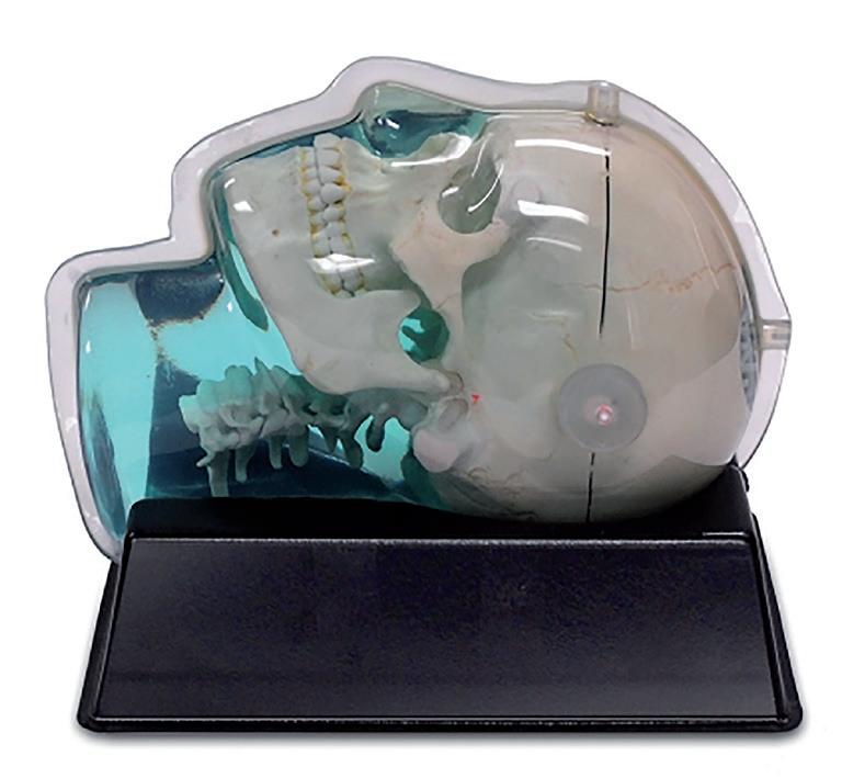
For the assessment of system- and sequence-related geometrical image distortions, we used a dedicated 3D anthropomorphic skull phantom (CIRS Model 603A, Computerized Imaging Reference Systems, Inc., Norfolk, Virginia, United States; figure 1 and 2a & 2b). This phantom is made up of a plastic-bone tissue substitute and soft-tissue equivalent material consisting of a water-based polyacrylamide (Zerdine®, CIRS). The entire phantom is encased in a clear vacuum-formed plastic shell to protect the gel from desiccation. The cranial portion of the skull volume is filled with an orthogonal 3D grid of 3 mm diameter rods (reinforced Nylon) spaced 15 mm apart. The maximum 3D grid-size is 15×12×13.5 cm3 (AP×LR×SI), resulting in 436 measurable grid-intersection points. Since Nylon shows magnetic susceptibility properties similar to water (i.e., difference in susceptibility <3 ppm), the phantom rods are expected not to induce artefacts and image abnormalities in either spinecho or gradient-echo sequences16. The phantom was placed in an ABS vacuumformed cradle to ensure reproducible placement within the scanner. Both CT and 7T MRI images were acquired in order to compare the geometrical distortions of both imaging modalities.
Fig. 1 Anthropomorphic skull phantom CIRS 603A. Source: Computerized Imaging Reference Systems, Inc (CIRS), viewed 22 November 2015 <http://www.cirsinc.com/ products/modality/99/mri-distortion-phantom-for-srs/>
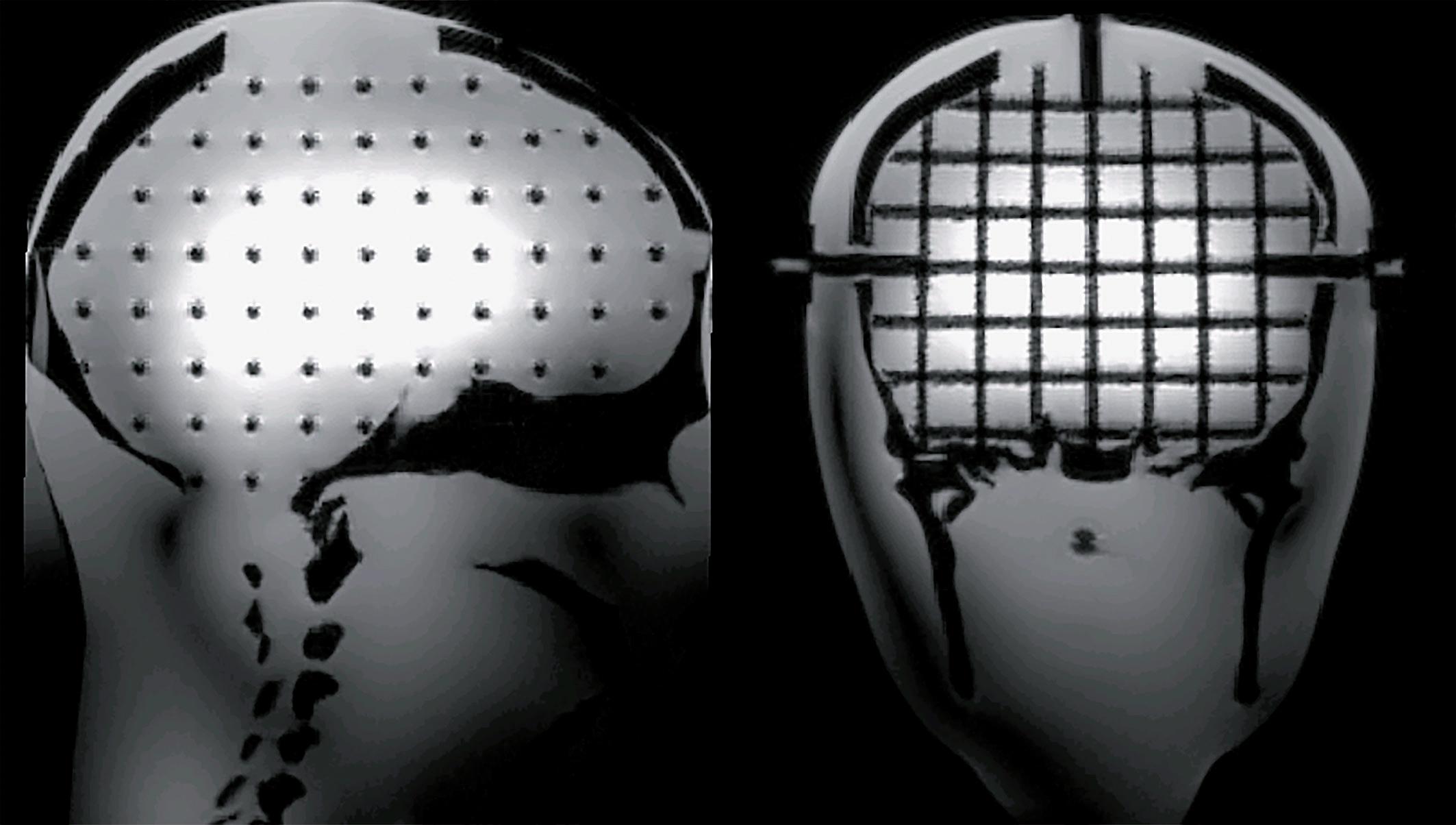
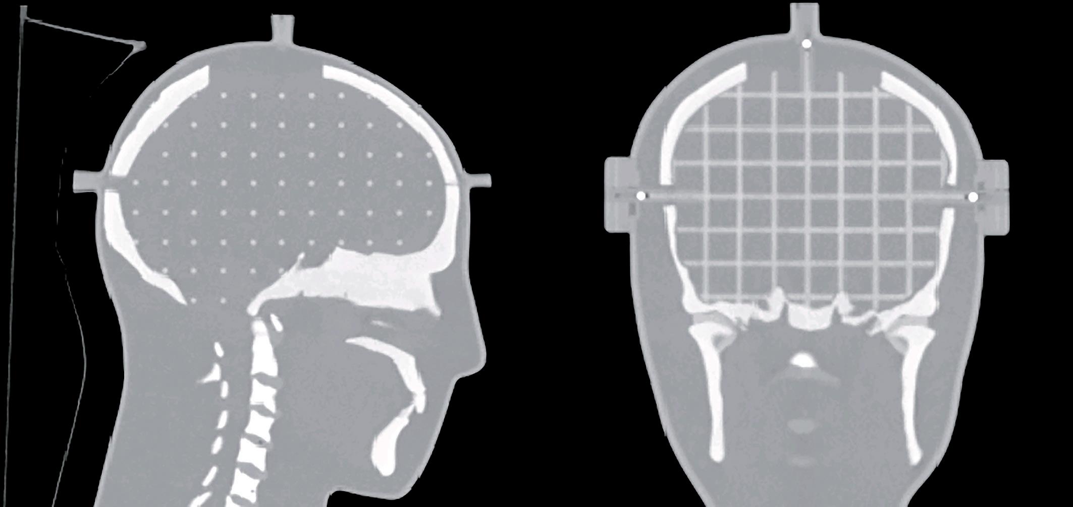
Fig. 2 Sagittal and coronal slice of the CIRS 603A phantom on CT (a), and 7T MRI (b) scan
Image acquisition
For the phantom study, CT images (SOMATOM Sensation 10, Siemens Healthcare, Erlangen, Germany) were acquired at MAASTRO clinic with a 1 mm slice thickness, 306 slices, 50×50 cm2 field of view, 140 kV, 400 mAs.
For the pilot and phantom study, MR images were acquired at the Scannexus facility with a 7T whole-body scanner (Magnetom 7T, Siemens Healthcare, Erlangen, Germany) in combination with a 32-channel head coil (Nova Medical, Wilmington, United States). Pulse sequences included multi-echo gradient echo (GRE), magnetization-prepared rapid gradient-echo (MP2RAGE)17, T2- sampling perfection with application optimized contrasts using different flip angle evolution (T2-SPACE), and sampling perfection with application optimized contrasts using different flip angle evolution FLAIR (SPACE-FLAIR) (table 1).
Table 1 Scan parameters per 7T MRI pulse sequence
MP2RAGE SPACE SPACE-FLAIR GRE
Repetition time TR (ms)
Echo time TE (ms)
Inversion time (ms)
5000
2.5
TI1 900
TI2 27500 4000
283
nvt 8000
302
2330 33
2.5
nvt
Field of View (mm)
223 x 223 192 x 192 193 x 206 160 x 223
Nominal flip angle (°)
5 & 3 variable variable 11
Acquisition matrix (pixel) 0\320\320\0 0\320\320\0 0\256\240\0 0\320\320\0
Bandwidth (Hz/pixel)
248 372 383 290
Slices (n)
240 288 208 208
Slice thickness (mm)
0.7
Acquisition time (minutes) 8.02 0.6 0.8 0.7
7.50 10.58 8.33
7T MRI sequence selection and optimization
Sequences were selected to highlight differences in tissue contrast18 and visualize microvascularization9,10. A standard method for obtaining T1-contrast is the MPRAGE (magnetization-prepared rapid gradient-echo) sequence, which provides good greywhite matter contrast19. Due to inhomogeneities in both the transmit and receive radiofrequency (RF) fields, a newer variant (MP2RAGE) was used that generates two different images at different inversion times and allows for self-correction of the bias fields20 .
The MP2RAGE sequence with optimized TR-FOCI inversion pulse was chosen for T1-weighted imaging instead of the MPRAGE because receive bias field could be corrected online (figure 3a)21-24. In particular, the T1-weighted images were obtained as the ratio of the two volumes acquired at different inversion times (INV1 and INV2), which minimizes the effect of B1-variations through space. A quantitative T1-map was calculated online by linear interpolation of the INV1 and INV2 images17. An optimized four-echo GRE was used instead of the typical single-echo GRE with a long echo time in order to correct for B1-inhomogeneities and obtain quantitative T2* images (figure 3b).
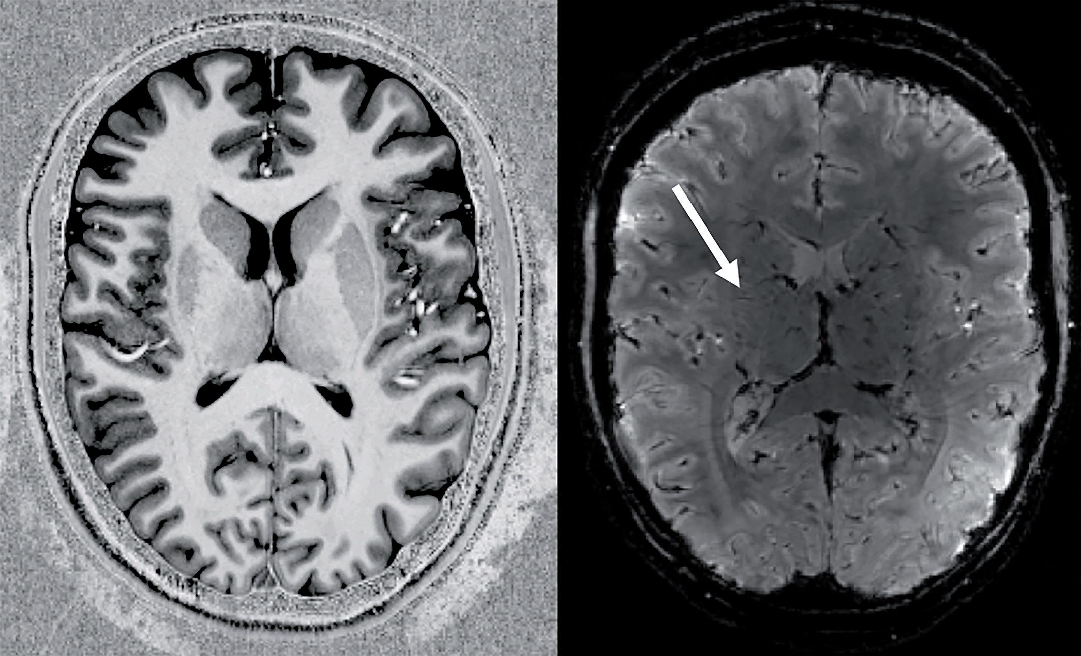
Fig. 3 Axial slices of 7T MRI T1-weighted image from MP2RAGE (a), and T2*weighted image from GRE (b) of a healthy volunteer. White arrow exemplifies the fine vascularisation detail that is observed
T2-weighted imaging using standard Turbo Spin Echo sequences becomes increasingly difficult at UHF due to relatively high SAR induced by multiple 180 degree refocussing pulses25. For this reason, a technique involving the use of a tailored RF flip angle train was used (T2-SPACE) to obtain T2-like contrast26-28. This same technique was also used as the basis for a FLAIR-type contrast (SPACE-FLAIR), where an inversion pulse, with inversion time of 2330 ms, was added to null fluid signals. For both T2-weighted sequences, a Works-In-Progress package [WIP 692] from Siemens was used and parameters were optimised to obtain excellent contrast, with reference to standard clinical imaging. All images were reconstructed with and without manufacturer-developed correction procedures to restore gradient uniformity.
Image transfer and processing workflow
Since MAASTRO clinic and Scannexus each operate independently from the Maastricht University Medical Centre (MUMC), a dedicated inter-institutional imaging workflow was developed to be able to collaboratively share the 7T MR images acquired at Scannexus with the department of Neurosurgery at MUMC and Radiation Oncology department at MAASTRO clinic. Our solution comprised a cloud-based medical image sharing environment (Quentry, version 3.0, Brainlab AG, Feldkirchen, Germany) to upload the 7T MR images into the neurosurgical cranial navigation software (iPlan Net, version 3.6, Brainlab AG, Feldkirchen, Germany) that will be used to obtain the biopsy specimen for the future clinical study. The MiTools software package was used to generate quantitative T2*-maps from the multi-echo GRE images (http://od1n. sourceforge.net/). The 7T MRI scans were imported directly as DICOM files into the radiation treatment planning system (Eclipse version 11, Varian, Palo Alto, United States of America) from the Scannexus facility.
Image quality assessment
Qualitative image analysis
During a multi-disciplinary session, a physicist, neuro-radiologist and radiation oncologist visually verified that the MR image quality was warranted after acquisition, transfer and processing of the images. Image quality was assessed by evaluating the images of all three volunteers on the MR scanner console and within the neurosurgical navigation and RTP software. The MP2RAGE, T2-SPACE and SPACE-FLAIR sequences were visually evaluated by an experienced neuro-radiologist for SNR, visualisation of the cerebral lobes, depiction of grey-white matter, basal ganglia, ventricles and CSF around the brain. Each of these factors was classified on a scale of 1 – 4 (1. Excellent, 2. Good, 3. Marginal, but still diagnostic, 4. Non-diagnostic). The GRE images were not assessed as these images were post-processed in order to obtain quantitative T2*-images.
To assess the geometrical reliability of the 7T MR images two clinically-relevant pulsesequences (GRE and MP2RAGE) were used to acquire images of the phantom, both with and without automatic geometric distortion correction. Unfortunately, it was found that the distortion correction processing was not compatible with the multiecho data, causing severe artefacts. For this reason, only the data from the MP2RAGE is presented. We aim to resolve this incompatibility in future clinical studies. The GRE and MP2RAGE sequences were selected to assess sequence-related geometrical image distortions as they will be primarily used for GTV delineation in the clinical study.
Next, the 3D coordinates of all 436 grid-intersection points were acquired after manual processing of the respective CT and MR images in Eclipse. The coordinates of the reference points were deduced from the phantom’s known geometry.
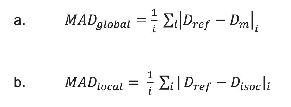
Two methods were used to measure the geometrical distortion at different levels of sophistication. The first method was used to assess the overall image distortion using an in-house developed MATLAB script (R2014b, MathWorks Inc, Natick, USA). All unique pairs of grid-intersection point coordinates were generated from which subsequently the Euclidean distance was determined, and absolute differences in distance were calculated between the reference (i.e., phantom-based) and measured (i.e., image-based) distances. In a second method, Euclidean distances were measured between a fixed reference point located at the magnetic field isocenter, and all other grid-intersection points in the image, and compared to corresponding distances within the reference frame. Hence, a measure of the geometrical dispersion relative to the magnetic field isocenter was obtained. Both methods were applied to the CT and MRI scans. The overall geometric distortion was quantified by the global mean absolute deviation (MADglobal) and its standard deviation (SD), while geometrical dispersion relative to the isocenter was quantified by local mean absolute deviation (MADlocal) and its SD:
where Dref represents the Euclidian distance between reference points, Dm
is the measured distance between image-based grid points, and Disoc is the measured distance between the magnetic field isocenter and surrounding grid points. For each measurement within a cluster of unique distances i, the MAD is then calculated as the average of the absolute difference in Euclidian distances.
Given the MAD and its standard deviation (SD), a 95% confidence interval (Δ) was calculated as:
Based on this confidence interval, a maximum acceptable tolerance level of 2.0 mm was defined. When MAD >1.0 mm and |∆| > 2.0 mm, the level of geometric deviation between the measured and reference dimensions were considered unacceptable.

Results
Image acquisition
The duration of the scanning sessions for the volunteers was 50 minutes. All three volunteers reported slight vestibular effects while being moved in and out of the magnetic field. One of the volunteers reported twitching of the nose tip during the scanning procedure. No other clinically relevant sensory effects were reported. The duration of the scanning session for the phantom was up to 150 minutes as it was scanned with the same sequences as the volunteers in three orthogonal planes (axial, sagittal, coronal).
Image quality
Qualitative image analysis
Images acquired with MP2RAGE (figure 3a), T2-SPACE and SPACE-FLAIR sequences all showed good to excellent SNR and visualization of the frontal and parietal lobes. Furthermore, there was a good to excellent reproduction of the ventricles and CSF surrounding the brain. All sequences showed marginal to good SNR and visualization of the cerebellum. The reproduction of CSF around the cerebellum was good, except for on T2-SPACE where it was judged to be non-diagnostic to marginal. All sequences demonstrated non-diagnostic to marginal image quality for SNR, visualization of the cerebral lobes, depiction of grey-white matter and ventricles and depiction of CSF around the brain at the frontobasal and temporal lobes. This decrease in image quality was primarily caused by a decrease in signal and susceptibility artefacts near the skull base (figure 4). The reproduction of the basal ganglia was good to excellent on T2-SPACE and MP2RAGE, respectively, but was considered marginal in one of the volunteers on SPACE-FLAIR due to flow-artefacts. The visualization of the cerebral vessels was good to excellent on both T2-SPACE and MP2RAGE and considered marginal to good on the SPACE-FLAIR. There was a signal inhomogeneity present in the T2-SPACE and SPACE- FLAIR images in both the medial-lateral and anteriorposterior direction. Moreover, the T2-SPACE showed a signal drop at both the skull base and the temporal lobes. Ghosting artefacts were anteriorly and posteriorly present in the T2-SPACE and SPACE-FLAIR. These may be the result of the relatively high amount of image acceleration used (2x2 GRAPPA) and it may be possible to reduce these artefacts through enhanced image reconstruction. In addition flowartefacts were observed near major intra-cranial vessels such as the carotid and basillary arteries. Image transfer and processing did not visually degrade the image quality.
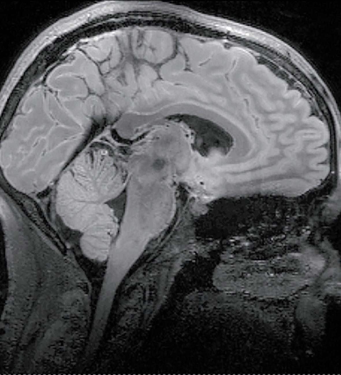
Fig. 4 Sagittal 7T MRI SPACE-FLAIR image of a healthy volunteer, showing increased geometrical distortion near the skull base, the ventral part of the temporal cortices, and the orbito-frontal cortex
Global distortion method
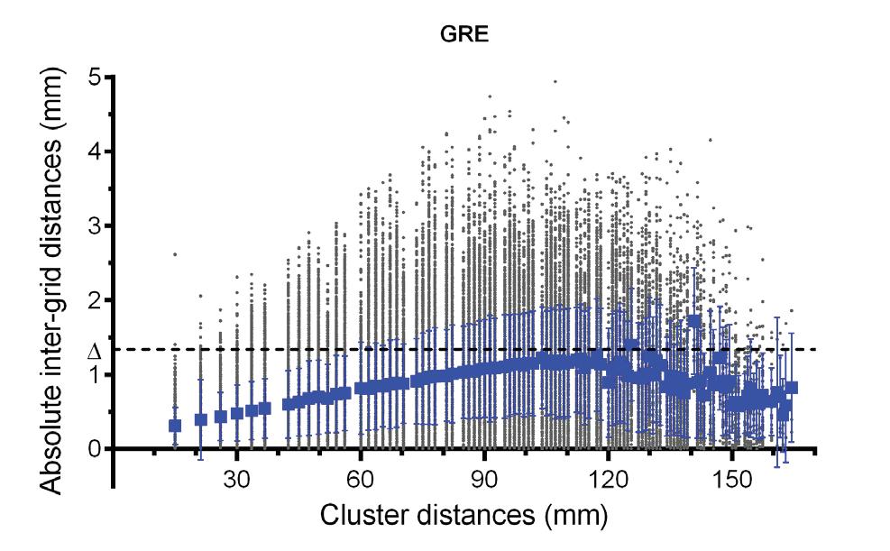
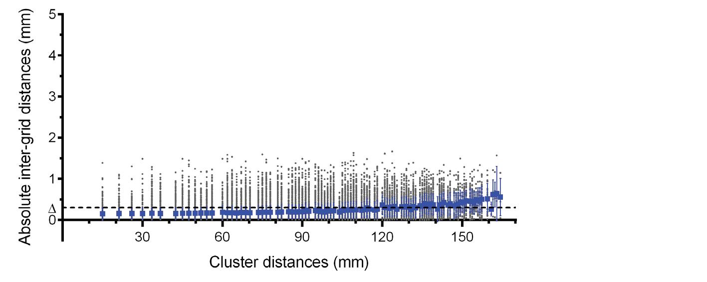
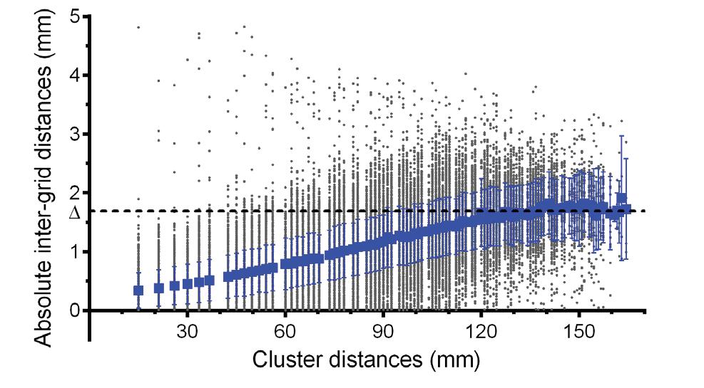
Based on the 436 reference points of the 3D grid, 95266 unique distances between grid-intersection points were calculated and paired into 100 unique distance clusters, ranging from 15 to 164.32 mm. The corresponding imagebased distances were paired into the same clusters for calculating MADglobal. Figure 5 presents the absolute difference between phantom- and image-based distances within each cluster, as well as the MADglobal of that cluster. The global geometric distortion within the MR-images is more pronounced than in CT images. However, CT is not completely free of distortion errors with average MADglobal of 0.20 mm (SD±0.05 mm) (fig. 5a). The GRE and MP2RAGE sequences present MADglobal ranges of 0.31−1.35 mm and 0.38−1.62 mm, respectively in distortion-uncorrected images (fig. 5b, 5c). Furthermore, the measured differences were less consistent in MP2RAGE images with various outliers above the 95% confidence interval (Δ = 1.29 mm), mostly at small intergrid distances. In the distortion-corrected MP2RAGE images, relatively similar patterns of MADglobal could be noted (fig. 5d) with values ranging from 0.34−1.91 mm (Δ = 1.68 mm). Table 3 provides a clear overview of MAD ranges, average MAD and MAD confidence intervals per selected image.

MP2RAGE
MP2RAGE (dc)
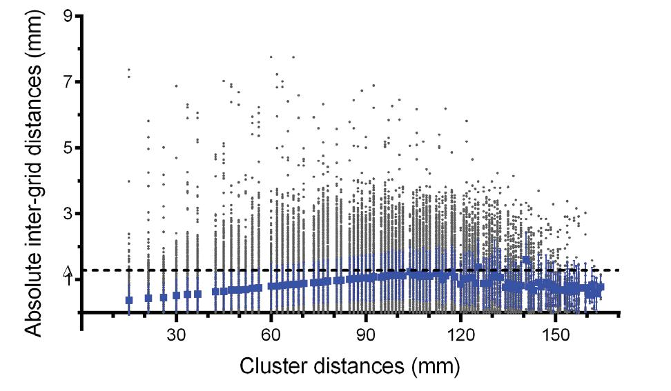
Fig. 5Absolute inter-grid distances (grey dot) measured within CT (a), distortion-uncorrected GRE images (b), distortion-uncorrected MP2RAGE images (c), and distortion-corrected MP2RAGE (d) relative to the possible cluster distances in mm. The overall geometric distortion was quantified by MADglobal (blue square) and its standard deviation. The 95% confidence interval (Δ) is shown as the dotted line
Local distortion method
Figure 6 shows the geometrical dispersion relative to the isocenter (MADlocal) and illustrates modality-and sequence-dependent distortion effects and the quality of the MR-distortion correction methods. In CT-images, MADlocal ranges from 0.06−0.27 mm indicating small spatial deviations. In the distortion-uncorrected 7T MR images, MADlocal ranges from 0.28−1.31 mm (Δ = 0.99 mm) in GRE images and 0.26−1.46 mm (Δ = 1.04 mm) in MP2RAGE images. In the distortion-corrected MR-images, MADlocal ranges from 0.22−1.82 mm (Δ = 1.56 mm) in MP2RAGE images. The value for MADlocal is observed with increasing distance from the magnetic field isocenter, with a maximum of 1.82 mm near the edges of the phantom. At equal distance from the isocenter, geometric distortion was found to be anisotropic. For distortion-corrected MP2RAGE images major spatial displacement could be noted in the superior-inferior (SI) direction (e.g., 0.82 mm at 45 mm isocenter distance).
Based on the average MAD-values for CT and 7T MRI, both modalities present tolerable levels of geometrical image distortion with average MAD ≤ 1.0 mm and Δ ≤ 2.0 mm (table 2 and 3). However, MADglobal and MADlocal of larger cluster distances often exceed this tolerance limit. For corrected MP2RAGE images, MADglobal was >1.0 mm for intergrid distances >77.9 mm. MADlocal measurements in distortion-corrected MP2RAGE images was <1.0 mm up until 68.7 mm from the isocenter.
Table 2 Geometric distortion measures for CT
CT
MADglobal (mm) MADLocal (mm)
Range Mean ± SD Δ Range Mean ± SD Δ 0.14-0.64 0.20 ± 0.05 0.30 0.06-0.27 0.16±0.04 0.24
Table 3 Geometric distortion measures for 7T MRI
MADglobal (mm) Sequence DC Range Mean ± SD Δ Range Mean ± SD Δ
GRE No 0.31-1.35 0.88 ±0.22 1.33 0.28-1.31 0.65 ±0.17 0.99
MP2RAGE No 0.38-1.62 0.88 ±0.21 1.29 0.26-1.46 0.64±0.20 1.04
MP2RAGE 3D 0.34-1.91 0.98 ± 0.35 1.68 0.22-1.82 0.85 ±0.36 1.56
MADLocal (mm)
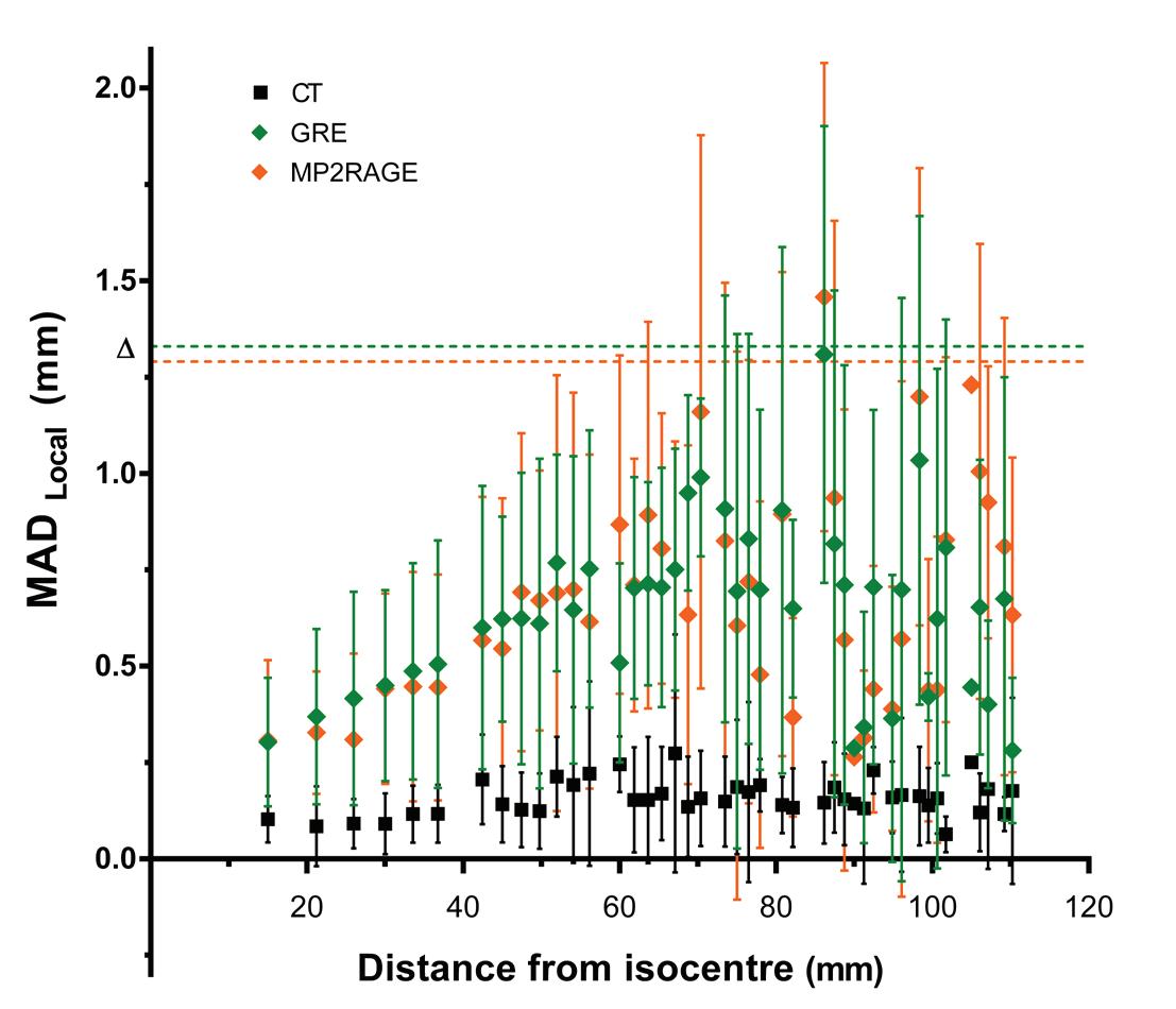
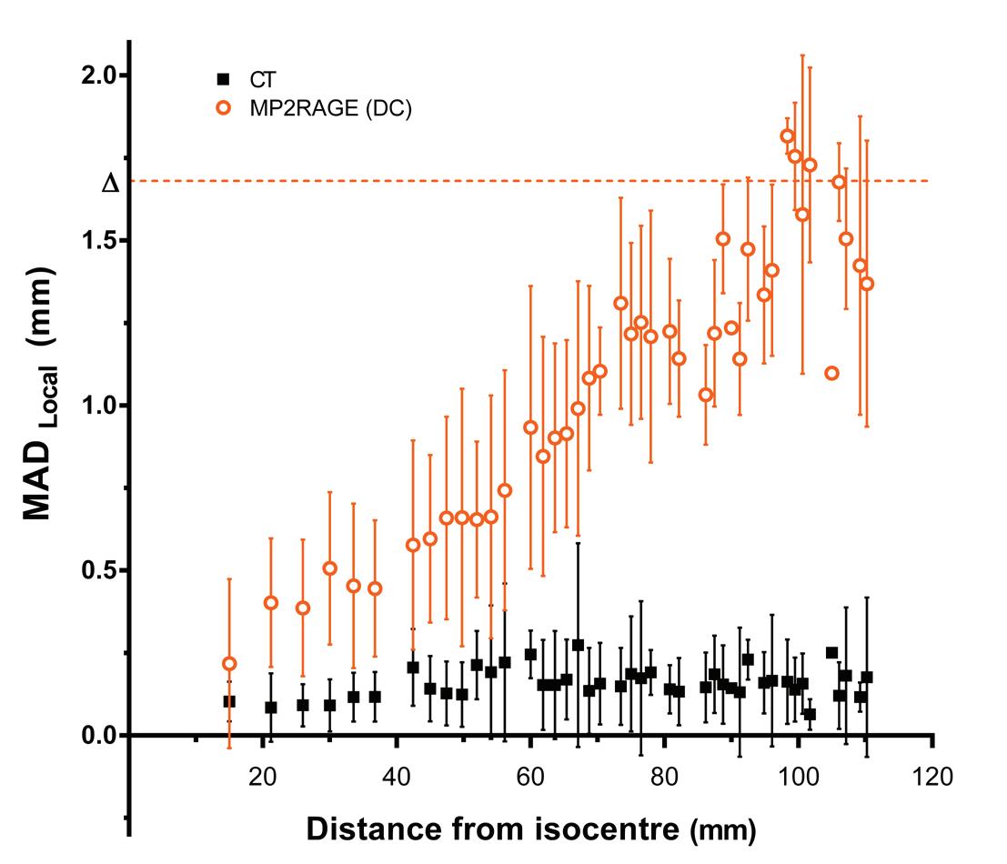
Fig. 6 Geometric dispersion relative to the magnetic field isocenter, quantified by MADlocal and its standard deviation within CT (black square) and distortion-uncorrected 7T MR images (a) acquired with GRE (green diamond) and MP2RAGE (orange diamond) pulse sequences. Measured MADlocal values for CT and distortion-corrected 7T MR images (b) acquired with MP2RAGE (orange circle) pulse sequence are shown relative to the distance
Discussion
This paper reports on the preparatory work for our clinical study to integrate 7T MR images into neurosurgical navigation and RTP systems. It includes a pilot study with healthy volunteers and a phantom study. The volunteers in our pilot study reported limited side effects of the 7T MRI. Similar to other studies on UHF MRI, mainly transient vestibular effects were noted29,30. In our study these effects were only present during a change in table position. The cause of transient vertigo and dizziness is not yet fully understood, but magnetic stimulation of the ear’s labyrinth has been suggested31 . There are currently still a few shortcomings in image quality that need to be resolved before clinical implementation of UHF MRI. Although image quality in our study was good to excellent in the frontal and parietal lobes, all sequences demonstrated a nondiagnostic to marginal image quality for all evaluated parameters at the frontobasal and temporal lobes. This is a well-known issue for UHF-MRI and is generally caused by a decrease in signal and susceptibility artefacts near the skull base. As a consequence, patients with tumours near the skull base will be excluded from our clinical trial. Many researchers have been working on improving the inversion contrast in MPRAGE, particularly trying to reduce the sensitivity to off-resonance and RF inhomogeneity effects19. RF transmit technology is a very active research field within UHF MRI, and better transmitter designs may become available. The application of dielectric pads improves the transmit field homogeneity, which in our case ensured obtaining sufficient signal from the temporal regions15,32. Concurrently, the application of dielectric pads also affects the receive B1 field and can lead to signal increases in their immediate vicinity. Image intensity inhomogeneities of this kind are also typical for multi-channel RF receive coils. They can be circumvented by image postprocessing techniques such as division by an image with the appropriate weighting or by model fitting of at least two images differing by a single parameter24. The latter approach is utilized when generating the T2* maps from the multi-echo GRE data and T1 maps from the MP2RAGE data, whereas the former approach is used to obtain the MP2RAGE T1-weighted images. It is worth mentioning that the inversion pulse of the MP2RAGE sequence has been specifically designed to deliver homogeneous T1-contrast across the whole brain despite B1-inhomogeneities22 . Yet further gains may be possible through the use of parallel transmit (pTx) technology, where 8 or 16 separate elements may be combined to produce a more uniform or targeted excitation. However, the use of such systems involves significant additional experimental setup (e.g., more complicated RF coils and pre-scans to determine per-subject transmit patterns, etc.), which may limit the applicability to patients. Additionally, SAR considerations are significantly more complicated in a pTx setup, as different excitation patterns will produce different SAR values within the subject. While considerable progress has been made, there is still significant work required to implement this in clinical studies.
The phantom we used provides a realistic anthropomorphic scenario for CT and MRI to assess spatial distortion in 7T MRI for clinical use. Although there is no standard procedure for assessing spatial distortion in 7T MRI, AAPM and the Institute of Physics and Engineering in Medicine recommend to measure the distance between two selected points in the MR-image and to calculate the deviation from the known distance33,34. Based on these recommendations we assessed the geometrical image distortion using two different methods, MADglobal and MADlocal. We selected two variable points in the image for MADglobal assessment and selected one variable and one fixed point in the image (e.g., magnetic field isocenter) for MADlocal assessment35,36 .
The assessment of geometrical distortion in our study is similar to the study by Cho et al. investigating the visualisation of targets for deep brain stimulation in Parkinson disease13. However, in the present study an updated version of the CIRS phantom was used. Furthermore, we did not consider the CT as a reference standard and therefore did not co-register the CT to the MRI scan. Instead, the known geometry of the phantom was considered the reference.
Although no geometrical distortion was expected, we do report minimal distortion from the CT images. This is probably caused by measurement uncertainties from manually assessing the grid coordinates and was not found to be correlated to inter-grid distances or distances from the isocenter. For MRI however, overall geometric distortion increases exponentially with increasing distances between grid-intersection points. In uncorrected GRE and MP2RAGE images, a plateau-stage starts to manifest at 58 mm from the isocenter. Presumably, this plateau-stage could be attributed to non-linearity of the gradient fields. Near the edges of the image, the assumed linear relationship between spin position and precession frequency in the local field is false, leading to a compression of the image. In distortioncorrected images, gradient uniformity was restored and as a result, the image was decompressed to its correct dimensions. Consequently, MP2RAGE sequences show a more linear increase of spatial deviation and higher MADlocal maxima are measured (table 3). The larger range of MAD-values is related to the increase in measurable distance between grid-intersection points and the residual system-related geometrical distortion factors, such as eddy currents and B0 inhomogeneity, which were not corrected for. Others have used a similar method as applied in this study to estimate the reliability of stereotactic coordinates on 1.5T and 3T MR images37. They reported a range of mean errors of 0.30−1.20 mm and 0.43−1.78 mm in T1w-images at 1.5T and 3T, respectively. For T2w-images, they have shown ranges of 0.29−0.58 mm and 0.31−1.85 mm, respectively.
Geometric distortion can occur to different extents depending on the imaging sequence parameter settings38. Differences in MADlocal between GRE and MP2RAGE could be attributed to differences in read-out bandwidth (rBW). With a lower rBW, more geometric distortion could be noted in the frequency-encoding or read-out
direction. For MP2RAGE, a low rBW was used (250 Hz/pixel) for measurements in the SI read-out direction, while the read-out direction for GRE images was AP. To minimise this type of geometric distortion, a larger rBW could be selected, however this in turn implies a loss in SNR. Increasing slice thickness could compensate for this loss in SNR, but is undesirable in clinical practise as resolution decreases.
Based on figure 5 and 6, we conclude that geometric distortion is anisotropic. When measuring local displacement at equal distances from the magnetic field isocenter, the geometric distortion correlates with the frequency-encoding direction. However, the exact quantification of geometric distortion anisotropy was not investigated and will be analysed in future work. When interpreting MADglobal and MADlocal, it is important to note that the phantom used in this study describes the dimensions of a realistic human head. This implies that larger measurable distances from the isocenter could be measured in AP and SI direction than are possible in LR direction, leading to a possible misinterpretation of MADglobal and MADlocal and their respective SD.
Geometrical distortion, image registration, and delineation inaccuracies should all be taken into account while establishing the PTV margin for the clinical application of the 7T MRI39. Although a larger PTV margin might be less relevant for RTP in GBM in which an isotropic CTV margin of 2 -3 cm is added. An increase in PTV margin is highly detrimental for stereotactic radiosurgery, in which a very high radiation dose is given to a small volume of the brain. Manufacturer’s distortion correction routines (i.e., gradient non-linearity correction and automated shimming) were applied to decrease geometric inaccuracies. However, Wang et al. showed that despite these measures, distortion can still be significantly present in UHF MR images40. As confirmed by our measurements (fig. 5), the spatial deformation in geometrically corrected MR images is non-negligible for RTP purposes, especially for tumours located far from the magnetic field isocenter. This finding corresponds to the results obtained by other studies using a different phantom12,13. Current use of MR images in radiotherapy planning requires co-registration to CT images as information of electron density is needed for dose calculation. Although CT shows negligible geometric distortion, co-registration of CT and MR images introduces registration errors41. MRI-only based treatment planning could provide an alternative to errors introduced through registration, but requires thorough investigation before clinical implementation42 .
Any technical and logistic challenges we encountered in the image transfer and processing workflow were primarily caused by the different image acquisition and processing platforms used at the different institutions and departments involved in this study, e.g. 7T MRI scanner at Scannexus, neurosurgical and RTP software. Not all image data formats (e.g. NIfTi, xBrain) were compatible with these software systems. Fortunately, we could solve these problems by converting all image datasets into
DICOM format and by using a cloud-based environment to collaboratively share the images. However, transfer and conversion of images could carry an inherent risk of data degradation and should be checked prior to clinical use.
The present study has several limitations and some factors that need to be considered when attempting to setup a similar study. First, the number of volunteers included in our pilot study was small. Moreover, sequence optimization was limited by the increase in SAR and scan duration and leaves room for future improvement. Second, unlike a human head, the anthropomorphic skull phantom did not contain any air cavities (i.e., sinuses) and only allowed assessment of the system-related geometrical distortion. Third, we only investigated geometrical distortion of 7T MRI in comparison to CT and did not yet evaluate lower field strength MR images. Geometrical distortion in relation to 1.5T and 3T field strengths will be addressed in a future study. Fourth, other hardware related geometrical distortions such as eddy currents and B0 inhomogeneity are not corrected and remain present in the images. In general, B0 inhomogeneity could be resolved with shimming and field map correction, although these corrections are mostly effective in EPI sequences43,44. Finally, the distortion corrected GRE images could not be assessed for geometrical distortion due to severe artefacts, therefore a comparison between distortion corrected pulse sequences was not possible.
Several investigators suggested the presence of microvasculature on UHF MRI may aid in improved identification of WHO tumour grade in GBM as compared to 3T MRI8,10. So far, these findings have not resulted in a systematic investigation of the potential value of 7T MRI for improved neurosurgical navigation and target volume definition in RTP. Therefore, we aim to demonstrate the potential benefit of UHF MRI in identifying tumour infiltration for the delineation of GBM in our upcoming clinical study. This study is expected to open for inclusion in the first quarter of 2016. During the preparatory phase of the current study image quality has significantly improved. However, further improvement of the image quality by reducing susceptibility artefacts in the frontobasal region and in the temporal lobe is required before clinical implementation.
Conclusion
Although the integration of high quality and geometrically reliable 7T anatomical MR images into neurosurgical navigation and RTP software is technically feasible and safe for the system and pulse sequences studied, image quality needs further improvement before it can be integrated into the clinical workflow. System-related geometrical distortions of the 7T sequences studied are clinically acceptable for image co-registration with CT prior to radiotherapy and for direct use in neurosurgical procedures.
References
1. Stupp R, Hegi ME, Mason WP, et al. Effects of radiotherapy with concomitant and adjuvant temozolomide versus radiotherapy alone on survival in glioblastoma in a randomised phase III study: 5-year analysis of the EORTC-NCIC trial. Lancet
Oncol. 2009;10(5):459-466. 2. Claes A, Idema AJ, Wesseling P. Diffuse glioma growth: a guerilla war. Acta neuropathologica. 2007;114(5):443-458. 3. Weller M, van den Bent M, Hopkins K, et al. EANO guideline for the diagnosis and treatment of anaplastic gliomas and glioblastoma. Lancet Oncol. 2014;15(9): e395-403. 4. Chang EL, Akyurek S, Avalos T, et al. Evaluation of peritumoral edema in the delineation of radiotherapy clinical target volumes for glioblastoma. Int J Radiat
Oncol Biol Phys. 2007;68(1):144-150. 5. Oppitz U, Maessen D, Zunterer H, Richter S, Flentje M. 3D-recurrence-patterns of glioblastomas after CT-planned postoperative irradiation. Radiother Oncol. 1999;53(1):53-57. 6. Kuroiwa T, Ueki M, Chen Q, Suemasu H, Taniguchi I, Okeda R. Biomechanical characteristics of brain edema: the difference between vasogenic-type and cytotoxic-type edema. Acta Neurochir Suppl (Wien). 1994;60:158-161. 7. Louis DN, International Agency for Research on Cancer, World Health
Organization. WHO classification of tumours of the central nervous system. 4th ed. Lyon: International Agency for Research on Cancer, 2007.; 2007. 8. Moenninghoff C, Maderwald S, Theysohn JM, et al. Imaging of adult astrocytic brain tumours with 7 T MRI: preliminary results. Eur Radiol. 2010;20(3):704-713. 9. Christoforidis GA, Grecula JC, Newton HB, et al. Visualization of microvascularity in glioblastoma multiforme with 8-T high-spatial-resolution MR imaging. AJNR
Am J Neuroradiol. 2002;23(9):1553-1556. 10. Christoforidis GA, Yang M, Abduljalil A, et al. “Tumoral pseudoblush” identified within gliomas at high-spatial-resolution ultrahigh-field-strength gradient-echo
MR imaging corresponds to microvascularity at stereotactic biopsy. Radiology. 2012;264(1):210-217. 11. Lupo JM, Li Y, Hess CP, Nelson SJ. Advances in ultra-high field MRI for the clinical management of patients with brain tumors. Curr Opin Neurol. 2011;24(6):605615. 12. Dammann P, Kraff O, Wrede KH, et al. Evaluation of hardware-related geometrical distortion in structural MRI at 7 Tesla for image-guided applications in neurosurgery. Acad Radiol. 2011;18(7):910-916. 13. Cho ZH, Min HK, Oh SH, et al. Direct visualization of deep brain stimulation targets in Parkinson disease with the use of 7-tesla magnetic resonance imaging.
J Neurosurg. 2010;113(3):639-647. 14. Duchin Y, Abosch A, Yacoub E, Sapiro G, Harel N. Feasibility of using ultra-high field (7 T) MRI for clinical surgical targeting. PLoS One. 2012;7(5):e37328.
15. Teeuwisse WM, Brink WM, Haines KN, Webb AG. Simulations of high permittivity materials for 7 T neuroimaging and evaluation of a new barium titanate-based dielectric. Magnetic resonance in medicine. 2012;67(4):912-918. 16. Schenck JF. The role of magnetic susceptibility in magnetic resonance imaging:
MRI magnetic compatibility of the first and second kinds. Medical physics. 1996;23(6):815-850. 17. Marques JP, Kober T, Krueger G, van der Zwaag W, Van de Moortele PF, Gruetter R.
MP2RAGE, a self bias-field corrected sequence for improved segmentation and
T1-mapping at high field. Neuroimage. 2010;49(2):1271-1281. 18. Mugler JP, 3rd, Brookeman JR. Three-dimensional magnetization-prepared rapid gradient-echo imaging (3D MP RAGE). Magnetic resonance in medicine. 1990;15(1):152-157. 19. Wrede KH, Johst S, Dammann P, et al. Caudal image contrast inversion in MPRAGE at 7 Tesla: problem and solution. Acad Radiol. 2012;19(2):172-178. 20. Marques JP, Gruetter R. New developments and applications of the MP2RAGE sequence--focusing the contrast and high spatial resolution R1 mapping. PLoS
One. 2013;8(7):e69294. 21. O’Brien KR, Kober T, Hagmann P, et al. Robust T1-weighted structural brain imaging and morphometry at 7T using MP2RAGE. PLoS One. 2014;9(6):e99676. 22. Hurley AC, Al-Radaideh A, Bai L, et al. Tailored RF pulse for magnetization inversion at ultrahigh field. Magnetic resonance in medicine. 2010;63(1):51-58. 23. Okubo G, Okada T, Yamamoto A, et al. MP2RAGE for deep gray matter measurement of the brain: A comparative study with MPRAGE. J Magn Reson Imaging. 2015. 24. Van de Moortele PF, Auerbach EJ, Olman C, Yacoub E, Ugurbil K, Moeller S. T1 weighted brain images at 7 Tesla unbiased for Proton Density, T2* contrast and
RF coil receive B1 sensitivity with simultaneous vessel visualization. Neuroimage. 2009;46(2):432-446. 25. Hennig J, Nauerth A, Friedburg H. RARE imaging: a fast imaging method for clinical MR. Magnetic resonance in medicine. 1986;3(6):823-833. 26. Mugler John P. KRB, Brookeman James R. . Three-dimensional T2-weighted imaging of the brain using very long spin-echo trains. . Proc 8th ISMRM 2000. 27. Mugler John P. WLL, Brookeman James R. . T2-weighted 3D spin-echo train imaging of the brain at 3 Tesla: reduced power deposition using low flip-angle refocusing RF pulses. . Proc 8th ISMRM. 2001. 28. Busse RF, Hariharan H, Vu A, Brittain JH. Fast spin echo sequences with very long echo trains: design of variable refocusing flip angle schedules and generation of clinical T2 contrast. Magnetic resonance in medicine. 2006;55(5):1030-1037. 29. Schaap K, Christopher-de Vries Y, Mason CK, de Vocht F, Portengen L, Kromhout
H. Occupational exposure of healthcare and research staff to static magnetic stray fields from 1.5-7 Tesla MRI scanners is associated with reporting of transient symptoms. Occupational and environmental medicine. 2014;71(6):423-429. 30. Heilmaier C, Theysohn JM, Maderwald S, Kraff O, Ladd ME, Ladd SC. A large-scale study on subjective perception of discomfort during 7 and 1.5 T MRI examinations.
Bioelectromagnetics. 2011;32(8):610-619.
31. Ward BK, Roberts DC, Della Santina CC, Carey JP, Zee DS. Vestibular stimulation by magnetic fields. Annals of the New York Academy of Sciences. 2015;1343:69-79. 32. Brink WM, van der Jagt AM, Versluis MJ, Verbist BM, Webb AG. High permittivity dielectric pads improve high spatial resolution magnetic resonance imaging of the inner ear at 7 T. Invest Radiol. 2014;49(5):271-277. 33. Price RR, Axel L, Morgan T, et al. Quality assurance methods and phantoms for magnetic resonance imaging: report of AAPM nuclear magnetic resonance Task
Group No. 1. Medical physics. 1990;17(2):287-295. 34. Lerski RA, Schad LR. The use of reticulated foam in texture test objects for magnetic resonance imaging. Magn Reson Imaging. 1998;16(9):1139-1144. 35. Wang D, Doddrell DM, Cowin G. A novel phantom and method for comprehensive 3-dimensional measurement and correction of geometric distortion in magnetic resonance imaging. Magn Reson Imaging. 2004;22(4):529-542. 36. Roue A, Ferreira IH, Van Dam J, Svensson H, Venselaar JL. The EQUAL-ESTRO audit on geometric reconstruction techniques in brachytherapy. Radiother Oncol. 2006;78(1):78-83. 37. Kim HY, Lee SI, Jin SJ, Jin SC, Kim JS, Jeon KD. Reliability of stereotactic coordinates of 1.5-tesla and 3-tesla MRI in radiosurgery and functional neurosurgery. Journal of
Korean Neurosurgical Society. 2014;55(3):136-141. 38. Walker A, Liney G, Metcalfe P, Holloway L. MRI distortion: considerations for MRI based radiotherapy treatment planning. Australasian physical & engineering sciences in medicine / supported by the Australasian College of Physical Scientists in Medicine and the Australasian Association of Physical Sciences in Medicine. 2014;37(1):103-113. 39. van Herk M. Errors and margins in radiotherapy. Seminars in radiation oncology. 2004;14(1):52-64. 40. Wang D, Strugnell W, Cowin G, Doddrell DM, Slaughter R. Geometric distortion in clinical MRI systems Part II: correction using a 3D phantom. Magn Reson Imaging. 2004;22(9):1223-1232. 41. Baldwin LN, Wachowicz K, Thomas SD, Rivest R, Fallone BG. Characterization, prediction, and correction of geometric distortion in 3 T MR images. Medical physics. 2007;34(2):388-399. 42. Schmidt MA, Payne GS. Radiotherapy planning using MRI. Physics in medicine and biology. 2015;60(22):R323-361. 43. Chang HC, Chuang TC, Lin YR, Wang FN, Huang TY, Chung HW. Correction of geometric distortion in Propeller echo planar imaging using a modified reversed gradient approach. Quantitative imaging in medicine and surgery. 2013;3(2):73-81. 44. Wang FN, Huang TY, Lin FH, et al. PROPELLER EPI: an MRI technique suitable for diffusion tensor imaging at high field strength with reduced geometric distortions.
Magnetic resonance in medicine. 2005;54(5):1232-1240.
- 106 -


